Temporal lobe
Model
Anatomical regions
Lobus temporalis
Temporal lobe
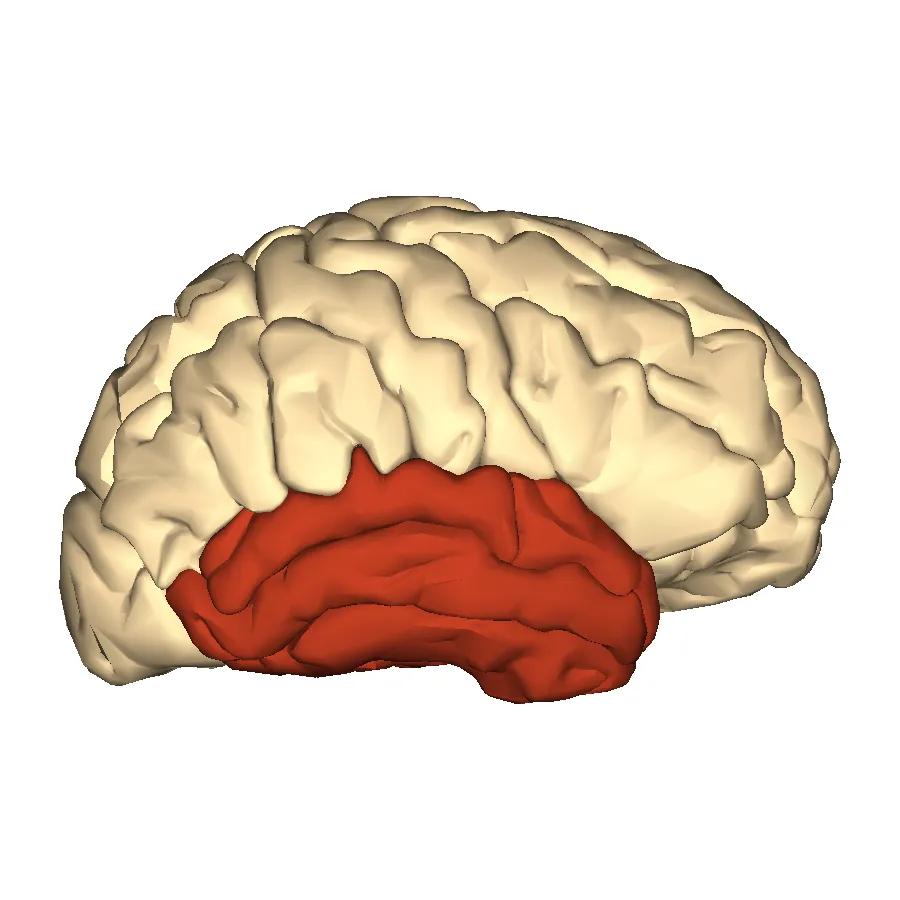
Lies inferior to the lateral sulcus. The transverse temporal gyri (Heschl) contain the primary auditory cortex (Brodmann areas 41, 42); its medial surface is closely related to the hippocampus and the amygdala.
Functions
- Hearing
- Language comprehension (Wernicke’s area, posterior superior temporal gyrus)
- Declarative memory
- Object and face recognition (fusiform gyrus)
Description
Lies inferior to the lateral sulcus. The transverse temporal gyri (Heschl) contain the primary auditory cortex (Brodmann areas 41, 42); its medial surface is closely related to the hippocampus and the amygdala.
Key structures
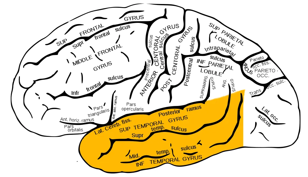
Gyri temporales transversi
- Name
- Transverse temporal gyri (Heschl)
- Role, notes
- Primary auditory cortex (Brodmann 41, 42)
Gyrus temporalis superior
- Name
- Superior temporal gyrus
- Role, notes
- Posterior part: Wernicke’s area, speech comprehension
Gyrus temporalis medius, inferior
- Name
- Middle and inferior temporal gyri
- Role, notes
- Semantics, object recognition (ventral visual stream)
Gyrus fusiformis
- Name
- Fusiform gyrus
- Role, notes
- Face and word-form recognition
Planum temporale
- Name
- Planum temporale
- Role, notes
- Usually larger on the left, linked to language
| Terminologia Anatomica | Name | Role, notes |
|---|---|---|
| Gyri temporales transversi | Transverse temporal gyri (Heschl) | Primary auditory cortex (Brodmann 41, 42) |
| Gyrus temporalis superior | Superior temporal gyrus | Posterior part: Wernicke’s area, speech comprehension |
| Gyrus temporalis medius, inferior | Middle and inferior temporal gyri | Semantics, object recognition (ventral visual stream) |
| Gyrus fusiformis | Fusiform gyrus | Face and word-form recognition |
| Planum temporale | Planum temporale | Usually larger on the left, linked to language |
Boundaries and structure
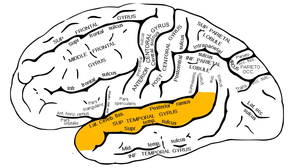
The temporal lobe lies below the lateral sulcus. It comprises the superior, middle and inferior temporal gyri laterally, the transverse temporal gyri (Heschl) on the floor of the lateral sulcus and, inferiorly, the lateral occipitotemporal (fusiform) and parahippocampal gyri. The perirhinal cortex sits at the boundary between the medial temporal lobe and the ventral visual pathway.[1][2]
Connections and function
Heschl’s gyrus is the primary auditory cortex; the pathway from the cochlea runs through the cochlear nuclei, superior olivary complex, inferior colliculus and medial geniculate body, splitting into a sound-localisation stream and a pattern-recognition stream. The fusiform gyrus is specialised for faces, objects and written words; the perirhinal cortex supports familiarity memory for objects.[3][4][2]
Functions
- 01Hearing
- 02Language comprehension (Wernicke’s area, posterior superior temporal gyrus)
- 03Declarative memory
- 04Object and face recognition (fusiform gyrus)
Regions
References
Sources for the values and descriptions on this page, indexed in PubMed (US National Library of Medicine).
- [1]Anatomy of the temporal lobe: From macro to microZachlod D et al., Handbook of clinical neurology 2022PMID 35964970
- [2]The perirhinal cortexSuzuki WA et al., Annual review of neuroscience 2014PMID 25032492
- [3]Auditory pathways: anatomy and physiologyPickles JO, Handbook of clinical neurology 2015PMID 25726260
- [4]The anatomical and functional specialization of the fusiform gyrusWeiner KS et al., Neuropsychologia 2016PMID 26119921
- [5]Automatic parcellation of human cortical gyri and sulci using standard anatomical nomenclatureDestrieux C et al., 2010PMID 20547229Supports: Nomenclature of cortical gyri and sulci
- [6]Neuroanatomy, Cerebral HemisphereStatPearls (NCBI Bookshelf)PMID 31747196
- [7]Neuroanatomy, CerebellumStatPearls (NCBI Bookshelf)PMID 30844194
- [8]Current Controversies on Wernicke's Area and its Role in LanguagePMID 28656532Supports: Wernicke’s area
- [9]The human brain in numbers: a linearly scaled-up primate brainHerculano-Houzel S, 2009PMID 19915731