Insula
Model
Anatomical regions
Lobus insularis
Insula
Lies deep within the lateral sulcus, covered by the frontal, parietal and temporal opercula and bounded by the circular sulcus of the insula.
Functions
- Interoception
- Gustation
- Emotional processing and empathy
Description
Lies deep within the lateral sulcus, covered by the frontal, parietal and temporal opercula and bounded by the circular sulcus of the insula.
Key structures
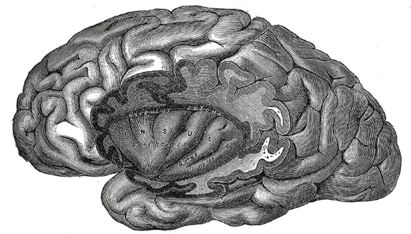
Gyri breves insulae
- Name
- Short insular gyri
- Role, notes
- Anterior insula: interoception, salience network
Gyrus longus insulae
- Name
- Long insular gyrus
- Role, notes
- Posterior insula: pain, temperature, visceral sensation
Sulcus circularis insulae
- Name
- Circular sulcus of insula
- Role, notes
- Boundary of the insula with the opercula
Limen insulae
- Name
- Limen insulae
- Role, notes
- Surgical landmark where the middle cerebral artery enters the insular cistern
| Terminologia Anatomica | Name | Role, notes |
|---|---|---|
| Gyri breves insulae | Short insular gyri | Anterior insula: interoception, salience network |
| Gyrus longus insulae | Long insular gyrus | Posterior insula: pain, temperature, visceral sensation |
| Sulcus circularis insulae | Circular sulcus of insula | Boundary of the insula with the opercula |
| Limen insulae | Limen insulae | Surgical landmark where the middle cerebral artery enters the insular cistern |
Boundaries and structure
The insula lies deep in the floor of the lateral sulcus, hidden by the frontal, parietal and temporal opercula; it is outlined by the circular sulcus and divided by the central insular sulcus into an anterior part (short gyri) and a posterior part (long gyri).
Connections and function
The insula is central to taste, interoception (awareness of the body’s internal state), emotion and decision-making; its position and wide connectivity make it a bridge between internal and external experience. It also regulates the autonomic nervous system with left–right asymmetry: right or left insular damage produces different cardiovascular changes after stroke.[1][2]
Functions
- 01Interoception
- 02Gustation
- 03Emotional processing and empathy
Regions
References
Sources for the values and descriptions on this page, indexed in PubMed (US National Library of Medicine).
- [1]The Insular Cortex: An Interface Between Sensation, Emotion and CognitionZhang R et al., Neuroscience bulletin 2024PMID 38722464
- [2]The insular cortex, autonomic asymmetry and cardiovascular control: looking at the right side of strokeFontes MAP et al., Clinical autonomic research 2024PMID 39316247
- [3]Automatic parcellation of human cortical gyri and sulci using standard anatomical nomenclatureDestrieux C et al., 2010PMID 20547229Supports: Nomenclature of cortical gyri and sulci
- [4]Neuroanatomy, Cerebral HemisphereStatPearls (NCBI Bookshelf)PMID 31747196
- [5]Neuroanatomy, CerebellumStatPearls (NCBI Bookshelf)PMID 30844194
- [6]Current Controversies on Wernicke's Area and its Role in LanguagePMID 28656532Supports: Wernicke’s area
- [7]The human brain in numbers: a linearly scaled-up primate brainHerculano-Houzel S, 2009PMID 19915731