Integumentary system
Model
Z-Anatomy anatomical model based on BodyParts3D, licensed CC BY-SA 4.0.
Overview
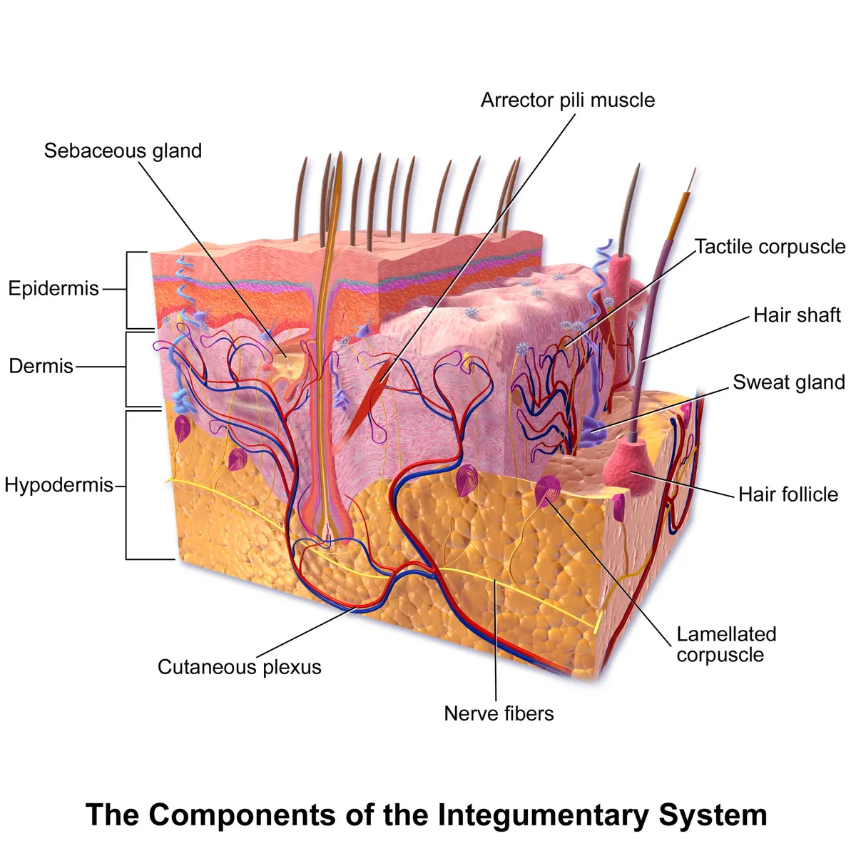
The integumentary system comprises the skin (epidermis and dermis), the subcutaneous tissue and the skin appendages: hair, nails, sweat and sebaceous glands, and the breast, a modified sweat gland. The skin is the largest organ, about 1.5–2 m² in adults, forming the boundary between the body and its environment.
Functions
- 01Mechanical, chemical, immune and water-loss barrier
- 02Thermoregulation through skin blood flow and sweating
- 03Touch, pressure, temperature and pain sensation; vitamin D synthesis
The barrier
The skin covers the whole body surface and is the barrier that keeps out pathogens, withstands chemical and physical insults and prevents uncontrolled loss of water and solutes. The physical barrier lies mainly in the stratum corneum, built like “bricks and mortar”: the bricks are protein-rich corneocytes wrapped in a cornified envelope and joined by corneodesmosomes; the mortar is the extracellular lipid lamellae.[1]
As keratinocytes differentiate they synthesise lipids and extrude them into the intercellular space. Ceramides A and B bind covalently to cornified-envelope proteins and form the scaffold on which free ceramides, free fatty acids and cholesterol assemble. Filaggrin is cross-linked to the envelope and aggregates keratin filaments into macrofibrils. The nucleated epidermis contributes through tight, gap and adherens junctions and desmosomes. The barrier is regulated by cytokines, cyclic AMP and calcium; when differentiation or lipid composition is disturbed, allergens penetrate and trigger inflammation, as in atopic dermatitis.[1]
The upper stratum corneum carries an “acid mantle”, a buffer system that keeps the skin surface acidic. This acidity stabilises the stratum corneum, shapes the microbiome and restrains inflammation; shifts in pH impair the barrier and contribute to many skin diseases, making pH a therapeutic target.[2]
The skin both excludes pathogens and hosts commensal bacteria and fungi. Sequencing shows a diverse, site-selective microbiome; some commensal strains tune the immune system, provide colonisation resistance and reinforce the epidermal barrier. Microbial shifts accompany common disorders such as atopic dermatitis.[3]
Appendages and innervation
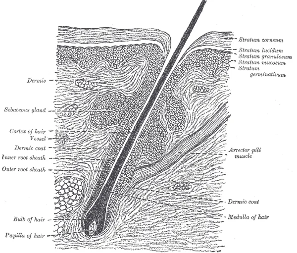
The hair follicle is a dynamic miniorgan: it forms in fetal skin through ectodermal–mesodermal interactions, anchors in the subcutis after birth and regenerates in cycles of growth (anagen), apoptosis-driven regression (catagen) and relative quiescence (telogen). The hair shaft is made of terminally differentiated keratinocytes; hair serves thermoregulation, protection, sensation and social signalling.[4]
Hair follicle stem cells sit in the bulge and are governed by a niche made of the dermal papilla, adipose tissue, vessels, nerves and immune cells, together with systemic signals such as hormones. The balance of activating and inhibitory cues decides when a follicle leaves quiescence and re-enters growth.[5]
All layers and appendages of the skin are densely innervated: afferent fibres carry touch, pain, temperature and itch, and efferent sympathetic fibres control skin perfusion and sweating. Sensory neurons and skin cells also signal in both directions, with trophic and immunomodulatory roles in disease.[6]
Ageing and wound healing
Aged skin shows wrinkles, laxity and pigmentary irregularity, driven by intrinsic factors (time, genetics) and extrinsic ones, of which sun exposure is the most harmful (photoageing). In the dermis, senescent fibroblasts accumulate because of macromolecular damage and inadequate clearance by NK cells; they release pro-inflammatory, matrix-degrading factors (the senescence-associated secretory phenotype, SASP) that spread ageing to their niche.[7][8]
Wound healing is a dynamic process (haemostasis, inflammation, proliferation, remodelling). Minimal scarring requires a clean, moist wound with little trauma or infection, and systemically avoiding drugs that impair healing, adequate nutrition, analgesia, UV protection and smoking cessation. In adults, skin injury always ends in a fibrotic, non-functional scar; heterogeneity among fibroblast populations is a key determinant of that scarring fate.[9][10]
The five layers of the epidermis
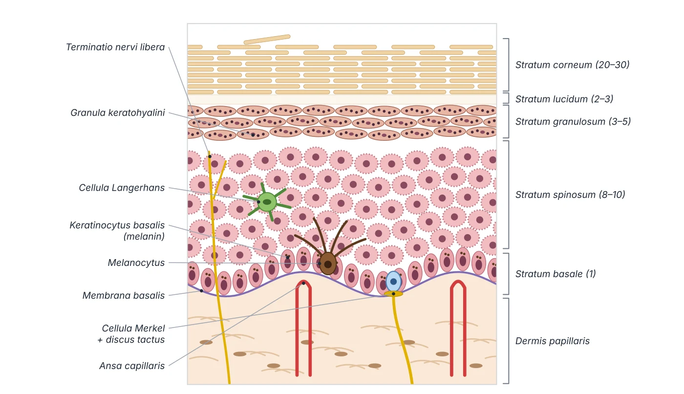
The epidermis is a keratinised stratified squamous epithelium with four layers in thin skin and five in the thick skin of the palms and soles. The stratum basale (germinativum) is a single row of cuboidal to low columnar cells anchored to the basement membrane by hemidesmosomes; it is the stem-cell layer that divides continuously to produce keratinocytes and contains melanocytes. The stratum spinosum has 8–10 rows of polyhedral cells joined by desmosomes, which look like spines in sections; dendritic (Langerhans) cells lie here. The stratum granulosum has 3–5 rows of diamond-shaped cells with keratohyalin granules (keratin precursors that aggregate into bundles) and lamellar granules that secrete glycolipids into the intercellular space as a “glue”; cells begin to lose their nuclei as they move away from nutrients. The stratum lucidum, 2–3 clear rows containing eleidin, exists only in thick skin. The stratum corneum has 20–30 rows of dead anucleate cells in a basket-weave pattern, holding keratin and lipids that prevent water loss and releasing defensins; it is the most variable in thickness, especially in calluses.[11][12][13]
Four main cell types live in the epidermis. Keratinocytes predominate, arise in the basal layer and make the keratin and lipids of the water barrier. Melanocytes, of neural crest origin, lie in the basal layer and hair follicles, synthesise melanin from tyrosine and pass it to keratinocytes in melanosomes to shield DNA from UV; differences in skin colour reflect the amount, size and density of melanin, not the number of melanocytes. Langerhans cells are antigen-presenting dendritic cells derived from bone marrow monocytes, recognised by electron microscopy by their tennis-racket Birbeck granules; both Langerhans cells and melanocytes are S-100 positive. Merkel cells are touch receptors at the base of the epidermis. The epidermis comes from surface ectoderm, while the dermis and hypodermis come from mesoderm. In sections, parakeratosis (retained nuclei in the stratum corneum) signals increased turnover as in psoriasis, while hyperkeratosis is seen in eczema, warts and corns; basal cell carcinoma, squamous cell carcinoma and melanoma are the three commonest skin cancers, all linked to UV light.[12][11][13]
Stratum basale
- Rows of cells
- 1
- Hallmark
- Dividing stem cells, melanocytes, hemidesmosomes
Stratum spinosum
- Rows of cells
- 8–10
- Hallmark
- Desmosomes (“spines”), Langerhans cells
Stratum granulosum
- Rows of cells
- 3–5
- Hallmark
- Keratohyalin and lamellar granules, nuclei begin to disappear
Stratum lucidum
- Rows of cells
- 2–3
- Hallmark
- Eleidin; thick skin only
Stratum corneum
- Rows of cells
- 20–30
- Hallmark
- Dead anucleate cells, keratin, lipids, defensins
| Layer (deep to superficial) | Rows of cells | Hallmark |
|---|---|---|
| Stratum basale | 1 | Dividing stem cells, melanocytes, hemidesmosomes |
| Stratum spinosum | 8–10 | Desmosomes (“spines”), Langerhans cells |
| Stratum granulosum | 3–5 | Keratohyalin and lamellar granules, nuclei begin to disappear |
| Stratum lucidum | 2–3 | Eleidin; thick skin only |
| Stratum corneum | 20–30 | Dead anucleate cells, keratin, lipids, defensins |
The dermis, vascular plexuses and thermoregulation
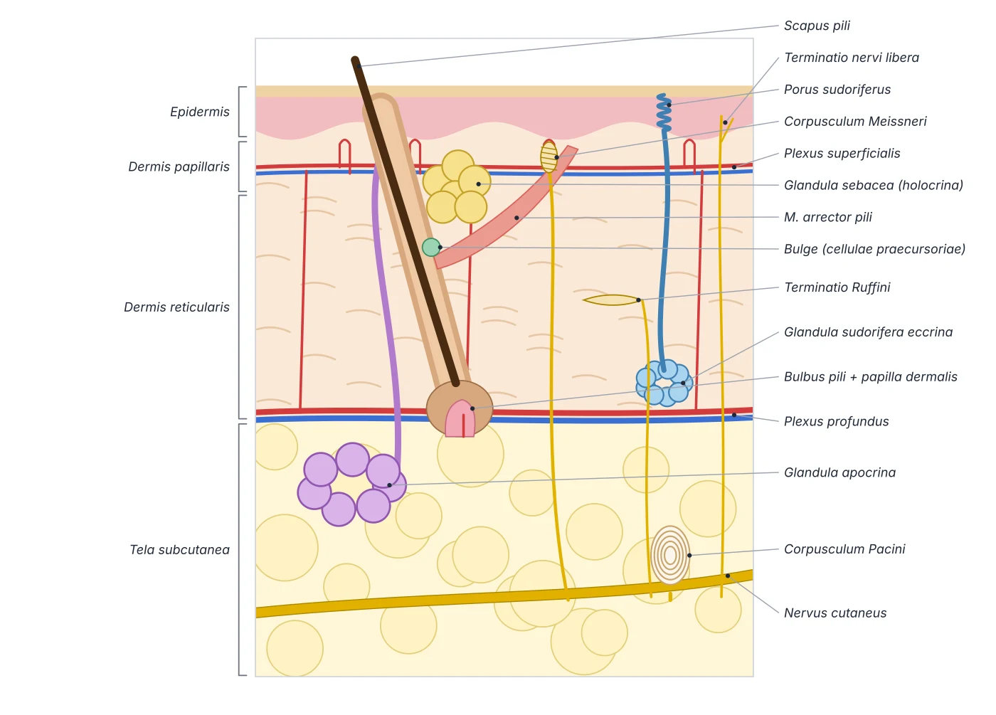
The dermis is thick connective tissue of collagen (for strength) and elastin (for elasticity), containing nerve endings, blood vessels, lymphatics and the skin appendages. The upper papillary dermis folds into small finger-like papillae that interlock with the epidermis; the lower reticular dermis is thicker and denser. Deepest is the hypodermis, mainly adipose tissue that stores lipid. Two vascular plexuses run parallel to the surface: a superficial one (beneath the epidermis, between papillary and reticular dermis) and a deep one (between dermis and hypodermis), joined by perpendicular capillaries that envelop the sweat glands and hair follicles; the epidermis is avascular and is fed from the superficial plexus. Blind-ended lymphatic capillaries begin in the dermal papillae and drain into deep lymphatic plexuses running alongside the blood vessels.[12][11][14]
Skin blood flow is the main tool of thermoregulation and is controlled by the autonomic nervous system. When body temperature rises, sympathetic centres in the posterior hypothalamus are inhibited, skin vessels dilate and heat passes to the environment; in the cold, sympathetic stimulation constricts them to retain heat. Skin thickness varies with region, age, sex and health: it is thickest on the palms and soles (which have a stratum lucidum), and among thin-skinned regions the back is thickest; it is thinnest on the eyelids, axillae and genitals. Because of androgens, men usually have thicker skin than women. Children have thin skin that thickens until the fourth decade and thins again from the fifth, mainly because the dermis loses appendages, elastic fibres and ground substance; occupational UV exposure causes premature ageing. The skin also initiates vitamin D production, needed for calcium absorption and bone metabolism.[11][13]
Sweat glands, sebaceous glands and the pilosebaceous unit
The skin appendages grow down from the epidermis from the third month of fetal life. The pilosebaceous unit comprises the hair shaft, hair follicle, sebaceous gland and arrector pili muscle; it is present almost everywhere except the lips, palms and soles, and is densest on the scalp. The follicle has three segments: the infundibulum (from the follicular opening to the entry of the sebaceous duct), the isthmus (down to the insertion of arrector pili, where the bulge holding stem cells lies) and the growing inferior segment, which expands into the bulb that cups a vascular dermal papilla; matrix cells in the bulb lengthen the hair. The hair cycle has growth (anagen), regression (catagen) and rest (telogen) phases. The shaft has a cuticle, a keratin cortex and, in thick hairs, a medulla. Sebaceous glands secrete sebum holocrinely: cells form at the basal layer, are pushed to the centre, accumulate fat and burst; sebum lubricates the skin, reduces friction and restrains bacteria and fungi. Sebaceous overactivity together with Cutibacterium acnes causes acne.[15]
There are two kinds of sweat gland, both secreting in a merocrine way, by exocytosis without damage to the cell. Eccrine glands are ectodermal, forming on the palms and soles around the fourth month and over the rest of the body a month later, complete at birth; they open directly onto the skin and cool the body by evaporation. The initial fluid contains water, sodium and potassium, but the duct reabsorbs most of the ions. They are supplied by sympathetic fibres that are mainly cholinergic: emotional (cortical) sweating affects the palms and soles, gustatory (medullary) sweating from spicy food the face, and thermal (hypothalamic) sweating the whole body. Apocrine glands arise from the follicular infundibulum around the fourth month, are present at birth but active only from puberty, and open into hair follicles in the axillae and genital region; their secretion is milky and odourless until skin bacteria break it down, and they respond to adrenaline and noradrenaline. Primary focal hyperhidrosis of the palms, soles and axillae is treated first with aluminium chloride, then botulinum toxin, and with endoscopic thoracic sympathectomy when refractory; widespread hypohidrosis or anhidrosis carries a risk of heat stroke.[14][15]
Mode of secretion
- Eccrine gland
- Merocrine
- Apocrine gland
- Merocrine
- Sebaceous gland
- Holocrine
Opens onto
- Eccrine gland
- Skin surface
- Apocrine gland
- Hair follicle
- Sebaceous gland
- Hair follicle
Distribution
- Eccrine gland
- Whole body, densest on palms and soles
- Apocrine gland
- Axillae, genital region
- Sebaceous gland
- Wherever there is hair, most on scalp and face
Active from
- Eccrine gland
- Birth
- Apocrine gland
- Puberty
- Sebaceous gland
- Puberty (marked increase)
Function
- Eccrine gland
- Thermoregulation, excretion, acid mantle
- Apocrine gland
- Body odour, possibly pheromones
- Sebaceous gland
- Lubricating, antimicrobial sebum
| Feature | Eccrine gland | Apocrine gland | Sebaceous gland |
|---|---|---|---|
| Mode of secretion | Merocrine | Merocrine | Holocrine |
| Opens onto | Skin surface | Hair follicle | Hair follicle |
| Distribution | Whole body, densest on palms and soles | Axillae, genital region | Wherever there is hair, most on scalp and face |
| Active from | Birth | Puberty | Puberty (marked increase) |
| Function | Thermoregulation, excretion, acid mantle | Body odour, possibly pheromones | Lubricating, antimicrobial sebum |
Sensory receptors and dermatomes
The skin has both somatic and autonomic nerves. Autonomic fibres control vascular tone, the arrector pili muscles and sweating. Somatic fibres carry pain, temperature, crude and discriminative touch, vibration, pressure and proprioception through specialised receptors. Free nerve endings enter the epidermis, most numerous in the stratum granulosum and around hair follicles, and sense pain, heat and cold. Merkel discs at the stratum basale sense sustained light touch. Meissner corpuscles in the dermal papillae sense light touch and low-frequency stimulation. Pacinian corpuscles lie deep and sense deep pressure and vibration. Ruffini endings sense deep pressure and stretch of collagen fibres.[11][13]
A dermatome is the area of skin whose sensation is supplied by one spinal nerve. There are 31 pairs of spinal nerves: 8 cervical, 12 thoracic, 5 lumbar, 5 sacral and 1 coccygeal; C1 usually has no dermatome. Nerves C1–C7 leave through the intervertebral foramen above the vertebra of the same number, C8 leaves between C7 and T1, and all the rest leave below their vertebra. Dermatomes on the trunk are arranged almost horizontally and on the limbs longitudinally; they overlap considerably, so maps are approximations. Some landmarks: T1 on the anteromedial forearm and arm, T2 on the medial arm up to the axilla, L4 over the front of the knee and medial malleolus, L5 on the dorsum of the foot and toes 1–3. Dermatomes help localise nerve lesions; sciatica, for example, radiates down the back of the lower limb. After chickenpox, varicella-zoster virus lies dormant in dorsal root ganglia; on reactivation (shingles), painful vesicles erupt in a single dermatome, most often on the trunk and then the face (herpes zoster ophthalmicus threatens sight).[16][13]
Free nerve endings
- Location
- Epidermis (stratum granulosum), around follicles
- Senses
- Pain, heat, cold
Merkel discs
- Location
- Stratum basale
- Senses
- Sustained light touch
Meissner corpuscles
- Location
- Dermal papillae
- Senses
- Light touch, low-frequency vibration
Pacinian corpuscles
- Location
- Deep dermis, hypodermis
- Senses
- Deep pressure, vibration
Ruffini endings
- Location
- Deep dermis
- Senses
- Pressure, skin stretch
| Receptor | Location | Senses |
|---|---|---|
| Free nerve endings | Epidermis (stratum granulosum), around follicles | Pain, heat, cold |
| Merkel discs | Stratum basale | Sustained light touch |
| Meissner corpuscles | Dermal papillae | Light touch, low-frequency vibration |
| Pacinian corpuscles | Deep dermis, hypodermis | Deep pressure, vibration |
| Ruffini endings | Deep dermis | Pressure, skin stretch |
Principal organs
The skin consists of the epidermis, a keratinised stratified squamous epithelium continuously renewed from its basal layer, and the dermis, connective tissue with collagen, elastic fibres, vessels, nerves and appendages. Thickness varies by region and is greatest on the palms and soles. Beneath lies the fat-rich subcutaneous tissue, which cushions and insulates.
Breast (mammary gland)Mamma (Glandula mammaria)An exocrine gland in the subcutaneous tissue of the anterior chest wall, usually from the second to the sixth rib, with an axillary tail extending towards the axilla. The glandular tissue has 15–20 lobes, each draining through a lactiferous duct with a sinus before opening on the nipple; suspensory (Cooper’s) ligaments tie the skin to the pectoral fascia. Lymph drains mainly to the axillary nodes.
Reference values
- The epidermis renews itself in about four weeks as keratinocytes travel from the basal layer to the stratum corneum and are shed.
References
Sources for the values and descriptions on this page, indexed in PubMed (US National Library of Medicine).
- [1]The skin: an indispensable barrierProksch E et al., Experimental dermatology 2008PMID 19043850
- [2]The Skin Acid Mantle: An Update on Skin pHBrooks SG et al., The Journal of investigative dermatology 2025PMID 39243251
- [3]Skin microbiome and dermatologic disordersScharschmidt TC et al., The Journal of clinical investigation 2025PMID 39895627
- [4]The hair follicle as a dynamic miniorganSchneider MR et al., Current biology 2009PMID 19211055
- [5]Local and systemic mechanisms that control the hair follicle stem cell nicheZhang B et al., Nature reviews. Molecular cell biology 2024PMID 37903969
- [6]Skin InnervationStänder S et al., The Journal of investigative dermatology 2024PMID 38402477
- [7]Aging skin: histology, physiology, and pathologyKhavkin J et al., Facial plastic surgery clinics of North America 2011PMID 21763983
- [8]Connective Tissue and Fibroblast Senescence in Skin AgingWlaschek M et al., The Journal of investigative dermatology 2021PMID 33563466
- [9]Skin: histology and physiology of wound healingGantwerker EA et al., Facial plastic surgery clinics of North America 2011PMID 21856533
- [10]Scarring Skin: Mechanisms and TherapiesLin X et al., International journal of molecular sciences 2024PMID 38338767
- [11]Anatomy, Skin (Integument), EpidermisYousef H et al., StatPearls 2024PMID 29262154
- [12]Histology, SkinAgarwal S et al., StatPearls 2023PMID 30726010
- [13]Anatomy, Skin (Integument)Lopez-Ojeda W et al., StatPearls 2022PMID 28723009
- [14]Anatomy, Skin, Sudoriferous GlandPatel BC et al., StatPearls 2023PMID 30020616
- [15]Histology, Skin AppendagesYousef H et al., StatPearls 2023PMID 29489291
- [16]Anatomy, Skin, DermatomesWhitman PA et al., StatPearls 2023PMID 30571022