Tongue
Model
Layers
Z-Anatomy anatomical model based on BodyParts3D, licensed CC BY-SA 4.0.
Description
A mass of striated muscle covered by mucosa on the floor of the mouth, with a body and a root separated by the V-shaped terminal sulcus. It takes part in chewing, swallowing and speech and carries the taste papillae.
Parts
Each part has its own card; press “Show on the model” to fly to it and select it in the 3D model above.
- 01
Apex, body and root
Apex, corpus et radix linguae
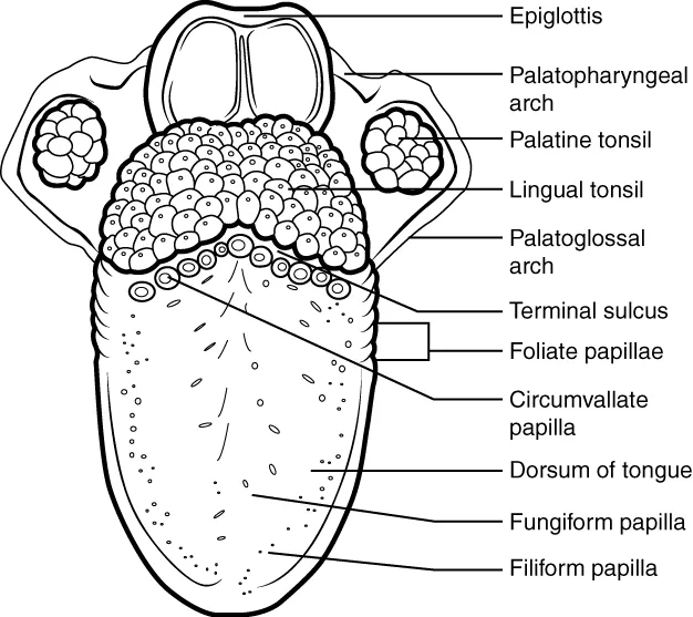
The V-shaped terminal sulcus, with the foramen caecum at its apex, divides the dorsum into an anterior two-thirds (oral part, from the first pharyngeal arch) and a posterior third (pharyngeal part, from the third and fourth arches). Their different embryological origins explain their different sensory nerves.
- 02
Lingual papillae
Papillae linguales
Filiform papillae (the most numerous, keratinised, without taste buds, giving roughness), fungiform papillae (scattered over tip and margins), vallate papillae (8–12 large ones in front of the terminal sulcus, richest in taste buds) and foliate papillae on the posterolateral margins.
- 03
Intrinsic muscles
Musculi intrinseci linguae
The superior and inferior longitudinal, transverse and vertical bundles lie wholly within the tongue without bony attachment; they change its shape (shortening, narrowing, flattening) for speech, chewing and swallowing. All are supplied by nerve XII.
- 04
Extrinsic muscles
Musculi extrinseci linguae
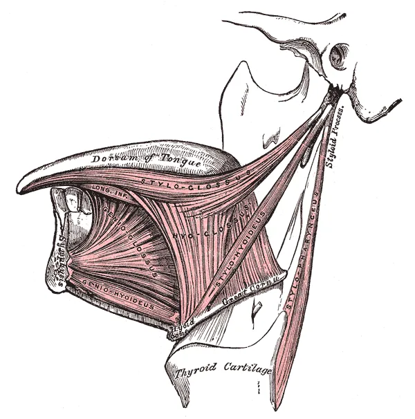
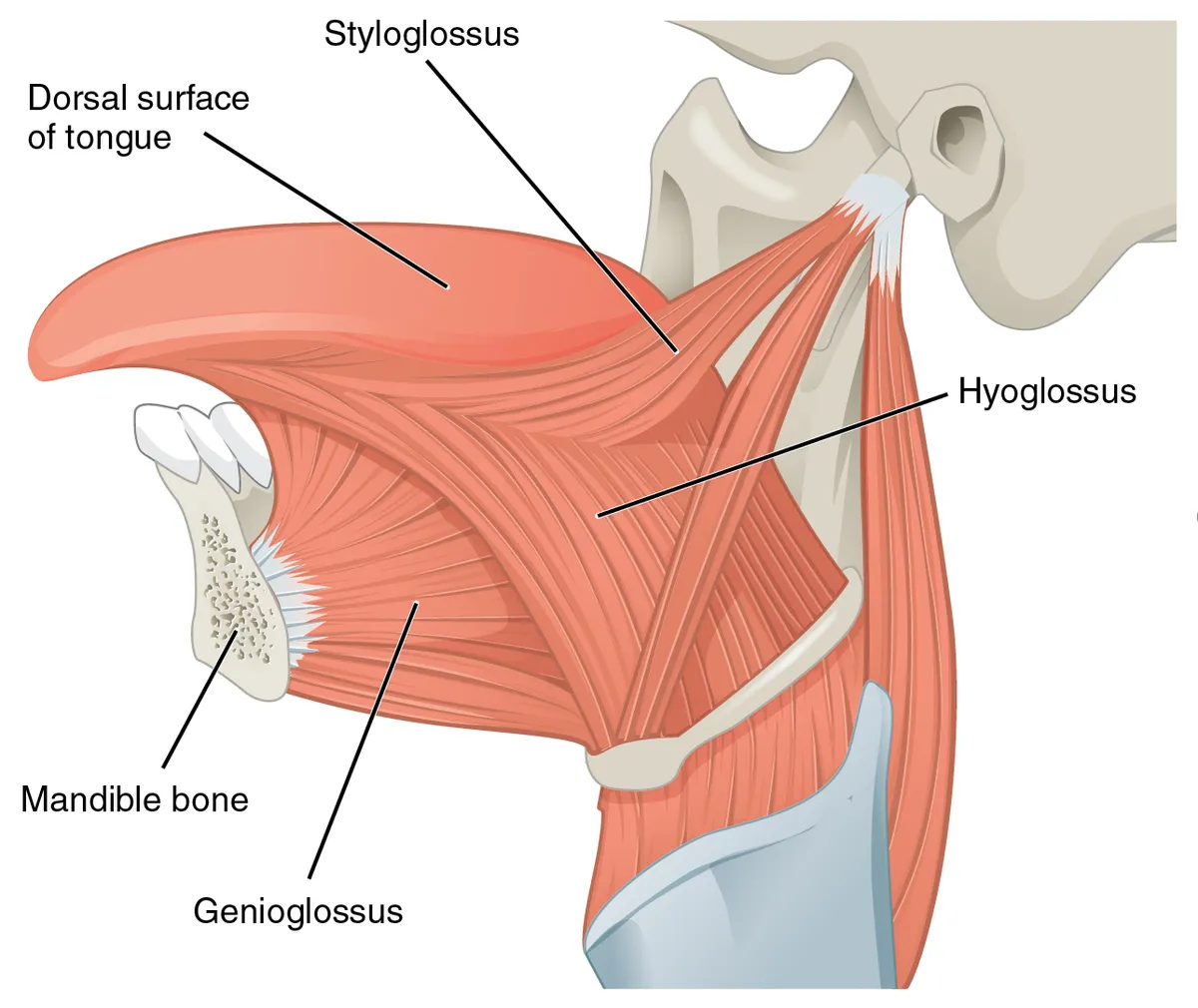
Four muscles from bone into the tongue that change its position: genioglossus (protrusion; keeps the airway open under anaesthesia), hyoglossus (depression), styloglossus (retraction and elevation) and palatoglossus (elevates the root; the only one supplied by the vagus).
- 05
Inferior surface and frenulum
Facies inferior linguae; Frenulum linguae
Covered by thin, smooth mucosa through which the deep lingual veins are visible, which is why sublingual drugs are absorbed rapidly. The frenulum joins it to the floor of the mouth, flanked by the sublingual caruncles where the submandibular ducts open; a short frenulum (tongue-tie) can restrict movement.
- 06
Lingual tonsil
Tonsilla lingualis
Lymphoid follicles beneath the mucosa of the posterior third of the dorsum, giving it a nodular surface; it forms the lower part of Waldeyer’s lymphoid ring around the entrance to the pharynx.
- 07
Nerve supply
Innervatio linguae
Motor: XII to all muscles except palatoglossus (X). Anterior two-thirds: general sensation by the lingual nerve (V3), taste by the chorda tympani (VII). Posterior third: both general sensation and taste by IX; the region near the valleculae by X.
Position and relations
The body (anterior two-thirds) lies in the oral cavity; the root (posterior third) faces the oropharynx and carries the lingual tonsil. The extrinsic muscles anchor the tongue to the hyoid, mandible and styloid process; the frenulum joins its underside to the floor of the mouth.
Form and structure
Intrinsic muscles (superior and inferior longitudinal, transverse, vertical) change its shape and extrinsic muscles (genioglossus, hyoglossus, styloglossus, palatoglossus) change its position, the halves divided by the lingual septum. The dorsum bears filiform, fungiform, foliate and vallate papillae (the vallate row lies in front of the terminal sulcus); taste buds sit mainly on fungiform, foliate and vallate papillae.
Vessels and nerves
Blood comes from the lingual artery (external carotid). The hypoglossal nerve (XII) supplies all tongue muscles except palatoglossus (vagus). General sensation of the anterior two-thirds is carried by the lingual nerve (V3) and taste by the chorda tympani (VII); the posterior third receives both from the glossopharyngeal nerve (IX), the area near the epiglottis from the vagus.
Muscles, papillae and taste
The tongue is a muscular organ with two groups of muscles: intrinsic muscles change its shape and extrinsic muscles move it in different directions. Through the extrinsic muscles it is attached to the hyoid bone, mandible, styloid process, soft palate and pharynx. The V-shaped sulcus terminalis divides it into an anterior two-thirds and a posterior third that differ in structure and in embryological origin. The foramen caecum at the apex of the sulcus marks where the thyroglossal duct began in the embryo. Besides chewing, swallowing and speech, the tongue also plays a role in breathing.[1]
Taste buds sit in the fungiform, foliate and circumvallate papillae; filiform papillae have none and serve only touch. There are five basic tastes. Sweet signals sugars and carbohydrates. Salty helps regulate sodium intake. Sour and bitter are protective, warning of spoiled food or harmful substances. Umami signals protein. Taste disorders can impair nutrition and may be early signs of systemic disease; testing taste helps separate peripheral from central causes.[2]
Each taste bud is a cluster of heterogeneous taste receptor cells. Taste information reaches the brain through gustatory neurons in the ganglia of cranial nerves VII and IX. Although taste perception is almost uninterrupted throughout life, all cells in a bud are replaced every one to two months, so diseases and treatments that disturb this renewal often cause taste dysfunction.[3][4]
The genioglossus is the main pharyngeal dilator that keeps the airway open during sleep. In obstructive sleep apnoea, reduced dilator tone lets the pharynx collapse repeatedly. Implanted stimulation of the hypoglossal nerve (XII) can prevent this collapse without waking the patient.[5]
Full-text reading (StatPearls)
The StatPearls chapters on this topic, reproduced verbatim. Their CC BY-NC-ND 4.0 licence allows unmodified copies only, so they are not translated or shortened.
Anatomy, Head and Neck, TongueDotiwala AK, Samra NS · StatPearls 2023 · NBK507782 · 18k characters
Loading…
Dotiwala AK, Samra NS. Anatomy, Head and Neck, Tongue. StatPearls [Internet]. Treasure Island (FL): StatPearls Publishing; 2023. NBK507782 · PMID 29939559 · CC BY-NC-ND 4.0. Reproduced verbatim, unmodified.
Anatomy, Head and Neck, Tongue Taste BudsGarg DR, Trillo G, Brizuela M · StatPearls 2025 · NBK539696 · 16k characters
Loading…
Garg DR, Trillo G, Brizuela M. Anatomy, Head and Neck, Tongue Taste Buds. StatPearls [Internet]. Treasure Island (FL): StatPearls Publishing; 2025. NBK539696 · PMID 30969518 · CC BY-NC-ND 4.0. Reproduced verbatim, unmodified.
Organs in this system
References
Sources for the values and descriptions on this page, indexed in PubMed (US National Library of Medicine).
- [1]Anatomy, Head and Neck, TongueDotiwala AK et al., StatPearls 2023PMID 29939559
- [2]Anatomy, Head and Neck, Tongue Taste BudsGarg DR et al., StatPearls 2025PMID 30969518
- [3]The sense of taste: Development, regeneration, and dysfunctionBarlow LA, WIREs mechanisms of disease 2022PMID 34850604
- [4]Taste buds: cells, signals and synapsesRoper SD et al., Nature reviews. Neuroscience 2017PMID 28655883
- [5]Electrical stimulation of the hypoglossal nerve: a potential therapySchwartz AR et al., Journal of applied physiology 2014PMID 23813525
- [6]Gastrointestinal mean transit times in young and middle-aged healthy subjectsPMID 11318834Supports: Gastrointestinal transit time
- [7]Revised Estimates for the Number of Human and Bacteria Cells in the BodySender R, Fuchs S, Milo R, 2016PMID 27541692Supports: Colonic bacterial count