Endocrine system
Model
Z-Anatomy anatomical model based on BodyParts3D, licensed CC BY-SA 4.0.
Overview
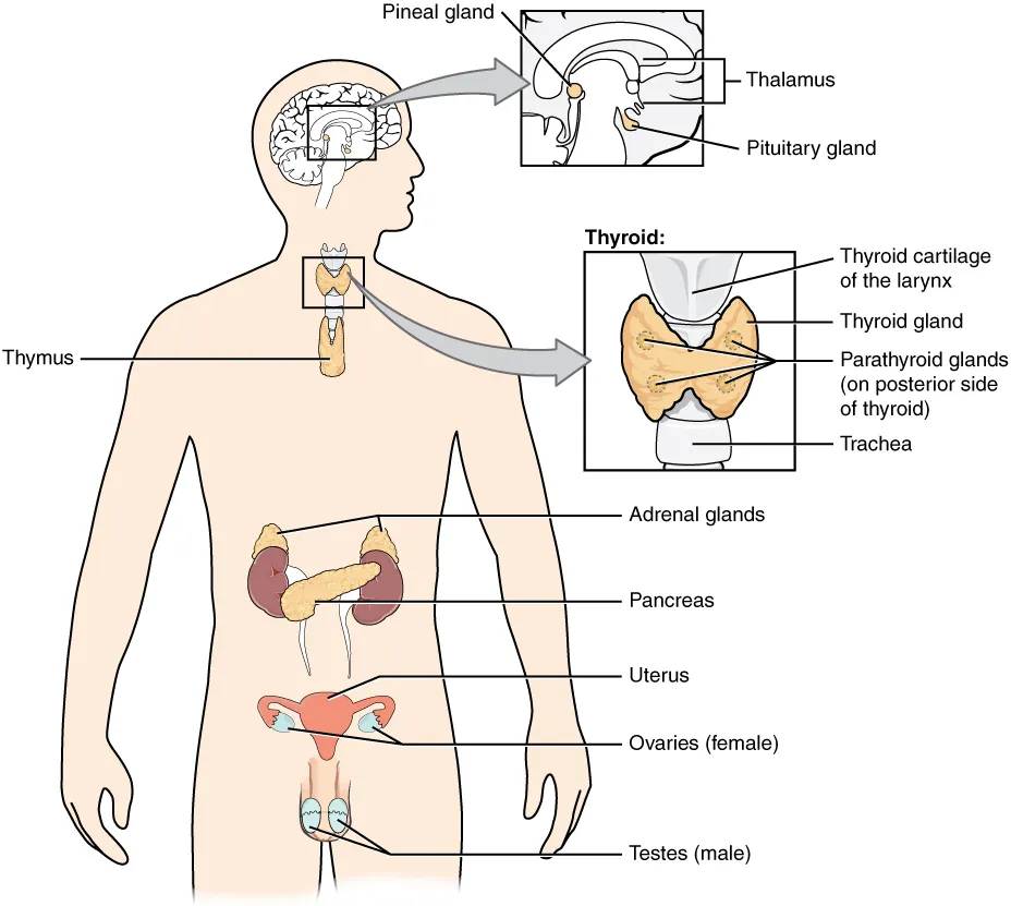
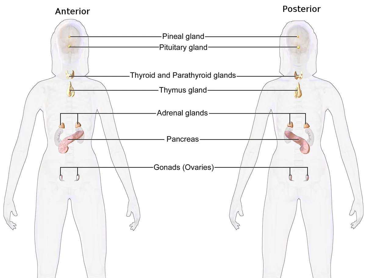
The endocrine system consists of ductless glands (pituitary, thyroid, parathyroids, adrenals, pancreatic islets, gonads, etc.) that secrete hormones directly into the bloodstream, regulating metabolism, growth, reproduction and the stress response.
Functions
- 01Regulation of energy metabolism
- 02Control of growth and development
- 03Regulation of reproductive function
- 04Homeostasis (glycaemia, calcaemia) and the stress response
Physiology
The thyroid traps iodide through the sodium/iodide symporter and synthesises thyroid hormone under the control of TSH acting on its receptor. Thyroid hormone acts on almost every organ; the fetus depends on maternal hormone until its own thyroid starts working in the second trimester, and the hormone is essential for growth and brain development in infancy.[1][2]
The pancreas has a dual function: its endocrine part (islets of Langerhans) secretes insulin and glucagon to regulate blood glucose, and its exocrine part secretes digestive enzymes. Both parts share an embryological origin and influence each other; in type 2 diabetes, β cells mainly lose their differentiated identity (dedifferentiation) rather than dying by apoptosis.[3][4]
The hypothalamo-pituitary axis
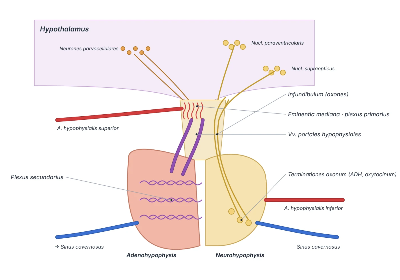
The adult pituitary weighs about 500 mg, measures 8 mm anteroposteriorly and 12 mm transversely, and almost doubles in size in pregnancy as the anterior lobe enlarges. It sits in the sella turcica of the sphenoid: its roof is the diaphragma sellae (a dural fold pierced by the stalk), its floor the roof of the sphenoid sinus, its sides the cavernous sinuses, with the optic chiasm anterosuperiorly. The stalk joins the gland to the tuber cinereum and median eminence of the hypothalamus. These relations explain why pituitary tumours can compress the chiasm or invade the cavernous sinus, and why surgery usually approaches through the sphenoid sinus.[5]
The pituitary has a dual origin. Rathke’s pouch, an upgrowth of oral ectoderm from week 4, forms the anterior lobe (adenohypophysis) and separates from the oral epithelium in weeks 6–8; a downgrowth of the ventral diencephalon forms the posterior lobe (neurohypophysis), an extension of the central nervous system. Transcription factors such as Prop-1, Pit-1 and Rpx, with BMP4, FGF8 and Notch signalling, direct this process; their mutations cause pituitary hypoplasia or malformation. The hypothalamus controls the anterior lobe through a portal system: the superior hypophyseal artery (from the internal carotid or posterior communicating artery) forms a primary capillary plexus in the median eminence, where hypothalamic neurons release their releasing hormones; portal hypophyseal veins carry them to a secondary plexus in the anterior lobe. The posterior lobe receives the inferior hypophyseal artery (from the meningohypophyseal trunk) and holds axon terminals that release ADH and oxytocin; the whole gland drains to the cavernous sinus.[5]
Embryological origins of the glands
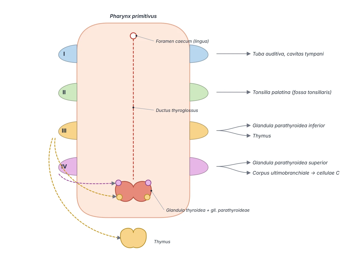
The thyroid parenchyma is endodermal, beginning at the foramen caecum at the junction of the anterior two-thirds and posterior third of the tongue. The bilobed diverticulum descends in front of the pharynx, the hyoid bone and the laryngeal cartilages to reach its final position in front of the upper trachea by week 7; the thyroglossal duct briefly links it to the tongue base and then involutes, remnants giving midline thyroglossal duct cysts. The ultimobranchial body (ventral part of the fourth pharyngeal pouch) fuses with the back of the gland and provides the parafollicular (C) cells that secrete calcitonin. The inferior parathyroids come from the third pharyngeal pouch and migrate further than the superior pair from the fourth pouch, which is why inferior glands are more often ectopic.[6][7]
The suprarenal gland is two tissues of different origin in one capsule. The cortex is mesodermal: in week 5 a cluster of cells appears in the urogenital ridge (the shared adrenogonadal primordium) and forms first the fetal and then the definitive cortex, under the control of SF-1 and β-catenin. The medulla comes from the neural crest: in week 7 neural crest cells migrate into the primordium from its medial side and differentiate into chromaffin cells arranged in cords, which the cortex then encloses. The parathyroids contain chief cells secreting parathyroid hormone (PTH) and larger oxyphil cells that increase with age; PTH is synthesised as a 115-amino-acid pre-prohormone, processed to a 90-amino-acid prohormone and finally to the active 84-amino-acid molecule.[8][7]
Anterior pituitary
- Origin
- Oral ectoderm (Rathke’s pouch)
- Main hormones
- GH, PRL, ACTH, TSH, FSH, LH
Posterior pituitary
- Origin
- Neuroectoderm (diencephalon)
- Main hormones
- ADH, oxytocin (made in the hypothalamus, stored here)
Thyroid (follicles)
- Origin
- Endoderm of the tongue base (foramen caecum)
- Main hormones
- T4, T3
Thyroid (C cells)
- Origin
- Ultimobranchial body, 4th pouch
- Main hormones
- Calcitonin
Inferior / superior parathyroids
- Origin
- 3rd / 4th pharyngeal pouch
- Main hormones
- PTH
Suprarenal cortex
- Origin
- Mesoderm (urogenital ridge)
- Main hormones
- Aldosterone, cortisol, androgens
Suprarenal medulla
- Origin
- Neural crest
- Main hormones
- Adrenaline, noradrenaline
| Gland (part) | Origin | Main hormones |
|---|---|---|
| Anterior pituitary | Oral ectoderm (Rathke’s pouch) | GH, PRL, ACTH, TSH, FSH, LH |
| Posterior pituitary | Neuroectoderm (diencephalon) | ADH, oxytocin (made in the hypothalamus, stored here) |
| Thyroid (follicles) | Endoderm of the tongue base (foramen caecum) | T4, T3 |
| Thyroid (C cells) | Ultimobranchial body, 4th pouch | Calcitonin |
| Inferior / superior parathyroids | 3rd / 4th pharyngeal pouch | PTH |
| Suprarenal cortex | Mesoderm (urogenital ridge) | Aldosterone, cortisol, androgens |
| Suprarenal medulla | Neural crest | Adrenaline, noradrenaline |
Suprarenal glands: form and blood supply
Each suprarenal gland is about 50 mm high, 30 mm wide and 10 mm thick and weighs about 5 g; it lies in the perirenal space within the upper renal fascia. The right gland is pyramidal, below the liver, behind the inferior vena cava and in front of the diaphragm; the left is crescentic, medial to the spleen, above the splenic vessels and lateral to the aorta. At birth the gland is about one-third the size of the kidney, in adults only one-thirtieth. The lipid-rich, yellow cortex makes up about 85% of the gland and has three zones: glomerulosa (aldosterone), fasciculata (cortisol) and reticularis (androgens); the reddish-brown medulla makes up about 15%.[8]
The gland has three arterial sources: the superior suprarenal arteries (small, often multiple, branches of the inferior phrenic artery), the middle suprarenal artery (directly from the aorta) and the inferior suprarenal artery (from the renal artery), with many variant origins. By contrast each gland has a single main vein: the right suprarenal vein is very short and drains straight into the inferior vena cava, so it is easily torn in right adrenalectomy; the left is longer, drains into the left renal vein and may communicate with the left gonadal and inferior phrenic veins. Blood flow through the gland is regulated by neuroendocrine and paracrine mechanisms, one way of adjusting circulating hormone levels.[8]
Principal organs
An H-shaped gland anterior to the trachea at the C5–T1 level, with two lobes joined by an isthmus. It secretes thyroxine (T4) and triiodothyronine (T3), which set the basal metabolic rate, as well as calcitonin.
Parathyroid glandsGlandulae parathyroideaeUsually four small glands (each about 30–50 mg) on the back of the thyroid that secrete parathyroid hormone (PTH), which regulates blood calcium and phosphate.
Suprarenal glandsGlandula suprarenalisPaired retroperitoneal glands capping the upper poles of the kidneys, pyramidal on the right and crescentic on the left. The cortex has three zones secreting aldosterone (glomerulosa), cortisol (fasciculata) and androgens (reticularis); the medulla secretes adrenaline and noradrenaline under sympathetic control.
Pituitary glandHypophysis (Glandula pituitaria)A small gland of about 0.5 g in the hypophysial fossa of the sphenoid, joined to the hypothalamus by the pituitary stalk. The anterior lobe (adenohypophysis) secretes GH, prolactin, ACTH, TSH, FSH and LH; the posterior lobe (neurohypophysis) releases ADH and oxytocin made by hypothalamic nuclei.
Pineal glandGlandula pinealisA small pine-cone-shaped gland about 8 mm long, attached to the roof of the third ventricle at the back of the diencephalon, between the superior colliculi. It secretes melatonin mainly at night, driven by light signals from the retina via the suprachiasmatic nucleus and the cervical sympathetic chain, helping to set circadian rhythm. It is often calcified in adults, serving as a midline landmark on imaging.
PancreasPancreasA mixed retroperitoneal gland. The exocrine part secretes enzyme-rich pancreatic juice; the islets of Langerhans (about 1–2% of pancreatic mass) secrete insulin, glucagon and somatostatin.
Reference values
- The pituitary weighs only about 0.5 g yet controls the thyroid, the adrenal cortex and the gonads.
References
Sources for the values and descriptions on this page, indexed in PubMed (US National Library of Medicine).
- [1]Thyroid physiologyStathatos N, The Medical clinics of North America 2012PMID 22443969
- [2]Thyroid Function During the Fetal and Neonatal PeriodsEng L et al., NeoReviews 2020PMID 31894080
- [3]Pancreatic endocrine and exocrine signaling and crosstalk in physiological and pathological statusHu C et al., Signal transduction and targeted therapy 2025PMID 39948335
- [4]Loss of β-cell identity and dedifferentiation, not an irreversible process?Patel S et al., Frontiers in endocrinology 2024PMID 38915897
- [5]Anatomy, Head and Neck, Pituitary GlandGanapathy MK et al., StatPearls 2023PMID 31855373
- [6]Anatomy, Head and Neck, ThyroidAllen E et al., StatPearls 2025PMID 29262169
- [7]Anatomy, Head and Neck, ParathyroidIlahi A et al., StatPearls 2023PMID 30725888
- [8]Anatomy, Abdomen and Pelvis: Adrenal Glands (Suprarenal Glands)Megha R et al., StatPearls 2022PMID 29489211
- [9]Physiology, Pituitary HormonesStatPearls (NCBI Bookshelf)PMID 32491488Supports: Pituitary and endocrine axes
- [10]Physiology, Anterior PituitaryStatPearls (NCBI Bookshelf)PMID 29763073