Uterus
Model
Hover or tap to see names, click to read a description; click a name in the legend to fly to it. Procedurally built to histological proportions; colours follow illustration conventions.
Description
A thick-walled, pear-shaped hollow organ in the lesser pelvis between the bladder and the rectum, usually anteverted and anteflexed; about 7–8 cm long in nulliparous women. It has a fundus, body and cervix, and its wall has three layers: endometrium, myometrium and perimetrium.
Parts
Each part has its own card; press “Show on the model” to fly to it and select it in the 3D model above.
- 01
Fundus
Fundus uteri
The rounded part above the entry of the uterine tubes (the cornua). Fundal height measured from the symphysis (in cm roughly equal to weeks of gestation from 20 to 36 weeks) is used to monitor pregnancy.
- 02
Body
Corpus uteri
The main part, flattened front to back, with a triangular cavity. Normally anteflexed on the cervix and anteverted on the vagina, it rests on the bladder. In a nulliparous woman it measures about 7.5 × 5 × 2.5 cm.
- 03
Isthmus
Isthmus uteri
A narrow segment about 1 cm long between body and cervix, at the internal os. In pregnancy it expands into the thin lower uterine segment, where the transverse incision for caesarean section is made.
- 04
Cervix
Cervix uteri
About 2.5 cm long, with supravaginal and vaginal parts (ectocervix). The ectocervix is covered by stratified squamous epithelium and the canal by mucus-secreting columnar epithelium; the transformation zone between them is where HPV-related cervical cancer arises and is the target of cytology screening.
- 05
Endometrium
Endometrium
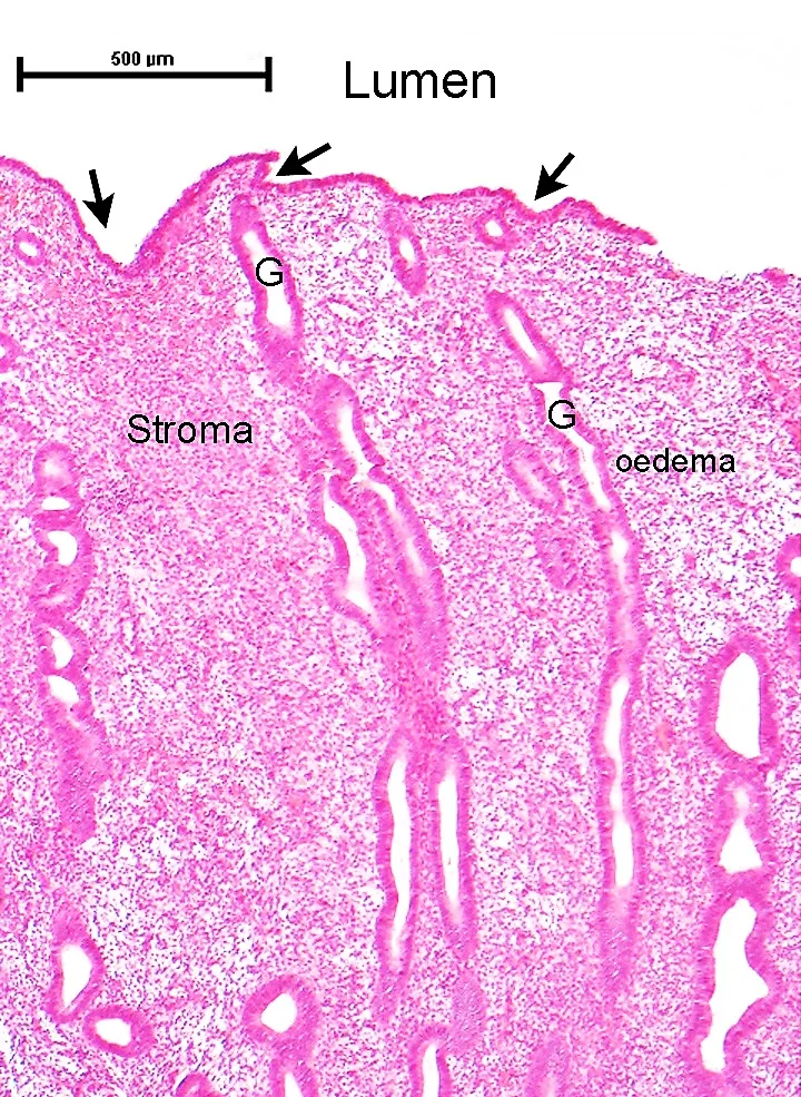
The mucosa, with a basal layer (persistent, regenerative) and a functional layer (shed at menstruation), changing through the cycle: proliferative under oestrogen, secretory under progesterone. Spasm of the spiral arteries when progesterone falls triggers menstruation; ectopic endometrium causes endometriosis.
- 06
Myometrium
Myometrium
The thickest layer, three interlacing layers of smooth muscle; the middle layer has figure-of-eight bundles round the vessels that clamp down after delivery of the placenta (“living ligatures”). Its cells hypertrophy and multiply greatly in pregnancy; fibroids (leiomyomas) are the commonest benign tumour in women.
- 07
Perimetrium and ligaments
Perimetrium; Ligamentum latum; Ligamentum teres uteri
The peritoneum covering the uterus extends laterally as the broad ligament, which holds the tube, ovary and uterine artery; the round ligament passes through the inguinal canal to the labium majus. The main supports are the cardinal (Mackenrodt) and uterosacral ligaments, whose weakening causes prolapse.
Position and relations
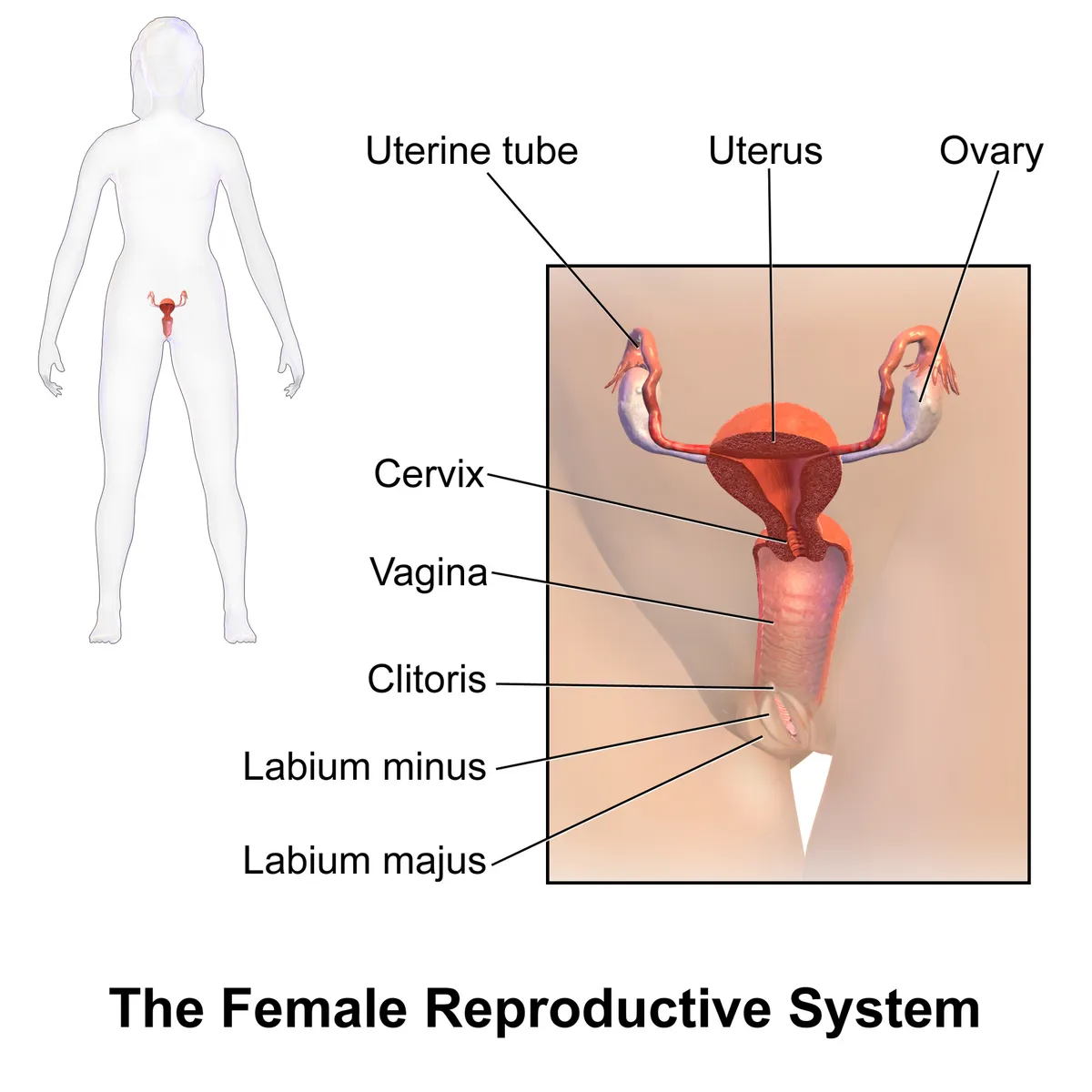
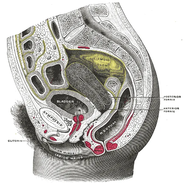
The uterus lies between the bladder in front and the rectum behind and is largely covered by peritoneum; the peritoneum reflected onto the bladder forms the vesico-uterine pouch, and onto the rectum the recto-uterine pouch (of Douglas), the lowest point of the female peritoneal cavity. On each side, the broad ligament encloses the uterine tube, ovary and uterine vessels.
Form and structure
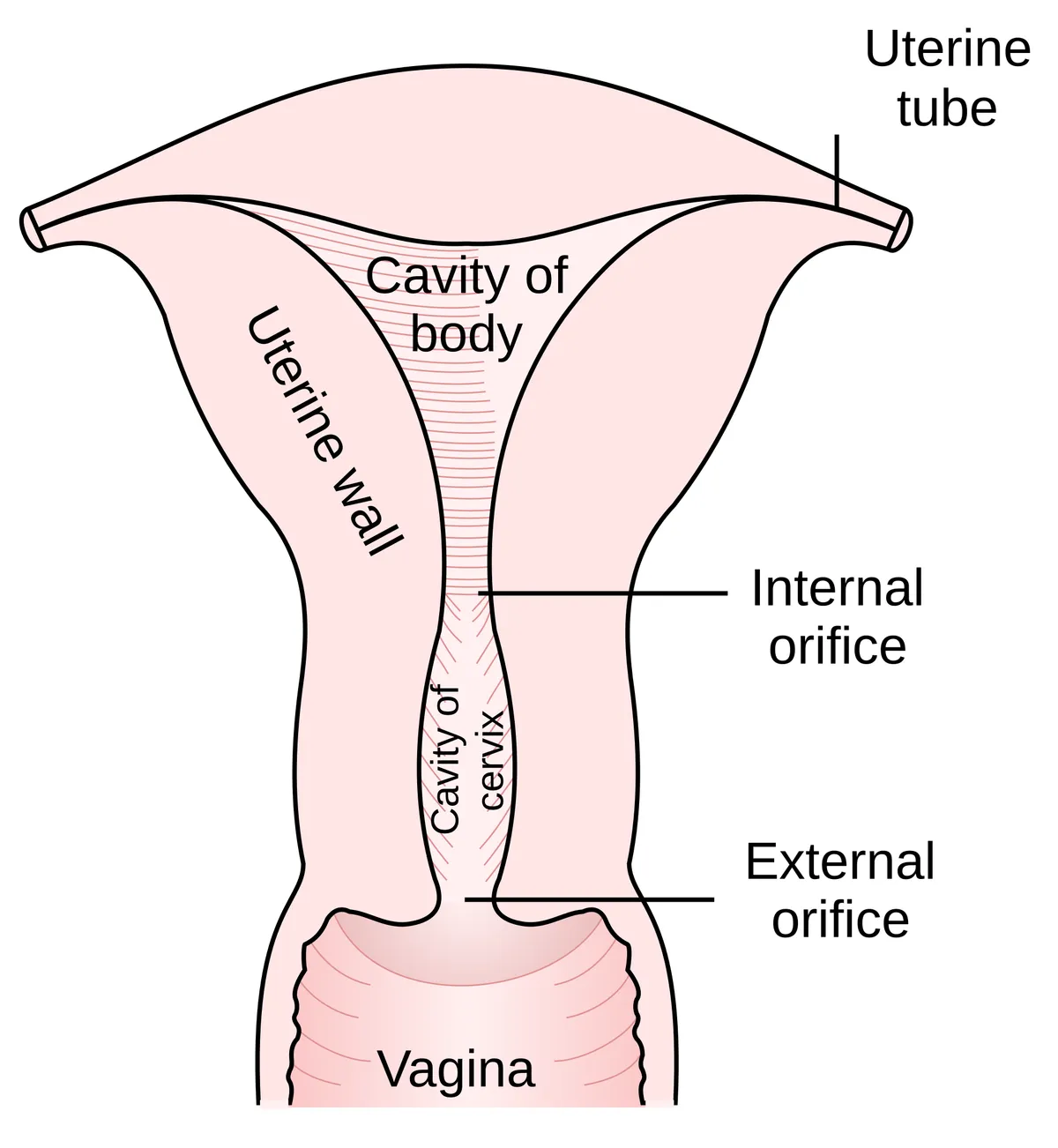
The uterus consists of the fundus (above the entry of the tubes), body, isthmus and cervix; the cervix has an internal os opening into the uterine cavity and an external os opening into the vagina. The wall comprises the endometrium (a functional layer shed at each menstruation and a regenerating basal layer), the myometrium, a thick smooth-muscle layer, and the serosa. Support comes mainly from the transverse cervical (cardinal) and uterosacral ligaments and the levator ani.
Vessels and nerves
The uterine artery (a branch of the internal iliac) runs in the base of the broad ligament and crosses above the ureter about 1.5–2 cm lateral to the cervix, a key landmark in hysterectomy for avoiding ureteric injury. It ascends along the lateral border of the uterus and anastomoses with the ovarian artery. Nerves come from the uterovaginal plexus, part of the inferior hypogastric plexus.
Physiology
The menstrual cycle is a coordinated sequence of events involving the hypothalamus, anterior pituitary, ovary and endometrium, and it is easily disturbed by stress, extreme exercise, eating disorders and obesity.[1]
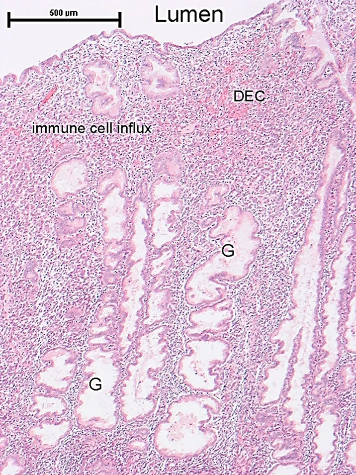
In humans, the endometrium begins to decidualise in the second half of every cycle, independently of an embryo. Adequate hormonal, biochemical and immunological decidualisation is the foundation for normal implantation and placentation. The endometrium regenerates repeatedly without scarring, but after some procedures or infections it may scar instead and cause infertility.[2][3]
Size, position, vessels and nerves
The uterus is a hollow, pear-shaped organ, on average 8 cm long, 5 cm wide and 4 cm thick, with a cavity of 80–200 mL that appears as an inverted triangle on coronal section. From above down it consists of the fundus (where the tubes enter), body, isthmus and cervix, which projects into the vagina. It is held by the utero-ovarian, round, broad, cardinal and uterosacral ligaments, together with the pelvic and urogenital diaphragms and the perineal body. The long axis of the cervix usually differs from that of the body: the body bends forwards over the cervix (anteflexion) or backwards (retroflexion), and the uterus tilts forwards relative to the vagina (anteversion, about 80%) or backwards (retroversion, about 20%). A retroverted or retroflexed uterus may cause pelvic pain, dyspareunia, mild incontinence and difficulty with tampons, may contribute to infertility and can rarely become incarcerated in pregnancy. The cervix-to-body ratio is 2:1 before puberty, reverses to 1:2 in the reproductive years and returns to 2:1 as the uterus atrophies after menopause; a nulliparous uterus is smaller than a multiparous one.[4]
The wall has three layers: the endometrium (a superficial functional layer that responds to hormones and is shed at menstruation, and a deep basal layer that regenerates; damage to the basal layer causes intrauterine adhesions and fibrosis, Asherman syndrome), the smooth-muscle myometrium that contracts in labour, and the serosa (perimetrium). The uterine artery, the main supply, divides in the myometrium into arcuate and then radial arteries, which on reaching the endometrium give basal and spiral arteries that sustain the endometrium through the cycle and the placental circulation in pregnancy. Lymph drains segmentally: the fundus to para-aortic nodes like the ovary and tube, some vessels along the round ligament to superficial inguinal nodes, and the lower segment along the uterine vessels to external and internal iliac nodes. Sympathetic supply comes from T11–T12 through the hypogastric plexus and parasympathetic from S2–S4. Uterus and cervix are sensitive to distension, and cervical dilatation contributes much of labour pain; uterine afferents run in the hypogastric nerves to T10–L1, so pain is referred to the lower back and abdomen, while the cervix signals pain through both hypogastric and pelvic nerves.[4]
Development and Müllerian anomalies
Around week 8 the primordia of both female and male internal genitalia, the paramesonephric (Müllerian) and mesonephric (Wolffian) ducts, are present. The paired Müllerian ducts run caudally to the urogenital sinus and project into its posterior wall as the Müllerian tubercle. Each duct has three segments: a first vertical part running parallel and lateral to the Wolffian duct, a horizontal part crossing in front of the ipsilateral Wolffian duct, and a second vertical part that fuses with its partner in the midline. The first two segments form the uterine tube; the fused third segment canalises to form the uterus, cervix and upper fifth of the vagina. In the female embryo, the absence of a Y chromosome and of testicular testosterone allows uninterrupted development of the Müllerian ducts, which fuse and canalise in the midline while the Wolffian ducts regress; disruption at this stage produces a spectrum of Müllerian anomalies.[4]
Clinically significant uterine anomalies markedly affect fertility and pregnancy, so early recognition and appropriate surgery help prevent miscarriage, preterm birth and infertility. A septate uterus is the commonest Müllerian anomaly, from incomplete resorption of the uterovaginal septum, leaving a single cavity divided by a fibrous or muscular septum; resecting the septum improves reproductive outcomes. A bicornuate uterus has a heart-shaped cavity from incomplete fusion of the ducts. Uterus didelphys is a rare anomaly from complete non-fusion, giving two cavities and two cervices, often with a longitudinal vaginal septum; many women have no symptoms, but the risk of miscarriage, preterm labour and breech presentation is higher. A unicornuate uterus results when one duct fails to develop, usually with a rudimentary horn that may or may not communicate with the main cavity; a non-communicating horn can cause dysmenorrhoea, haematometra and endometriosis. Segmental hypoplasia leaves a small uterus with prepubertal proportions, and fetal exposure to DES produces a T-shaped cavity.[4]
Septate uterus
- Mechanism
- Incomplete septal resorption
- Main consequences
- Miscarriage, preterm birth, infertility; septum resectable
Bicornuate uterus
- Mechanism
- Incomplete fusion
- Main consequences
- Recurrent miscarriage, preterm labour
Uterus didelphys
- Mechanism
- Complete non-fusion
- Main consequences
- Two cervices, vaginal septum; breech, preterm labour
Unicornuate uterus
- Mechanism
- One duct fails to develop
- Main consequences
- Rudimentary horn: dysmenorrhoea, haematometra
T-shaped uterus
- Mechanism
- DES exposure
- Main consequences
- Infertility, miscarriage, preterm labour
| Anomaly | Mechanism | Main consequences |
|---|---|---|
| Septate uterus | Incomplete septal resorption | Miscarriage, preterm birth, infertility; septum resectable |
| Bicornuate uterus | Incomplete fusion | Recurrent miscarriage, preterm labour |
| Uterus didelphys | Complete non-fusion | Two cervices, vaginal septum; breech, preterm labour |
| Unicornuate uterus | One duct fails to develop | Rudimentary horn: dysmenorrhoea, haematometra |
| T-shaped uterus | DES exposure | Infertility, miscarriage, preterm labour |
Common uterine disorders
Hysterectomy is the second commonest operation in women in the United States after caesarean section. An endometrial polyp is a localised overgrowth of endometrial glands and stroma projecting into the cavity; it may be silent or cause painless abnormal bleeding, small polyps can regress, progestins and GnRH agonists relieve symptoms, and removal is definitive. Leiomyoma (fibroid) is the commonest benign tumour in women, often multiple, oestrogen-sensitive, growing in pregnancy and shrinking after menopause, with a small risk of transformation to leiomyosarcoma; fibroids can cause abnormal bleeding, miscarriage and iron-deficiency anaemia, distort the uterus and compress the rectum causing constipation. Oral drugs are usually ineffective, so myomectomy or hysterectomy is the standard treatment. Endometrial hyperplasia from excessive oestrogen stimulation raises the risk of endometrial carcinoma and needs close follow-up.[4]
Adenomyosis is the extension of endometrial glands into the myometrium through hyperplasia of the basal layer; it causes dysmenorrhoea and menorrhagia with a uniformly enlarged, globular uterus on imaging, and is treated with NSAIDs and hormones (combined oestrogen–progestin pills, patches or vaginal rings), with hysterectomy when these fail. Endometritis usually follows retained products of conception after delivery, miscarriage or abortion, or a foreign body, when vaginal or intestinal bacteria colonise the retained material. Endometriosis is endometrial glands or stroma outside the uterine cavity, most often on the ovaries, pelvis and peritoneum, and can form a blood-filled ovarian endometrioma (“chocolate cyst”); retrograde menstruation is the favoured explanation. Endometrial cancer is the commonest gynaecological cancer in developed countries and is strongly linked to excess oestrogen, whether exogenous (tamoxifen, unopposed oestrogen therapy) or endogenous (polycystic ovary syndrome, obesity, granulosa-cell tumours); most patients present with abnormal vaginal bleeding, especially after menopause.[4]
Full-text reading (StatPearls)
The StatPearls chapters on this topic, reproduced verbatim. Their CC BY-NC-ND 4.0 licence allows unmodified copies only, so they are not translated or shortened.
Anatomy, Abdomen and Pelvis: UterusAmeer MA, Peterson DC · StatPearls 2025 · NBK470297 · 14k characters
Loading…
Ameer MA, Peterson DC. Anatomy, Abdomen and Pelvis: Uterus. StatPearls [Internet]. Treasure Island (FL): StatPearls Publishing; 2025. NBK470297 · PMID 29262069 · CC BY-NC-ND 4.0. Reproduced verbatim, unmodified.
Anatomy, Abdomen and Pelvis: Uterus Round LigamentChaudhry SR, Chaudhry K · StatPearls 2023 · NBK499970 · 8k characters
Loading…
Chaudhry SR, Chaudhry K. Anatomy, Abdomen and Pelvis: Uterus Round Ligament. StatPearls [Internet]. Treasure Island (FL): StatPearls Publishing; 2023. NBK499970 · PMID 29763145 · CC BY-NC-ND 4.0. Reproduced verbatim, unmodified.
Organs in this system
References
Sources for the values and descriptions on this page, indexed in PubMed (US National Library of Medicine).
- [1]The menstrual cycle: basic biologyHawkins SM et al., Annals of the New York Academy of Sciences 2008PMID 18574203
- [2]Endometrial Decidualization: The Primary Driver of Pregnancy HealthNg SW et al., International journal of molecular sciences 2020PMID 32521725
- [3]Mechanisms of Regeneration and Fibrosis in the EndometriumAng CJ et al., Annual review of cell and developmental biology 2023PMID 37843929
- [4]Anatomy, Abdomen and Pelvis: UterusAmeer MA et al., StatPearls 2025PMID 29262069