Head and neck
Model
Layers
Z-Anatomy anatomical model based on BodyParts3D, licensed CC BY-SA 4.0.
Overview
A regional model of the head and neck in nine layers, from skin to brain: facial and masticatory muscles, skull and cervical spine, the carotid and vertebral arterial systems, jugular veins, cranial nerves, brain and meninges, eye and ear, and the cervical viscera and glands (salivary glands, thyroid, larynx, lymph nodes). Toggle or fade each layer to dissect.
Boundaries and subdivisions
The head comprises the cranial vault with the scalp, the face and its cavities (orbits, nasal cavity, oral cavity), and the temporal and infratemporal fossae. The neck runs from the lower border of the mandible and the superior nuchal line down to the clavicle, the jugular notch and the C7 spinous process. Sternocleidomastoid divides each side of the neck into anterior and posterior triangles; the digastric and omohyoid further divide the anterior triangle into submandibular, submental, carotid and muscular triangles, and the inferior belly of omohyoid splits the posterior triangle into occipital and supraclavicular triangles. A useful bony landmark is the pterion, where the frontal, parietal, temporal and sphenoid bones meet just above the auricle.[1][2]
Layers from superficial to deep
The scalp has five layers: skin, a dense vascular connective tissue, the epicranial aponeurosis (galea) joining frontalis to occipitalis, a loose areolar layer (a “danger space” through which blood and pus spread freely), and the pericranium. In the face the muscles of facial expression lie within the subcutaneous tissue and are supplied by the facial nerve (VII); the muscles of mastication (masseter, temporalis, the two pterygoids) are supplied by the mandibular nerve (V3). In the neck, platysma is superficial, and beneath it the cervical fascia forms three layers (the investing layer around sternocleidomastoid and trapezius, the pretracheal layer around the viscera, the prevertebral layer around the deep muscles) and the carotid sheath. The parotid gland spreads from in front of the ear down to the angle of the jaw; the facial nerve passes through it and divides it into superficial and deep lobes.[3][4][5][6]
Vessels, nerves and lymphatics
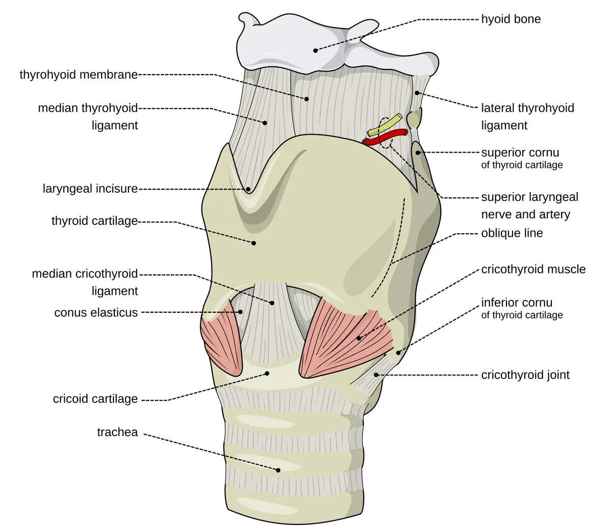
The common carotid artery runs in the carotid sheath with the internal jugular vein and the vagus nerve and divides at the level of the upper border of the thyroid cartilage; the external carotid gives branches to the thyroid, tongue, face and occiput and ends as the maxillary and superficial temporal arteries, whereas the internal carotid gives no branches in the neck. The maxillary artery gives off the middle meningeal artery, which enters the skull through the foramen spinosum. The thyroid gland overlies C5–T1, its isthmus crossing the second and third tracheal rings, and is supplied by the superior and inferior thyroid arteries. Lymph from the head and neck drains to cervical node chains (grouped as levels I–VI in oncology); the Virchow node belongs to left level IV, near the junction of the internal jugular and subclavian veins.[7][8][9][10]
Embryology
The face forms in weeks 4 to 6 from five swellings around the primitive mouth: the frontonasal prominence (forehead, nasal bridge and the medial and lateral nasal prominences) and the paired maxillary and mandibular prominences of the first pharyngeal arch. The medial nasal prominences give the primary palate, philtrum and four upper incisors; the lateral ones form the alae of the nose. The oropharyngeal membrane breaks down in week 4 to open the oral cavity. The first arch also gives rise to the mandible, the maxillary artery and the muscles of mastication, so the contents of the infratemporal fossa share a common origin. The maxilla, zygomatic bone and squamous temporal bone ossify in membrane from neural crest mesenchyme. The scalp epidermis comes from ectoderm and its deeper layers from mesoderm; the carotid sheath appears late, at about 20 weeks, as part of the deep cervical fascia.[11][2][5][6]
Anatomical variants
The arterial network of the scalp varies considerably: when the superficial temporal and occipital arteries are small, the posterior auricular artery enlarges to compensate and may anastomose directly with the supraorbital artery to supply the forehead. The occipital emissary vein may be absent, and the parietal emissary vein usually communicates with the frontal branch of the superficial temporal vein. The carotid sheath itself has few true variants, but it may lie in an unusual fascial plane when the common carotid artery takes an atypical course (for example, a right common carotid arising directly from the aortic arch). The extracranial course of the cranial nerves related to the sheath (IX, X, XI, XII) differs markedly between individuals, so dissection here calls for care and sometimes ultrasound or preoperative imaging.[5][12][6]
Clinical correlations
Blunt trauma to the temple at the pterion can tear the middle meningeal artery and cause an epidural haematoma. In thyroid surgery the recurrent and superior laryngeal nerves must be identified and monitored, because injury causes hoarseness or airway compromise. Parotid surgery requires preservation of the facial nerve branches that run through the gland. In the posterior triangle the accessory nerve (XI) lies very superficially and is easily injured during lymph node biopsy, causing trapezius weakness and a drooping shoulder. An enlarged, hard left supraclavicular node (Virchow node) suggests metastatic abdominal cancer.[1][13][3][10]
Surgical and procedural considerations
In carotid endarterectomy (indicated for internal carotid stenosis above roughly 70–80%), the surgeon retracts sternocleidomastoid and opens the carotid sheath: the artery lies medial, the internal jugular vein lateral and the vagus behind; the hypoglossal nerve crosses horizontally above the bifurcation and must be preserved. The loose areolar layer of the scalp separates easily and is the standard plane for craniofacial surgery and scalp flaps; because of the rich anastomoses, after a complete scalp avulsion, repairing a single artery can be enough to revascularise the replanted scalp. Surgery in the infratemporal fossa risks hearing loss, facial weakness, temporomandibular dysfunction and, above all, laceration of the internal carotid artery with torrential bleeding. Sinonasal tumours may spread perineurally into the pterygopalatine fossa, so clear margins are sought through an endoscopic transmaxillary approach.[6][5][2][14]
Key structures
Trigonum cervicale anterius
- Name
- Anterior triangle of the neck
- Position, role
- Between the midline, sternocleidomastoid and the mandible; holds the carotid sheath, thyroid, larynx and trachea
Trigonum cervicale posterius
- Name
- Posterior triangle of the neck
- Position, role
- Between sternocleidomastoid, trapezius and the clavicle; contains nerve XI, the cervical plexus and brachial plexus trunks
Vagina carotica
- Name
- Carotid sheath
- Position, role
- Encloses the common/internal carotid artery, internal jugular vein and nerve X
Glandula parotidea
- Name
- Parotid gland
- Position, role
- Largest salivary gland; nerve VII divides it into superficial and deep lobes
Glandula thyroidea
- Name
- Thyroid gland
- Position, role
- Two lobes and an isthmus in front of the trachea, at C5–T1
Pterion
- Name
- Pterion
- Position, role
- Junction of frontal, parietal, temporal and sphenoid bones; overlies the middle meningeal artery
Fossa infratemporalis
- Name
- Infratemporal fossa
- Position, role
- Below the zygomatic arch, behind the maxilla; holds the pterygoids, maxillary artery and V3
| Terminologia Anatomica | Name | Position, role |
|---|---|---|
| Trigonum cervicale anterius | Anterior triangle of the neck | Between the midline, sternocleidomastoid and the mandible; holds the carotid sheath, thyroid, larynx and trachea |
| Trigonum cervicale posterius | Posterior triangle of the neck | Between sternocleidomastoid, trapezius and the clavicle; contains nerve XI, the cervical plexus and brachial plexus trunks |
| Vagina carotica | Carotid sheath | Encloses the common/internal carotid artery, internal jugular vein and nerve X |
| Glandula parotidea | Parotid gland | Largest salivary gland; nerve VII divides it into superficial and deep lobes |
| Glandula thyroidea | Thyroid gland | Two lobes and an isthmus in front of the trachea, at C5–T1 |
| Pterion | Pterion | Junction of frontal, parietal, temporal and sphenoid bones; overlies the middle meningeal artery |
| Fossa infratemporalis | Infratemporal fossa | Below the zygomatic arch, behind the maxilla; holds the pterygoids, maxillary artery and V3 |
What each layer contains
SkinCutis52
- Angle of mouth
- Antihelix
- Antitragus
- Apex of auricle
- Cavity of concha
- Eyelashes
- Labial commissure
- Concha of auricle
- Crura of antihelix
- Cymba conchae
- Eminentia conchae
- Eminentia fossae triangularis
- Eminentia scaphae
- Fossa antihelica
- Greater supraclavicular fossa
- Lesser supraclavicular fossa
- Triangular fossa
- Helix
- Anterior notch of auricle
- Intertragic incisure
- Lobule of auricle
- Philtrum
- Hairs of eyebrow
- Auricular region
- Buccal region
- Frontal region
- Infra-orbital region
- Lateral region of neck
- Mastoid region
- Mental region
- Nasal region
- Occipital region
- Oral region
- Orbital region
- Parietal region
- Parotideomasseteric region
- Posterior region of neck
- Sternocleidomastoid region
- Temporal region
- Zygomatic region
- Scapha
- Mentolabial sulcus
- Nasolabial sulcus
- Posterior auricular groove
- Eyebrow
- Tragus
- Carotid triangle
- Muscular triangle
- Submandibular triangle
- Submental triangle
- Auricular tubercle
- Tubercle of upper lip
MusclesMusculi84
- Common tendinous ring
- Bucinator
- Inferior head of lateral pterygoid muscle
- Superior head of lateral pterygoid muscle
- Inferior pharyngeal constrictor
- Middle pharyngeal constrictor
- Superior pharyngeal constrictor
- Depressor anguli oris
- Depressor labii inferioris
- Depressor septi nasi
- Epicranial aponeurosis
- Levator anguli oris
- Levator labii superioris
- Levator nasolabialis
- Levator palpebrae superioris
- Interspinales colli muscles
- Transverse arytenoid muscle
- Corrugator supercilii
- Lateral crico-arytenoid muscle
- Posterior crico-arytenoid muscle
- Frontalis muscle
- Genioglossus muscle
- Geniohyoid muscle
- Hyoglossus muscle
- Longissimus capitis muscle
- Longissimus colli muscle
- Longus capitis muscle
- Longus colli muscle
- Mentalis muscle
- Multifidus colli muscle
- Mylohyoid muscle
- Nasalis muscle
- Inferior oblique muscle
- Obliquus inferior capitis muscle
- Superior oblique muscle
- Obliquus superior capitis muscle
- Occipitalis muscle
- Omohyoid muscle
- Orbicularis oris muscle
- Palatopharyngeus muscle
- Procerus muscle
- Medial pterygoid muscle
- Rectus anterior capitis muscle
- Inferior rectus muscle
- Lateral rectus muscle
- Rectus lateralis capitis muscle
- Medial rectus muscle
- Rectus posterior major capitis muscle
- Rectus posterior minor capitis muscle
- Superior rectus muscle
- Risorius muscle
- Scalenus anterior muscle
- Scalenus medius muscle
- Scalenus posterior muscle
- Semispinalis colli muscle
- Spinalis capitis muscle
- Spinalis colli muscle
- Splenius capitis muscle
- Sternocleidomastoid muscle
- Sternohyoid muscle
- Sternothyroid muscle
- Stylohyoid muscle
- Stylopharyngeus muscle
- Temporalis muscle
- Temporoparietalis muscle
- Thyrohyoid muscle
- Zygomaticus major muscle
- Zygomaticus minor muscle
- Ary-epiglottic part of oblique arytenoid muscle
- External part of thyro-arytenoid muscle
- Oblique part of cricothyroid muscle
- Orbital part of orbicularis oculi
- Palpebral part of orbicularis oculi
- Deep part of masseter
- Straight part of cricothyroid muscle
- Superficial part of masseter
- Thyro-epiglottic part of thyro-arytenoid muscle
- Platysma
- Inferior tarsus
- Superior tarsus
- Intermediate tendon of digastric muscle
- Trochlea of superior oblique muscle
- Anterior belly of digastric muscle
- Posterior belly of digastric muscle
Viscera and glandsViscera et glandulae46
- (Accessory parotid gland)
- Adenohypophysis
- Arytenoid cartilage
- Corniculate cartilage
- Cricoid cartilage
- Thyroid cartilage
- Parotid duct
- Submandibular duct
- Epiglottis
- Gingiva
- Inferior parathyroid gland
- Superior parathyroid gland
- Parotid gland
- Pineal gland
- Sublingual gland
- Submandibular gland
- Thyroid gland
- Tongue
- Neurohypophysis
- Superficial anterior cervical nodes
- Superficial lateral cervical nodes
- Infra-auricular nodes
- Anterior inferior jugular nodes
- Mastoid nodes
- Occipital nodes
- Paratracheal cervical nodes
- Intraglandular parotid nodes
- Superficial parotid nodes
- Pre-auricular nodes
- Pretracheal nodes
- Retropharyngeal nodes
- Submandibular nodes
- Submental nodes
- Thyroid nodes
- Bucinator node
- Lateral superior jugular node
- Jugulodigastric node
- Mandibular node
- Nasolabial node
- Soft palate
- Laryngopharynx
- Nasopharynx
- Oropharynx
- Palatine tonsil
- Mucosa of nasal cavity
- Uvula of palate
ArteriesArteriae86
- ?x
- Anterior temporal branch
- Inferior alveolar artery
- Posterior superior alveolar artery
- Angular artery
- Basilar artery
- Buccal artery
- Callosomarginal artery
- Artery of pterygoid canal
- Central retinal artery
- Anterior inferior cerebellar artery
- Posterior inferior cerebellar artery
- Superior cerebellar artery
- Anterior cerebral artery
- Middle cerebral artery (M1-segment)
- Middle cerebral artery (M3-segment)
- Posterior cerebral artery
- Deep cervical artery
- Anterior communicating artery
- Posterior communicating artery
- Anterior ethmoidal artery
- Posterior ethmoidal artery
- Facial artery
- Infra-orbital artery
- Supreme intercostal artery
- Inferior labial artery
- Superior labial artery
- Lacrimal artery
- Maxillary artery
- Middle meningeal artery
- Occipital artery
- Lateral occipital artery
- Medial occipital artery
- Ophthalmic artery
- Descending palatine artery
- Greater palatine artery
- Parieto-occipital artery
- Pericallosal artery
- Ascending pharyngeal artery
- Anterior spinal artery
- Submental artery
- Supra-orbital artery
- Supratrochlear artery
- Anterior deep temporal artery
- Posterior deep temporal artery
- Superficial temporal artery
- Inferior thyroid artery
- Transverse cervical artery
- Transverse facial artery
- Vertebral artery
- Short posterior ciliary arteries
- Long posterior ciliary arteries
- Artery of central sulcus
- Artery of precentral sulcus
- Branch to angular gyrus
- Right common carotid artery
- Left common carotid artery
- External carotid artery
- Internal carotid artery
- Distal lateral striate branches
- Insular branches of middle cerebral artery (M2-segment)
- Lateral frontobasal artery
- Middle cerebral artery (M3 segment)
- Posterior lateral nasal branches of sphenopalatine artery.
- Posterior parietal artery
- Posterior temporal branch
- Prefrontal artery
- Proximal lateral striate branches
- Frontal branches of callosomarginal artery
- Insular branches of middle cerebral artery (M2)
- Lateral pontine branches of basilar artery
- Medial pontine branches of basilar artery
- Orbitofrontal branches of anterior cerebral artery
- Parietal branches of middle cerebral artery
- Anterior septal branches of anterior ethmoidal artery
- Septal branches of posterior ethmoidal artery
- Posterior septal branches of sphenopalatine artery
- Temporal branches of middle cerebral artery
- Accessory branch of middle meningeal artery
- Postcentral arterial branch
- Frontal branch of superficial temporal artery
- Mental branch of inferior alveolar artery
- Mylohyoid branch of inferior alveolar artery
- Temporo-occipital branch
- Costocervical trunk
- Thyrocervical trunk
VeinsVenae31
- Anterior division of retromandibular vein
- Posterior division of retromandibular vein
- Basilar venous plexus
- Cavernous sinus
- Anterior intercavernous sinus
- Posterior intercavernous sinus
- Occipital sinus
- Inferior petrosal sinus
- Superior petrosal sinus
- Straight sinus
- Inferior sagittal sinus
- Superior sagittal sinus
- Sigmoid sinus
- Transverse sinus
- Angular vein
- Posterior auricular vein
- Facial vein
- Common facial vein
- Anterior jugular vein
- External jugular vein
- Internal jugular vein
- Superior labial vein
- Lingual vein
- Inferior ophthalmic vein
- Superior ophthalmic vein
- Retromandibular vein
- Submental vein
- Superior thyroid vein
- Inferior labial veins
- Maxillary veins
- Superficial temporal veins
Cranial nervesNervi craniales26
- Chorda tympani
- Anterior division of mandibular nerve
- Posterior division of mandibular nerve
- Abducens nerve (VI)
- Inferior alveolar nerve
- Buccal nerve
- Cochlear nerve
- Facial nerve (VII)
- Glossopharyngeal nerve (IX)
- Hypoglossal nerve (XII)
- Lingual nerve
- Maxillary nerve
- Mental nerve
- Nerve to mylohyoid muscle
- Oculomotor nerve (III)
- Olfactory nerve (I)
- Ophthalmic nerve
- Optic nerve (II)
- Trigeminal nerve (V)
- Trochlear nerve (IV)
- Vagus nerve (X)
- Vestibular nerve
- Vestibulocochlear nerve (VIII)
- Motor root of trigeminal nerve
- Sensory root of trigeminal nerve
- Meningeal branch of maxillary nerve
Eye and earOrgana sensuum25
- Ampulla of lacrimal canaliculus
- Anterior chamber of eyeball
- Lacrimal canaliculus
- Anterior semicircular canal
- Lateral semicircular canal
- Posterior semicircular canal
- Cornea
- Vitreous body
- Common bony limb
- Nasolacrimal duct
- Zonular fibres
- Lacrimal gland
- Iris
- Lens
- Suspensory ligament of eyeball
- Tympanic membrane
- Retina
- Lacrimal sac
- Scala tympani
- Scala vestibuli
- Sclera
- Anterior segment of eyeball
- Posterior segment of eyeball
- Auditory tube
- Vestibule
Bones and teethOssa et dentes46
- Atlas (C1)
- Axis (C2)
- Major alar cartilage
- Nasal septal cartilage
- Anterior cells of ethmoid bone
- Middle cells of ethmoid bone
- Posterior cells of ethmoid bone
- Inferior nasal concha bone
- Lower canine
- Upper canine
- Lower lateral incisor
- Upper lateral incisor
- Lower medial incisor
- Upper medial incisor
- Lower first molar tooth
- Upper first molar tooth
- Lower second molar tooth
- Upper second molar tooth
- Lower first premolar
- Upper first premolar
- Lower second premolar
- Upper second premolar
- Mandible
- Maxilla
- Ethmoid bone
- Frontal bone
- Hyoid bone
- Lacrimal bone
- Nasal bone
- Occipital bone
- Palatine bone
- Parietal bone
- Sphenoid bone
- Temporal bone
- Zygomatic bone
- Lateral process of nasal septal cartilage
- Sinus of frontal bone
- Sinus of sphenoid bone
- Vertebra C3
- Vertebra C4
- Vertebra C5
- Vertebra C6
- Vertebra C7
- Vertebra T1
- Vertebra T2
- Vomer
Brain and meningesEncephalon et meninges132
- Wing of central lobule
- Anterior occipital sulcus*
- Aqueduct of midbrain
- Base of peduncle
- Optic chiasm
- Cingulate gyrus (Posteroventral part*)
- Cingulate gyrus and sulcus (Middle anterior part)
- Cingulate gyrus and sulcus (Middle posterior part)
- Cingulate gyrus and sulcus (Posterior dorsal part)
- Cingulate sulcus (Marginal part*)
- Inferior colliculus
- Superior colliculus
- Posterior commissure
- Amygdaloid body
- Lateral geniculate body
- Medial geniculate body
- Mamillary body
- Culmen
- Cuneus
- Declive
- Falx cerebri
- Flocculus
- Folium of vermis
- Interpeduncular fossa
- Globus pallidus
- Superior occipital gyri
- Orbital gyri
- Transverse temporal gyri
- Angular gyrus
- Middle frontal gyrus
- Superior frontal gyrus
- Lingual gyrus
- Lateral occipitotemporal gyrus
- Postcentral gyrus
- Precentral gyrus
- Supramarginal gyrus
- Inferior temporal gyrus
- Middle temporal gyrus
- Habenula
- Hippocampus
- Hypothalamus
- Inferior occipital gyrus and sulcus*
- Insula (Subcentral gyrus and ant. and post. sulci*)
- Lat_Fis-ant-Horizont
- Lat_Fis-ant-Vertical
- Lat_Fis-post
- Lateral occipital gyrus (Middle occipital gyrus*)
- Lingula of cerebellum
- Biventral lobule
- Central lobule
- Gracile lobule
- Superior parietal lobule
- Anterior quadrangular lobule
- Posterior quadrangular lobule
- Inferior semilunar lobule
- Superior semilunar lobule
- Lunate sulcus
- Medial occipitotemporal gyrus (Parahippocampal*)
- Medulla oblongata
- Midbrain
- Nodule of vermis
- Septal nuclei
- Vestibular nuclei
- Accessory nucleus of oculomotor nerve
- Nucleus ambiguus
- Caudate nucleus
- Anterior cochlear nucleus
- Posterior cochlear nucleus
- Lentiform nucleus
- Nucleus of abducens nerve
- Motor nucleus of facial nerve
- Nucleus of hypoglossal nerve
- Nucleus of oculomotor nerve
- Nucleus of trochlear nerve
- Posterior nucleus of vagus nerve
- Red nucleus
- Inferior salivatory nucleus
- Superior salivatory nucleus
- Nucleus of solitary tract
- Occipitotemporal sulcus (Lateral part*)
- Olive
- Orbital gyri (Frontomarginal gyrus and sulcus*)
- Orbital sulci (H-shaped orbital sulci*)
- Orbital sulci (Lateral Orbital sulcus*)
- Paracentral gyrus and sulcus*
- Opercular part of inferior frontal gyrus
- Orbital part of inferior frontal gyrus
- Triangular part of inferior frontal gyrus
- Superior cerebellar peduncle
- Peduncle of flocculus
- Temporal plane
- Choroid plexus
- Occipital pole
- Temporal pole
- Pons
- Posterior transverse collateral sulcus
- Precentral sulcus (inferior part)*
- Precentral sulcus (Superior part)*
- Precuneus
- Putamen
- Pyramid of medulla oblongata
- Pyramis of vermis
- Septum pellucidum
- Straight gyrus (Gyrus rectus)
- Stria medullaris thalami
- Calcarine sulcus
- Central sulcus
- Circular sulcus of insula
- Collateral sulcus
- Inferior frontal sulcus
- Superior frontal sulcus
- Sulcus interm_prim-Jensen
- Intraparietal sulcus
- Transverse occipital sulcus
- Olfactory sulcus
- Paracentral sulcus
- Parieto-occipital sulcus
- Postcentral sulcus
- Subparietal sulcus
- Inferior temporal sulcus
- Superior temporal sulcus
- Superior temporal gyrus (Lateral part)
- Tentorium cerebelli
- Thalamus
- Tonsil of cerebellum
- Optic tract
- Transverse frontopolar gyrus and sulcus*
- Tuber of vermis
- Uvula of vermis
- Lateral ventricle
- Fourth ventricle
- Third ventricle
Full-text reading (StatPearls)
The StatPearls chapters on this topic, reproduced verbatim. Their CC BY-NC-ND 4.0 licence allows unmodified copies only, so they are not translated or shortened.
Anatomy, Head and Neck: FaceNguyen JD, Duong H · StatPearls 2023 · NBK551530 · 24k characters
Loading…
Nguyen JD, Duong H. Anatomy, Head and Neck: Face. StatPearls [Internet]. Treasure Island (FL): StatPearls Publishing; 2023. NBK551530 · PMID 31855374 · CC BY-NC-ND 4.0. Reproduced verbatim, unmodified.
Anatomy, Head and Neck: Carotid SheathGarner DH, Kortz MW, Baker S · StatPearls 2023 · NBK519577 · 14k characters
Loading…
Garner DH, Kortz MW, Baker S. Anatomy, Head and Neck: Carotid Sheath. StatPearls [Internet]. Treasure Island (FL): StatPearls Publishing; 2023. NBK519577 · PMID 30137861 · CC BY-NC-ND 4.0. Reproduced verbatim, unmodified.
Anatomy, Head and Neck, ScalpTajran J, Gosman AA · StatPearls 2023 · NBK551565 · 13k characters
Loading…
Tajran J, Gosman AA. Anatomy, Head and Neck, Scalp. StatPearls [Internet]. Treasure Island (FL): StatPearls Publishing; 2023. NBK551565 · PMID 31855392 · CC BY-NC-ND 4.0. Reproduced verbatim, unmodified.
Anatomy, Head and Neck: Infratemporal FossaCasale J, Bordoni B · StatPearls 2023 · NBK537034 · 13k characters
Loading…
Casale J, Bordoni B. Anatomy, Head and Neck: Infratemporal Fossa. StatPearls [Internet]. Treasure Island (FL): StatPearls Publishing; 2023. NBK537034 · PMID 30725719 · CC BY-NC-ND 4.0. Reproduced verbatim, unmodified.
Anatomy, Head and Neck, Scalp VeinsGermann AM, Jamal Z, Al Khalili Y · StatPearls 2023 · NBK540961 · 12k characters
Loading…
Germann AM, Jamal Z, Al Khalili Y. Anatomy, Head and Neck, Scalp Veins. StatPearls [Internet]. Treasure Island (FL): StatPearls Publishing; 2023. NBK540961 · PMID 31082005 · CC BY-NC-ND 4.0. Reproduced verbatim, unmodified.
Anatomy, Pterygopalatine FossaCappello ZJ, Arbor TC, Potts KL · StatPearls 2024 · NBK513269 · 8k characters
Loading…
Cappello ZJ, Arbor TC, Potts KL. Anatomy, Pterygopalatine Fossa. StatPearls [Internet]. Treasure Island (FL): StatPearls Publishing; 2024. NBK513269 · PMID 30020641 · CC BY-NC-ND 4.0. Reproduced verbatim, unmodified.
Regional anatomy
References
Sources for the values and descriptions on this page, indexed in PubMed (US National Library of Medicine).
- [1]Anatomy, Head and Neck, SkullAnderson BW et al., StatPearls 2023PMID 29763009
- [2]Anatomy, Head and Neck: Infratemporal FossaCasale J et al., StatPearls 2023PMID 30725719
- [3]Anatomy, Head and Neck, Parotid GlandChason HM et al., StatPearls 2024PMID 30480958
- [4]Anatomy, Head and Neck, Mastication MusclesBasit H et al., StatPearls 2023PMID 31082071
- [5]Anatomy, Head and Neck, ScalpTajran J et al., StatPearls 2023PMID 31855392
- [6]Anatomy, Head and Neck: Carotid SheathGarner DH et al., StatPearls 2023PMID 30137861
- [7]Anatomy, Head and Neck, ThyroidAllen E et al., StatPearls 2025PMID 29262169
- [8]Anatomy, Head and Neck, Thyroid ArteriesWhite AM et al., StatPearls 2023PMID 32809501
- [9]Anatomy, Head and Neck, Maxillary ArteryBrizuela M et al., StatPearls 2025PMID 31194441
- [10]Anatomy, Head and Neck, Supraclavicular Lymph NodeBanjar FK et al., StatPearls 2023PMID 31335020
- [11]Anatomy, Head and Neck: FaceNguyen JD et al., StatPearls 2023PMID 31855374
- [12]Anatomy, Head and Neck, Scalp VeinsGermann AM et al., StatPearls 2023PMID 31082005
- [13]Anatomy, Head and Neck, Larynx Recurrent Laryngeal NerveAllen E et al., StatPearls 2025PMID 29261997
- [14]Anatomy, Pterygopalatine FossaCappello ZJ et al., StatPearls 2024PMID 30020641