Spleen
Model
Hover or tap to see names, click to read a description; click a name in the legend to fly to it. Procedurally built to histological proportions; colours follow illustration conventions.
Description
The largest lymphoid organ, lying in the left hypochondrium behind ribs IX–XI, about 12 cm long and weighing about 150 g in adults. The red pulp filters blood and removes ageing red cells; the white pulp is lymphoid tissue sheathing the arterioles. It is supplied by the splenic artery, a branch of the coeliac trunk.
Parts
Each part has its own card; press “Show on the model” to fly to it and select it in the 3D model above.
- 01
Capsule and trabeculae
Tunica fibrosa splenica et trabeculae splenicae
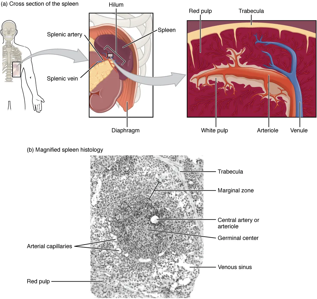
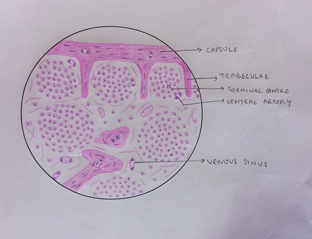
The spleen is almost entirely covered by visceral peritoneum, beneath which lies a thin fibrous capsule with a few smooth muscle fibres. Trabeculae run inwards from it carrying the trabecular arteries and veins, dividing the parenchyma into communicating compartments. The thin, friable capsule explains why the spleen ruptures easily in left-sided abdominal trauma.
- 02
Splenic hilum
Hilum splenicum
A longitudinal fissure on the visceral surface where the terminal branches of the splenic artery enter and the splenic vein leaves. It is joined to the stomach by the gastrosplenic ligament (carrying the short gastric arteries) and to the left kidney by the splenorenal ligament (carrying the splenic vessels and the tail of the pancreas).
- 03
Diaphragmatic and visceral surfaces
Facies diaphragmatica et facies visceralis
The convex diaphragmatic surface lies against the diaphragm opposite ribs 9 to 11, so a normal spleen is not palpable below the costal margin. The concave visceral surface bears the gastric, renal, colic (splenic flexure) and pancreatic impressions. The superior border is notched, a clue that a mass felt in the left upper quadrant is an enlarged spleen.
- 04
White pulp
Pulpa alba
Lymphoid tissue sheathing the central arteries: the periarteriolar lymphoid sheath (PALS), rich in T cells, and the lymphoid follicles (Malpighian corpuscles), rich in B cells and showing germinal centres during an immune response. It is where the spleen mounts immune responses to blood-borne antigens.
- 05
Marginal zone
Zona marginalis
The transition between white and red pulp, where arterioles open into the marginal sinuses. Its macrophages and marginal-zone B cells respond rapidly to the polysaccharide antigens of encapsulated bacteria (pneumococcus, meningococcus, Hib), which is why asplenic patients must be vaccinated against them.
- 06
Red pulp
Pulpa rubra
About 75% of the splenic volume, made of splenic cords (of Billroth) and venous sinuses. Red cells must squeeze through narrow slits between the rod-shaped endothelial cells of the sinuses; old or poorly deformable cells are held back and phagocytosed by macrophages. The red pulp also holds about one third of the body’s platelets.
- 07
Splenic segments
Segmenta splenica
The splenic artery divides at the hilum into two (sometimes three) branches, each supplying its own segment with few anastomoses to its neighbours. These relatively avascular planes allow partial splenectomy, preserving immune function.
Position and relations
The spleen lies in the left hypochondrium, its long axis along the tenth rib, behind the stomach and above the left colic flexure. Its convex diaphragmatic surface faces the diaphragm; its visceral surface bears gastric, renal and colic impressions and the hilum. It is linked to the stomach by the gastrosplenic ligament and to the kidney by the splenorenal ligament; a normal spleen is not palpable below the costal margin.
Form and structure
The spleen is enclosed in a fibrous capsule that sends trabeculae inwards. The white pulp consists of periarteriolar lymphoid sheaths (T cells) and lymphoid follicles (B cells); the red pulp consists of splenic cords (of Billroth) and venous sinuses, where aged or rigid red cells are trapped and removed by macrophages. The marginal zone between the two captures blood-borne antigens.
Vessels and nerves
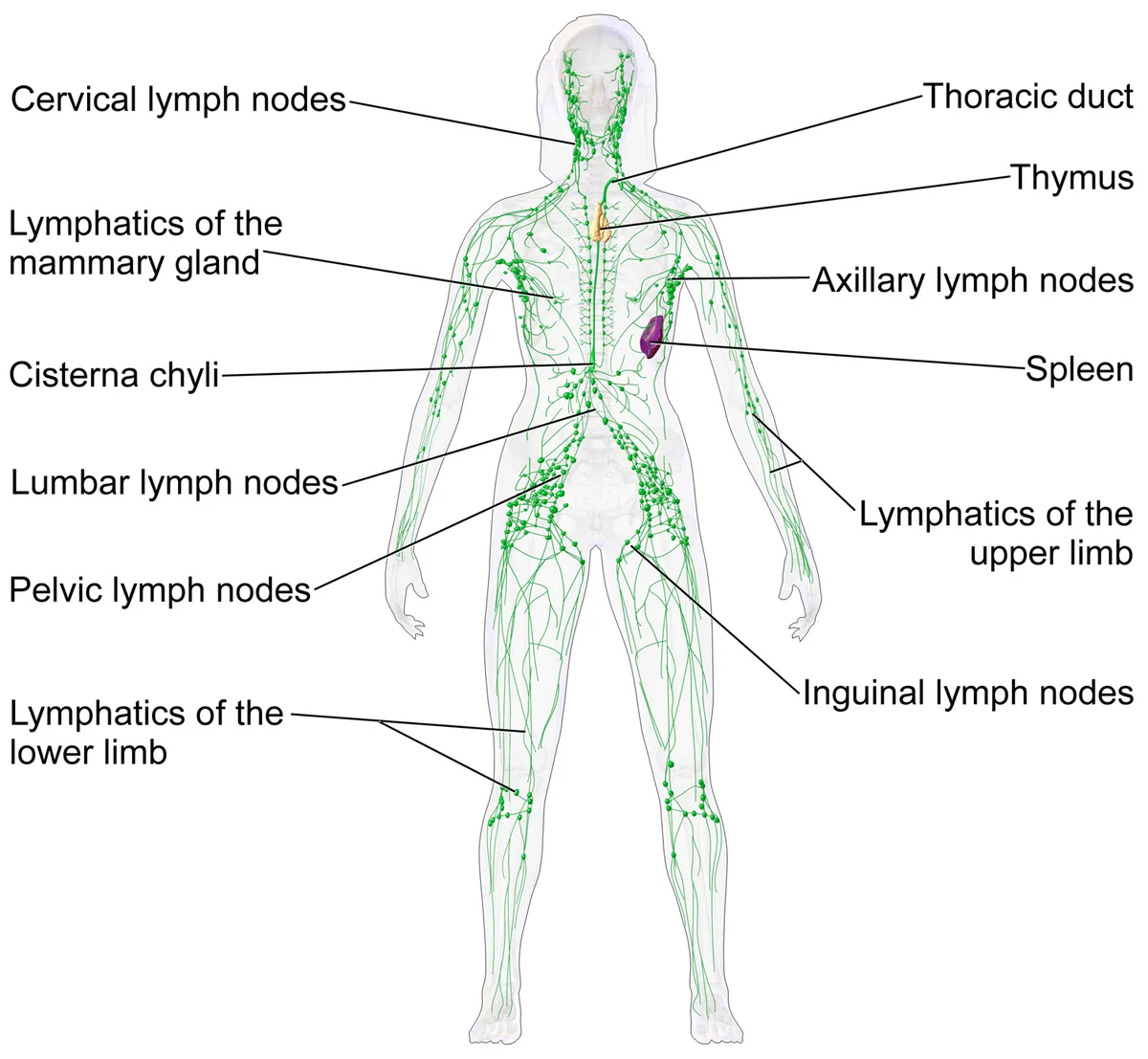
The splenic artery, the largest branch of the coeliac trunk, runs a tortuous course along the upper border of the pancreas and divides at the hilum; its intrasplenic branches are end arteries, so occlusion causes wedge-shaped infarcts. The splenic vein runs behind the pancreas and joins the superior mesenteric vein to form the portal vein, which is why portal hypertension enlarges the spleen.
Physiology
The spleen is the largest secondary lymphoid organ and combines innate and adaptive immunity: its structure lets it remove old red cells, filter microorganisms and debris from the blood, and bring antigen-presenting cells together with rare cognate lymphocytes. This makes it the most important organ for immunity against blood-borne bacteria and fungi.[1][2]
Borders, surfaces, vascular segments and splenic injury
The spleen is reddish-purple and spongy because of its rich vascularity, lies between the gastric fundus and the diaphragm at the level of ribs 9–11, measures 10–12 cm, weighs 150–200 g and is normally not palpable. It has three borders, a superior border with a notch at its anterior end (a clue to splenic enlargement on examination), an inferior and an intermediate border, and two surfaces, diaphragmatic and visceral. On the visceral surface the gastric impression is the deepest, the left kidney marks the intermediate and inferior borders, the splenic flexure produces the colic impression and the pancreatic tail leaves an impression between the hilum and the colic area. The splenic artery runs in the splenorenal ligament and divides before the hilum into about five non-anastomosing branches, creating separate vascular segments that make partial splenectomy possible. The spleen has only efferent lymphatics, draining to the pancreaticosplenic nodes; its sympathetic nerves come from the coeliac plexus and its parasympathetic fibres from the vagus.[3]
The spleen is not vital, because red marrow, liver and lymph nodes can take over its functions, yet splenic dysfunction can still be life-threatening. It is among the most frequently injured abdominal organs; rupture means disruption of the capsule and parenchyma by blunt or penetrating trauma and should be suspected with left rib fractures, since a rib fragment can pierce it. Splenomegaly (from viral or bacterial infection, venous obstruction with raised venous pressure, malignancy or abnormal blood-cell activity) increases the risk of rupture and may be felt below the costal margin. Management ranges from repair to partial or total splenectomy. People without a spleen are prone to infection by pneumococcus, meningococcus and Haemophilus influenzae and need vaccination.[3]
Accessory spleens
Around week 5 the primitive spleen forms from the mesoderm of the dorsal mesogastrium; as this mesentery rotates to the left with the foregut, the mesenchyme migrates to the splenic position between ribs 9 and 11. If mesenchymal buds leaving the midline fail to fuse, they form accessory spleens. Accessory tissue can also follow the descent of the gonads: the spleen first lies near the urogenital ridge, so splenic tissue can fuse with a gonad and be dragged down with it (splenogonadal fusion), leaving splenic tissue in the pelvis or groin. About 75% of accessory spleens lie at the splenic hilum, 20% near the pancreatic tail and 5% elsewhere (gastrosplenic and splenorenal ligaments, greater omentum, mesentery, gut wall, pelvis and rarely the scrotum). They have the structure and function of a normal spleen and are supplied by the splenic artery.[4]
Accessory spleens matter in two situations. When the spleen is removed to treat a blood disorder such as haemolytic anaemia or chronic immune thrombocytopenic purpura, accessory spleens must be removed too, otherwise the remaining functional tissue lets the symptoms return. On CT they can mimic lymph nodes, tumours or splenosis; scintigraphy with 99mTc-nanocolloid confirms them because splenic tissue takes up the tracer far more avidly. An accessory spleen is distinguished from splenosis by its thick capsule, smooth-muscle elements positive for smooth muscle actin, and its supply from the splenic artery.[4]
Full-text reading (StatPearls)
The StatPearls chapters on this topic, reproduced verbatim. Their CC BY-NC-ND 4.0 licence allows unmodified copies only, so they are not translated or shortened.
Anatomy, Abdomen and Pelvis, SpleenOmole AE, Launico MV · StatPearls 2026 · NBK482235 · 11k characters
Loading…
Omole AE, Launico MV. Anatomy, Abdomen and Pelvis, Spleen. StatPearls [Internet]. Treasure Island (FL): StatPearls Publishing; 2026. NBK482235 · PMID 29489170 · CC BY-NC-ND 4.0. Reproduced verbatim, unmodified.
Anatomy, Abdomen and Pelvis: Accessory SpleenBajwa SA, Kasi A · StatPearls 2023 · NBK519040 · 7k characters
Loading…
Bajwa SA, Kasi A. Anatomy, Abdomen and Pelvis: Accessory Spleen. StatPearls [Internet]. Treasure Island (FL): StatPearls Publishing; 2023. NBK519040 · PMID 30085582 · CC BY-NC-ND 4.0. Reproduced verbatim, unmodified.
Organs in this system
References
Sources for the values and descriptions on this page, indexed in PubMed (US National Library of Medicine).
- [1]Structure and function of the spleenMebius RE et al., Nature reviews. Immunology 2005PMID 16056254
- [2]Structure and function of the immune system in the spleenLewis SM et al., Science immunology 2019PMID 30824527
- [3]Anatomy, Abdomen and Pelvis, SpleenOmole AE et al., StatPearls 2026PMID 29489170
- [4]Anatomy, Abdomen and Pelvis: Accessory SpleenBajwa SA et al., StatPearls 2023PMID 30085582