Autonomic nervous system
Model
Layers
Z-Anatomy anatomical model based on BodyParts3D, licensed CC BY-SA 4.0.
Description
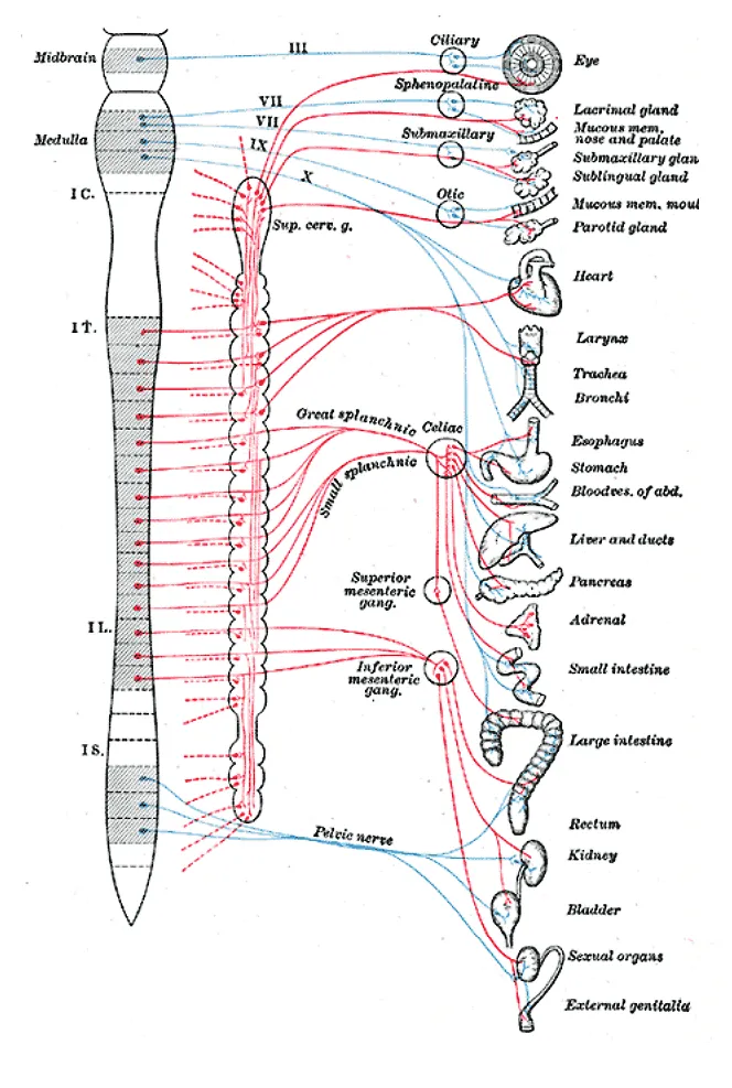
The division of the nervous system that controls smooth muscle, cardiac muscle and glands involuntarily. The sympathetic part arises from the thoracolumbar cord (T1–L2) through the paravertebral sympathetic trunk; the parasympathetic part arises from the brainstem (nerves III, VII, IX, X) and the sacral cord (S2–S4).
Parts
Each part has its own card; press “Show on the model” to fly to it and select it in the 3D model above.
- 01
Sympathetic trunk
Truncus sympathicus
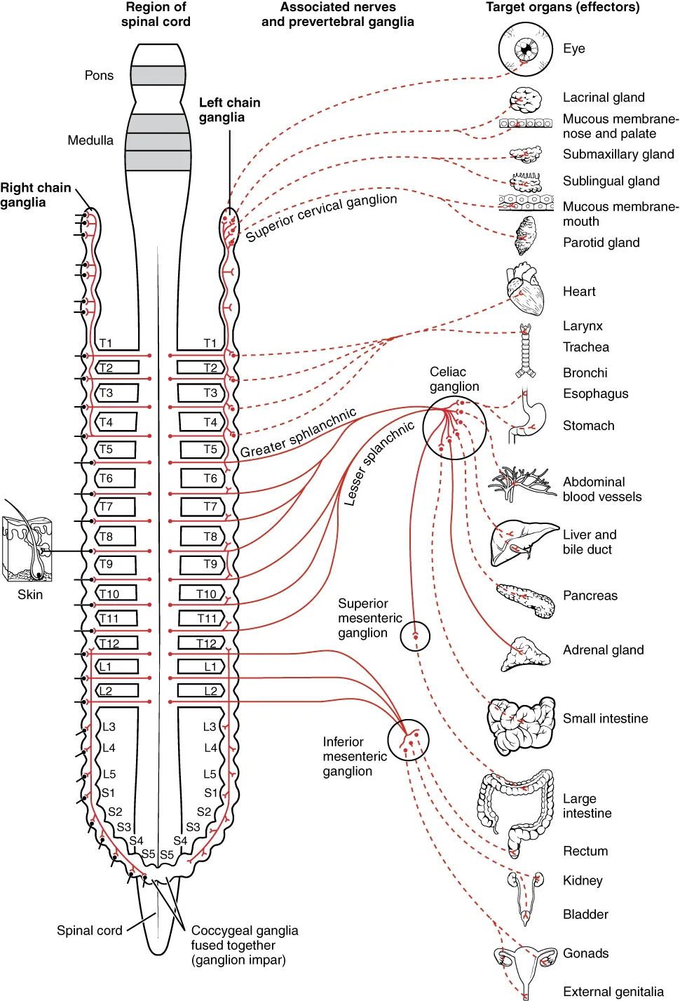
Two paravertebral chains of ganglia from the skull base to the coccyx (3 cervical, about 11 thoracic, 4 lumbar and 4 sacral, joining at the ganglion impar). They receive preganglionic fibres through white rami (T1–L2) and return postganglionic fibres to every spinal nerve through grey rami; the inferior cervical ganglion often fuses with the first thoracic as the stellate ganglion.
- 02
Splanchnic nerves and prevertebral ganglia
Nervi splanchnici; Ganglia prevertebralia
The greater (T5–T9), lesser (T10–T11) and least (T12) splanchnic nerves pass through the trunk without synapsing to prevertebral ganglia around the aortic branches (coeliac, superior mesenteric, aorticorenal, inferior mesenteric), which send postganglionic fibres to the abdominal viscera. The adrenal medulla alone receives preganglionic fibres directly.
- 03
Cranial parasympathetic outflow
Pars cranialis partis parasympathicae
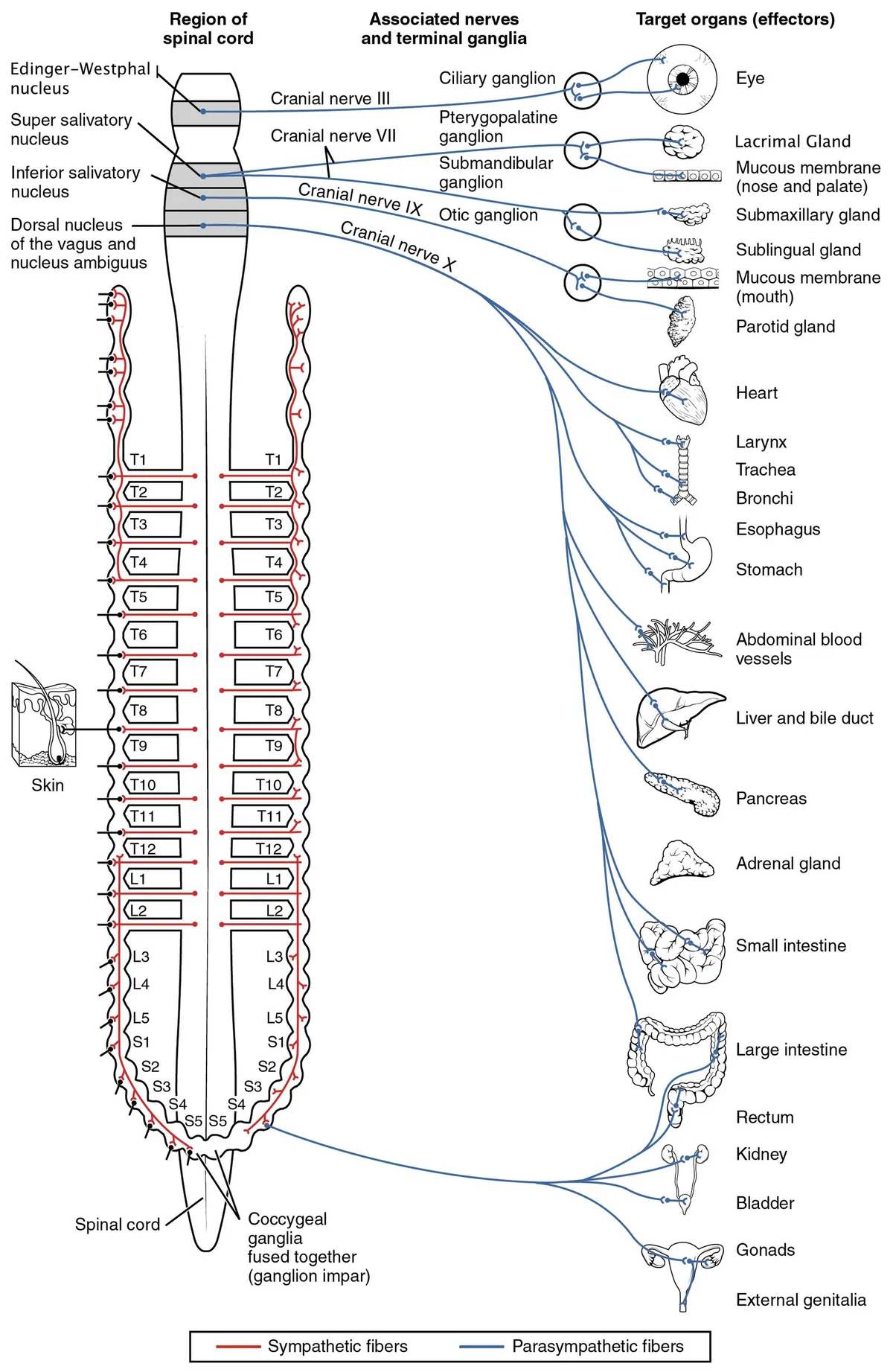
Preganglionic fibres travel with nerves III (ciliary ganglion: pupillary constriction, accommodation), VII (pterygopalatine ganglion: lacrimal and nasal glands; submandibular ganglion: submandibular and sublingual glands), IX (otic ganglion: parotid) and X (ganglia in the walls of thoracic and abdominal organs).
- 04
Sacral parasympathetic outflow
Pars sacralis; Nervi splanchnici pelvici
Cell bodies lie in the S2–S4 cord; their fibres run in the pelvic splanchnic nerves to the inferior hypogastric plexus. They drive detrusor contraction, the descending and sigmoid colon and rectum, and erection; conus or cauda equina lesions disturb bladder, bowel and sexual function.
- 05
Autonomic plexuses
Plexus autonomici
Mixed sympathetic and parasympathetic networks around viscera and large arteries: the cardiac, pulmonary and oesophageal plexuses in the thorax, and the coeliac (solar), mesenteric and superior and inferior hypogastric plexuses in abdomen and pelvis. Visceral afferents travel in them too, which explains referred pain.
- 06
Enteric nervous system
Systema nervosum entericum
Some 400–600 million neurons in the gut wall: the myenteric (Auerbach) plexus between the muscle layers controls peristalsis, and the submucosal (Meissner) plexus controls secretion and blood flow. It works even when disconnected from the CNS; congenital absence of ganglion cells causes Hirschsprung disease.
Sympathetic division
Preganglionic neurons lie in the lateral horn of the cord from T1 to L2. Their axons leave in the anterior root and pass through the white rami communicantes into the paravertebral sympathetic trunk, which runs alongside the vertebral bodies from the skull base to the coccyx. There they may synapse at once, ascend or descend in the trunk, or pass straight through as splanchnic nerves to the prevertebral ganglia (coeliac, superior mesenteric, inferior mesenteric).
In the neck there are three ganglia (superior, middle and the stellate ganglion, formed by fusion of the inferior cervical and first thoracic ganglia) supplying the head, face, upper limb and heart. Postganglionic fibres return through the grey rami to every spinal nerve to reach blood vessels, sweat glands and arrector pili muscles. The adrenal medulla is a special case: it receives preganglionic fibres directly and releases adrenaline into the blood.
Parasympathetic division
The parasympathetic outflow is craniosacral. The cranial part travels with nerve III (ciliary ganglion: pupil constriction, accommodation), VII (pterygopalatine and submandibular ganglia: lacrimal, submandibular and sublingual glands), IX (otic ganglion: parotid gland) and X (heart, lungs and gut as far as about two-thirds of the transverse colon). The sacral part (S2–S4) runs in the pelvic splanchnic nerves to the descending colon, rectum, bladder and genital organs.
Unlike sympathetic ganglia, parasympathetic ganglia lie close to or within the target organ, so preganglionic fibres are long and postganglionic fibres very short. Both divisions use acetylcholine at the ganglionic synapse; sympathetic postganglionic fibres mostly release noradrenaline (sweat glands, which use acetylcholine, are the exception), while parasympathetic postganglionic fibres release acetylcholine onto muscarinic receptors.
Example: autonomic control of the heart
Vagal tone slows the sinus node and atrioventricular conduction. A strong vagal surge can cause paroxysmal block within the AV node together with sinus slowing; this vagally mediated block is benign, is managed as neurally mediated syncope, and does not call for a pacemaker when it is asymptomatic.[1]
After heart transplantation the donor heart is completely denervated, losing both afferent and efferent connections. Most patients remain fully denervated for the first 6–12 months; reinnervation of the myocardium, sinus node and coronary vessels usually appears from the second year but remains incomplete and regionally limited many years later.[2]
Clinical correlations
Interruption of the cervical sympathetic pathway (for example by an apical lung tumour invading the stellate ganglion, or a carotid dissection) causes Horner syndrome: mild ptosis, miosis and reduced sweating of the same side of the face. Autonomic failure shows as orthostatic hypotension and bladder, bowel and sexual dysfunction.
Functional organisation
The autonomic nervous system comprises the sympathetic, parasympathetic and enteric divisions and controls every part of the body except skeletal muscle. Its main task is to keep cells, tissues and organs in homeostasis despite external and internal perturbations. Many common drugs and toxins act by altering autonomic transmission, and autonomic dysfunction is a hallmark of many neurological diseases.[3]
The vagus nerve (X) is the largest and longest cranial nerve and the only one whose territory lies mostly outside the head (neck, thorax, abdomen). Although part of the parasympathetic division it is primarily sensory: most axons are afferents with cell bodies in the nodose ganglion ending in the nucleus of the solitary tract; efferents arise from the dorsal motor nucleus and nucleus ambiguus.[4]
Vagal sensory neurons are highly diverse, each type devoted to respiratory, cardiovascular or digestive organs; they govern breathing, heart rate, blood pressure, gut motility, cough and swallowing reflexes, and behaviours such as feeding, drinking and sickness responses. In the gut, the vagus with serotonin and enteroendocrine cells forms the gut–brain axis that influences food intake, fat metabolism and mood.[5][6][7]
The inflammatory reflex and applications
Efferent vagal cholinergic signalling controls immune function and pro-inflammatory responses through the “inflammatory reflex”. In obesity, chronic inflammation is a critical step towards insulin resistance and type 2 diabetes; this cholinergic mechanism can attenuate obesity-related inflammation, opening therapeutic avenues such as vagus nerve stimulation.[8]
Under the “brain-first versus body-first” hypothesis, α-synuclein pathology in some patients with Parkinson’s disease may begin in the enteric nervous system and spread to the brain via the vagus, whereas in others it starts in the brain and only later reaches the peripheral autonomic system.[9]
Full-text reading (StatPearls)
The StatPearls chapters on this topic, reproduced verbatim. Their CC BY-NC-ND 4.0 licence allows unmodified copies only, so they are not translated or shortened.
Anatomy, Autonomic Nervous SystemWaxenbaum JA, Reddy V, Das JM · StatPearls 2025 · NBK539845 · 26k characters
Loading…
Waxenbaum JA, Reddy V, Das JM. Anatomy, Autonomic Nervous System. StatPearls [Internet]. Treasure Island (FL): StatPearls Publishing; 2025. NBK539845 · PMID 30969667 · CC BY-NC-ND 4.0. Reproduced verbatim, unmodified.
Anatomy, Head and Neck: Anterior Vagus NerveBaquiran M, Bordoni B · StatPearls 2023 · NBK547696 · 11k characters
Loading…
Baquiran M, Bordoni B. Anatomy, Head and Neck: Anterior Vagus Nerve. StatPearls [Internet]. Treasure Island (FL): StatPearls Publishing; 2023. NBK547696 · PMID 31613476 · CC BY-NC-ND 4.0. Reproduced verbatim, unmodified.
Neuroanatomy, Cranial Nerve 10 (Vagus Nerve)Kenny BJ, Bordoni B · StatPearls 2022 · NBK537171 · 10k characters
Loading…
Kenny BJ, Bordoni B. Neuroanatomy, Cranial Nerve 10 (Vagus Nerve). StatPearls [Internet]. Treasure Island (FL): StatPearls Publishing; 2022. NBK537171 · PMID 30725856 · CC BY-NC-ND 4.0. Reproduced verbatim, unmodified.
Neuroanatomy, Autonomic Nervous System Visceral Afferent Fibers and PainSanvictores T, Jozsa F, Tadi P · StatPearls 2023 · NBK560843 · 8k characters
Loading…
Sanvictores T, Jozsa F, Tadi P. Neuroanatomy, Autonomic Nervous System Visceral Afferent Fibers and Pain. StatPearls [Internet]. Treasure Island (FL): StatPearls Publishing; 2023. NBK560843 · PMID 32809678 · CC BY-NC-ND 4.0. Reproduced verbatim, unmodified.
Organs in this system
References
Sources for the values and descriptions on this page, indexed in PubMed (US National Library of Medicine).
- [1]Vagally mediated atrioventricular block: pathophysiology and diagnosisAlboni P et al., Heart 2013PMID 23286970
- [2]Reinnervation post-heart transplantationGrupper A et al., European heart journal 2018PMID 28087606
- [3]Overview of the Anatomy, Physiology, and Pharmacology of the Autonomic Nervous SystemWehrwein EA et al., Comprehensive physiology 2016PMID 27347892
- [4]Structure and Functions of the Vagus Nerve in MammalsOttaviani MM et al., Comprehensive physiology 2022PMID 35950655
- [5]Internal senses of the vagus nervePrescott SL et al., Neuron 2022PMID 35051375
- [6]Vagus Nerve and Gut-Brain CommunicationWang Y et al., The Neuroscientist 2025PMID 39041416
- [7]Interaction of the Vagus Nerve and Serotonin in the Gut-Brain AxisHwang YK et al., International journal of molecular sciences 2025PMID 39940928
- [8]The vagus nerve and the inflammatory reflex--linking immunity and metabolismPavlov VA et al., Nature reviews. Endocrinology 2012PMID 23169440
- [9]Brain-First versus Gut-First Parkinson's Disease: A HypothesisBorghammer P et al., Journal of Parkinson's disease 2019PMID 31498132
- [10]The human brain in numbers: a linearly scaled-up primate brainHerculano-Houzel S, 2009PMID 19915731Supports: Neuron count
- [11]Determinants of conduction velocity in myelinated nerve fibersWaxman SG, 1980PMID 6245357Supports: Conduction velocity
- [12]Quantitative imaging of energy expenditure in human brainPMID 22487547Supports: Cerebral energy expenditure