Uterine tubes
Model
Human Reference Atlas (HuBMAP), CC BY 4.0, meshopt-compressed. Drag “Section” to look inside.
Description
Paired ducts about 10–12 cm long in the upper border of the broad ligament, linking the uterine cornua to the ovaries. From lateral to medial they have four parts: the fimbriated infundibulum, the ampulla (the usual site of fertilisation), the isthmus and the intramural part within the uterine wall. Cilia and peristalsis carry the oocyte and embryo towards the uterine cavity.
Parts
Each part has its own card; press “Show on the model” to fly to it and select it in the 3D model above.
- 01
Intramural (uterine) part
Pars uterina tubae uterinae
About 1 cm long, passing through the wall at the uterine cornu, with the narrowest lumen (about 1 mm). Interstitial pregnancy here is rare but ruptures late with heavy bleeding because the area is very vascular.
- 02
Isthmus
Isthmus tubae uterinae
The medial third, narrow with a thick muscular wall, joining the uterine cornu. It is the usual site of tubal ligation for sterilisation.
- 03
Ampulla
Ampulla tubae uterinae
The longest and widest part (about two-thirds of the tube), with an intricately folded mucosa; it is the usual site of fertilisation and also the commonest site of ectopic pregnancy (about 70%).
- 04
Infundibulum and fimbriae
Infundibulum; Fimbriae tubae uterinae
The funnel opens into the peritoneal cavity through the abdominal ostium, fringed by 20–25 fimbriae, the longest (ovarian fimbria) attached to the ovary. At ovulation the fimbriae sweep over the ovary and gather the oocyte; this is also the route by which genital infection reaches the peritoneum.
- 05
Tubal wall and mesosalpinx
Tunica mucosa tubae uterinae; Mesosalpinx
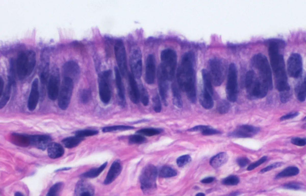
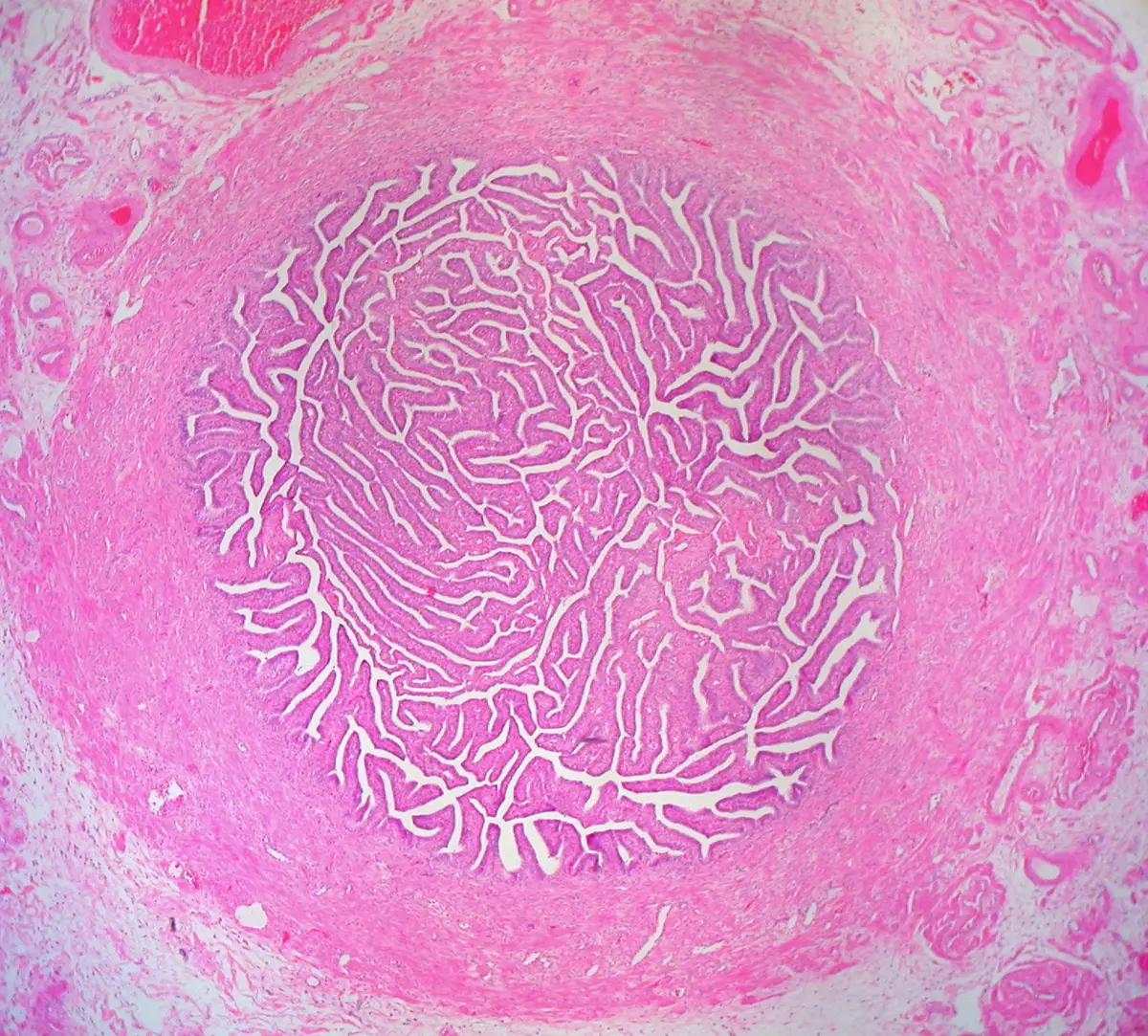
A simple columnar mucosa of ciliated cells (moving the oocyte towards the uterus) and secretory peg cells (nourishing gametes), with peristaltic smooth muscle. The tube hangs in the upper edge of the broad ligament by the mesosalpinx; salpingitis causes adhesions and narrowing, leading to infertility and ectopic pregnancy.
Position and relations
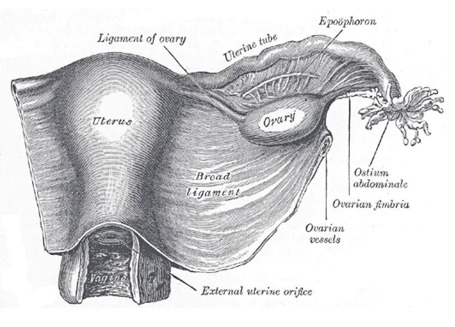
The uterine tube runs in the free upper border of the broad ligament; the part of the ligament between tube and ovary is the mesosalpinx. Its lateral end (infundibulum) opens into the peritoneal cavity through the abdominal ostium and embraces the upper pole of the ovary with its fimbriae, the longest of which (the ovarian fimbria) is attached to the ovary; its medial end opens into the uterine cavity at the cornu.
Form and structure
The tubal wall has three layers: a mucosa, most richly folded in the ampulla and lined by simple columnar epithelium of ciliated and secretory cells; a smooth-muscle layer that thickens towards the isthmus; and a serosa. The wide, long ampulla is where fertilisation usually occurs; the isthmus is narrow and thick-walled. Tubal ectopic pregnancy most often implants in the ampulla.
Vessels and nerves
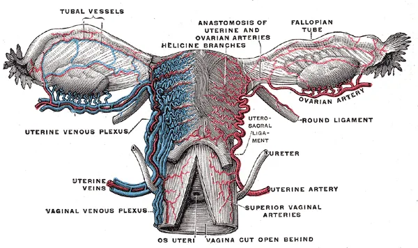
The tube is supplied by tubal branches of the uterine and ovarian arteries, which anastomose in the mesosalpinx; veins accompany them. Lymph follows the ovarian vessels to the para-aortic nodes. Autonomic nerves come from the ovarian and uterovaginal plexuses.
Clinical correlations
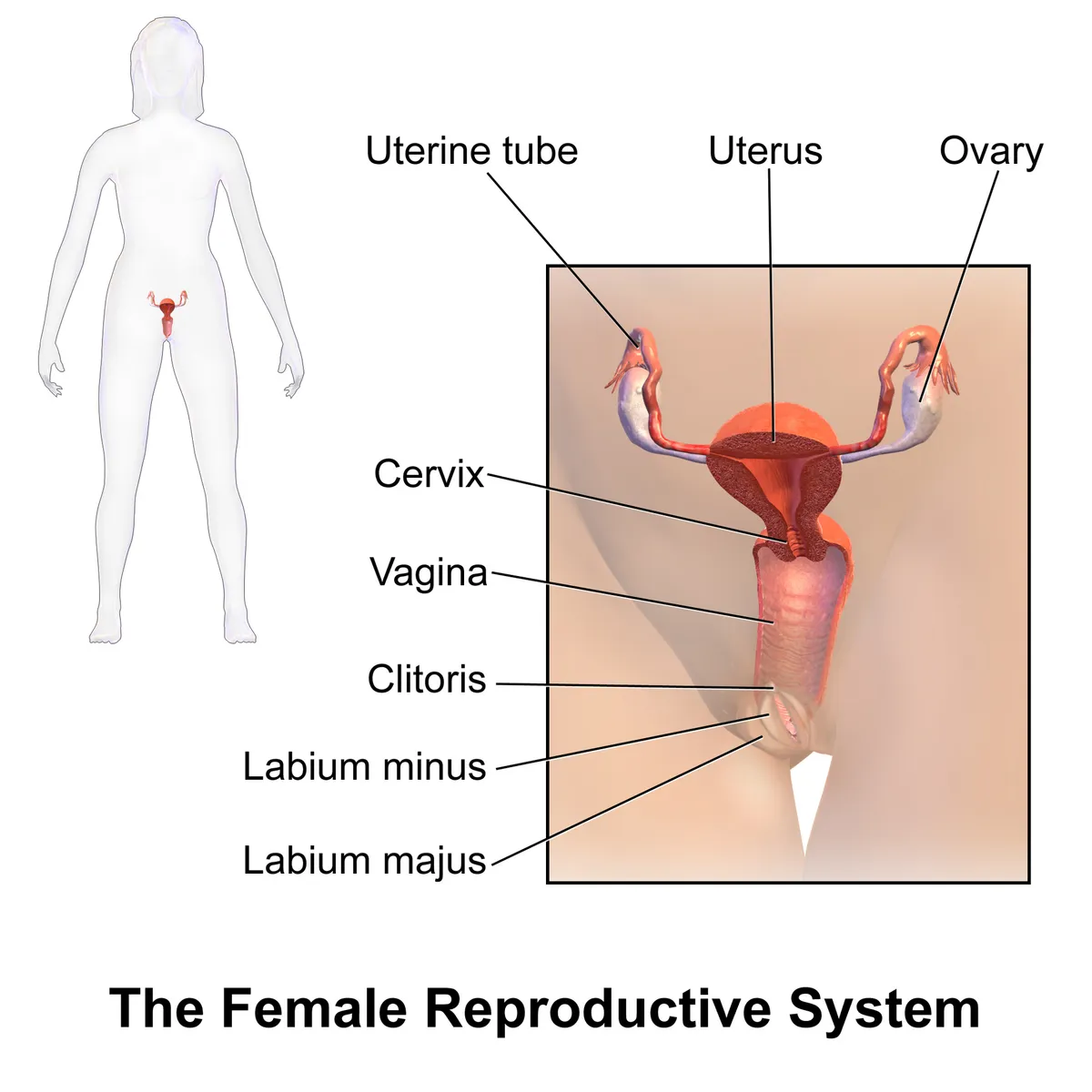
The uterine tube is where sperm complete capacitation, fertilisation occurs and the embryo spends its first days; its epithelium of ciliated and secretory cells produces the tubal fluid. About 30% of infertile women have tubal disease, most often from Chlamydia trachomatis, gonorrhoea, genital tuberculosis, endometriosis or complications of abdominal surgery.[1][2]
Over 98% of ectopic pregnancies implant in the uterine tube, and tubal pregnancy remains the leading cause of maternal death in the first trimester. Risk factors include tubal damage from surgery or infection, smoking and in vitro fertilisation; current evidence points to retention of the embryo in the tube combined with changes in the tubal environment that favour implantation.[3]
Four segments, muscle layer, vessels and nerves
The uterine (fallopian) tube is a seromuscular tube 11–12 cm long with a lumen under 1 mm across, running laterally from the uterine horn in the upper edge of the mesosalpinx of the broad ligament and ending beside the ipsilateral ovary. From medial to lateral it has four segments: the intramural (uterine) part, which crosses the uterine wall and bears the uterine ostium; the narrow isthmus; the wide ampulla, the commonest site of fertilisation; and the infundibulum, which opens into the peritoneal cavity through the abdominal ostium and is fringed by fimbriae that catch the oocyte each cycle, one of them (the ovarian fimbria) joining it to the ovary. The muscle layer lies between the mucosa, whose cilia project into the lumen, and the serosa, and consists of an inner circular and an outer longitudinal layer. Short, frequent contractions mix the tubal fluid; continuous tonic contraction, together with ciliary beating and secretions, moves the oocyte or gametes towards the uterus and admits the embryo across the uterotubal junction at the hormonally most favourable point of the cycle.[4]
The tube develops from the cranial end of the paramesonephric (Müllerian) duct, which forms in the female embryo around weeks 5–6 because anti-Müllerian hormone is absent; under oestrogen the cranial ends become the tubes, while the caudal ends fuse in the midline to form the uterus and upper vagina. A remnant at the very top may persist as the vesicular appendage (hydatid of Morgagni). Tubal branches of the ovarian artery (from the abdominal aorta below the renal arteries) supply the lateral half and the ascending branch of the uterine artery (from the internal iliac) the medial half; because the two anastomose, compromise of either rarely makes any part of the tube ischaemic. The right ovarian vein drains to the inferior vena cava and the left to the left renal vein. Lymph passes to para-aortic (lumbar) and pelvic nodes. Pain fibres follow the sympathetic route from T11–L1; parasympathetic supply comes from vagal fibres in the ovarian plexus laterally and from the pelvic splanchnic nerves (S1–S3) medially. The isthmus is the most densely innervated part, and both nerves and muscle become sparser distally.[4]
Variants, surgery and the tubal origin of ovarian cancer
Benign paratubal cysts are often found incidentally on ultrasound or at surgery, usually in middle age and without symptoms. Rarer variants are diverticula, agenesis and duplication; diverticula and duplications raise the risk of hydrosalpinx, torsion and ectopic pregnancy, while occlusion or agenesis can cause infertility, and in utero exposure to diethylstilbestrol (DES) has been linked to abnormal tubal development. Tubal abnormalities are implicated in up to a third of infertility cases, through congenital malformation, infection with Chlamydia trachomatis, gonorrhoea or genital tuberculosis, or adhesions from endometriosis; recurrent pelvic inflammatory disease or previous abdominal surgery raises the risk, and more infections mean more severe damage. Tubal occlusion is investigated by hysterosalpingography or hysteroscopy and may be treated by selective salpingography, recanalisation, re-anastomosis or neosalpingostomy.[4]
Common operations on the tube are salpingo-oophorectomy, salpingectomy, salpingostomy and tubal sterilisation (by ligation, occlusion or excision). The ampulla is the commonest site of ectopic pregnancy, treated medically or surgically by salpingostomy or salpingectomy; salpingectomy is also needed for hydrosalpinx, tubo-ovarian abscess and cancers of the ovary, tube or uterus. The tube has a special place in ovarian cancer: high-grade serous carcinoma, the commonest type, usually diagnosed late with a poor prognosis, is now thought in most cases to arise in the tube years before it presents in the ovary. Tubal sterilisation is very safe and effective but permanent; risk factors for later regret include age under 30, low parity, sterilisation immediately after delivery and low socioeconomic status.[4]
Full-text reading (StatPearls)
The StatPearls chapters on this topic, reproduced verbatim. Their CC BY-NC-ND 4.0 licence allows unmodified copies only, so they are not translated or shortened.
Anatomy, Abdomen and Pelvis: Fallopian TubeHan J, Sadiq NM · StatPearls 2023 · NBK547660 · 8k characters
Loading…
Han J, Sadiq NM. Anatomy, Abdomen and Pelvis: Fallopian Tube. StatPearls [Internet]. Treasure Island (FL): StatPearls Publishing; 2023. NBK547660 · PMID 31613440 · CC BY-NC-ND 4.0. Reproduced verbatim, unmodified.
Organs in this system
References
Sources for the values and descriptions on this page, indexed in PubMed (US National Library of Medicine).
- [1]Oviductal response to gametes and early embryos in mammalsMaillo V et al., Reproduction 2016PMID 27512123
- [2]Fallopian tubes--literature review of anatomy and etiology in female infertilityBriceag I et al., Journal of medicine and life 2015PMID 25866566
- [3]Current knowledge of the aetiology of human tubal ectopic pregnancyShaw JL et al., Human reproduction update 2010PMID 20071358
- [4]Anatomy, Abdomen and Pelvis: Fallopian TubeHan J et al., StatPearls 2023PMID 31613440