Pharynx
Model
Layers
Z-Anatomy anatomical model based on BodyParts3D, licensed CC BY-SA 4.0.
Description
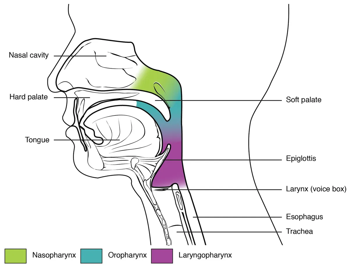
A musculomembranous tube about 12–15 cm long from the skull base to the lower border of the cricoid cartilage (C6), lying behind the nose, mouth and larynx and divided into the nasopharynx, oropharynx and laryngopharynx. It is the crossroads of the airway and the food passage: during swallowing the constrictors drive the bolus into the oesophagus while the larynx is raised and closed.
Parts
Each part has its own card; press “Show on the model” to fly to it and select it in the 3D model above.
- 01
Nasopharynx
Pars nasalis pharyngis
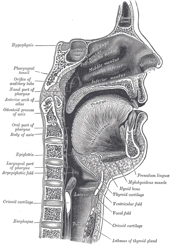
The upper part behind the choanae and above the soft palate, always open for breathing. Its lateral wall bears the pharyngeal opening of the auditory tube with the torus tubarius and tubal tonsil, behind which lies the pharyngeal recess (fossa of Rosenmüller, the usual origin of nasopharyngeal carcinoma); the pharyngeal tonsil (adenoid) is on the posterosuperior wall.
- 02
Oropharynx
Pars oralis pharyngis
Extends from the soft palate to the upper border of the epiglottis and opens forward into the mouth through the oropharyngeal isthmus. Its anterior wall is the posterior third of the tongue with the valleculae; the palatine tonsils lie on the lateral walls between the palatoglossal and palatopharyngeal arches.
- 03
Laryngopharynx (hypopharynx)
Pars laryngea pharyngis
Extends from the upper border of the epiglottis to the lower border of the cricoid (C6), where it joins the oesophagus, lying behind the larynx. On each side of the laryngeal inlet is a piriform recess, where fish bones lodge and hypopharyngeal cancers often arise.
- 04
Pharyngeal constrictors
Musculi constrictores pharyngis
The superior, middle and inferior constrictors overlap like stacked cups, insert behind into the pharyngeal raphe and contract in sequence to drive the bolus down. Between the two parts of the inferior constrictor (thyropharyngeus and cricopharyngeus) lies Killian’s weak triangle, where a Zenker diverticulum forms.
- 05
Longitudinal muscles
Musculi longitudinales pharyngis
Stylopharyngeus (IX), palatopharyngeus and salpingopharyngeus (X) run vertically, elevating and shortening the pharynx and lifting the larynx in swallowing. All pharyngeal muscles except stylopharyngeus are supplied by the pharyngeal plexus (mainly the vagus).
Position and relations
The pharynx hangs from the skull base in front of the cervical spine and prevertebral muscles, separated from them by the retropharyngeal space. The nasopharynx opens into the nasal cavity through the choanae and into the middle ear through the auditory tube; the oropharynx opens into the mouth through the oropharyngeal isthmus; the laryngopharynx opens into the larynx in front and has the piriform recesses on each side, where foreign bodies lodge.
Form and structure
The wall comprises the mucosa, the pharyngobasilar fascia, the muscle coat and the buccopharyngeal fascia. The outer muscle layer is the three overlapping constrictors (superior, middle, inferior); the inner layer consists of longitudinal muscles (stylopharyngeus, salpingopharyngeus, palatopharyngeus) that elevate the pharynx in swallowing. The lowest part of the inferior constrictor (cricopharyngeus) is the upper oesophageal sphincter; the weak area above it (Killian’s triangle) is where a Zenker diverticulum forms.
Vessels and nerves
Arteries come mainly from the ascending pharyngeal artery with branches of the facial, superior thyroid and maxillary arteries. The pharyngeal plexus is formed by the glossopharyngeal nerve (sensory) and the vagus (motor to most muscles, stylopharyngeus being supplied by the glossopharyngeal), so the gag reflex tests both nerves. Lymph drains to the retropharyngeal and deep cervical nodes.
Swallowing and speech
The pharynx is a complex muscular tube shared by the airway and the food passage, allowing breathing, swallowing and speech through a common airspace. On imaging, the pharynx and cervical oesophagus are difficult because of numerous interleaved muscles and connections; certain landmarks (parapharyngeal and retropharyngeal spaces, piriform sinus) govern how disease spreads.[1]
The velopharyngeal mechanism is a muscular valve from the posterior edge of the hard palate to the posterior pharyngeal wall, made up of the velum (soft palate), the lateral walls and the posterior wall. During speech and swallowing it seals the oral from the nasal cavity through the levator veli palatini, musculus uvulae, superior constrictor, palatopharyngeus, palatoglossus and salpingopharyngeus; the tensor veli palatini mainly opens the auditory tube. Understanding it is the first step in managing velopharyngeal insufficiency in children with cleft lip and palate.[2]
Swallowing requires precise coordination of the central and peripheral nervous systems with the muscles of the mouth, pharynx and airway. The brainstem houses the swallowing central pattern generator and cranial nerve nuclei; the cortex initiates and coordinates the swallow after receiving afferent input, while the basal ganglia and thalamus regulate the movement through the cortico–basal ganglia–thalamo–cortical loop. Dysphagia is therefore common in neurological disease such as stroke or Parkinson’s disease.[3][4]
In newborns, sucking, swallowing and breathing must be synchronised for safe oral feeding; each function has several components that mature at different times and rates, which is why preterm infants are often discharged late for want of competent feeding.[5]
Clinical correlations
The pharyngeal tonsil (adenoid) sits at the junction of the roof and posterior wall of the nasopharynx and belongs to Waldeyer’s ring; as the first lymphoid tissue to meet respiratory and digestive antigens it matters for immune development in children. Adenoid hypertrophy is very common in childhood and causes mouth breathing, snoring, sleep apnoea and otitis media with effusion.[6][7]
Chronic mouth breathing in children (often due to adenoid hypertrophy) alters tongue, mandible and head posture and can lead to a long face, a narrow palate and malocclusion.[8]
Full-text reading (StatPearls)
The StatPearls chapters on this topic, reproduced verbatim. Their CC BY-NC-ND 4.0 licence allows unmodified copies only, so they are not translated or shortened.
Anatomy, Head and Neck, PharynxAlbahout KS, Lopez RA · StatPearls 2023 · NBK544271 · 12k characters
Loading…
Albahout KS, Lopez RA. Anatomy, Head and Neck, Pharynx. StatPearls [Internet]. Treasure Island (FL): StatPearls Publishing; 2023. NBK544271 · PMID 31334991 · CC BY-NC-ND 4.0. Reproduced verbatim, unmodified.
Organs in this system
References
Sources for the values and descriptions on this page, indexed in PubMed (US National Library of Medicine).
- [1]Anatomy of the Pharynx and Cervical EsophagusAkgoz Karaosmanoglu A et al., Neuroimaging clinics of North America 2022PMID 36244724
- [2]Anatomy and physiology of the velopharyngeal mechanismPerry JL, Seminars in speech and language 2011PMID 21948636
- [3]The Cortical and Subcortical Neural Control of Swallowing: A Narrative ReviewWei KC et al., Dysphagia 2024PMID 37603047
- [4]Dysphagia: Evaluation and Collaborative ManagementWilkinson JM et al., American family physician 2021PMID 33448766
- [5]Development of Suck and Swallow Mechanisms in InfantsLau C, Annals of nutrition & metabolism 2015PMID 26226992
- [6]Adenoid hypertrophy in children: a narrative review of pathogenesis and clinical relevanceNiedzielski A et al., BMJ paediatrics open 2023PMID 37045541
- [7]Prevalence of adenoid hypertrophy: A systematic review and meta-analysisPereira L et al., Sleep medicine reviews 2018PMID 29153763
- [8]The impact of mouth breathing on dentofacial development: A concise reviewLin L et al., Frontiers in public health 2022PMID 36159237
- [9]Gastrointestinal mean transit times in young and middle-aged healthy subjectsPMID 11318834Supports: Gastrointestinal transit time
- [10]Revised Estimates for the Number of Human and Bacteria Cells in the BodySender R, Fuchs S, Milo R, 2016PMID 27541692Supports: Colonic bacterial count