Oesophagus
Model
Layers
Z-Anatomy anatomical model based on BodyParts3D, licensed CC BY-SA 4.0.
Description
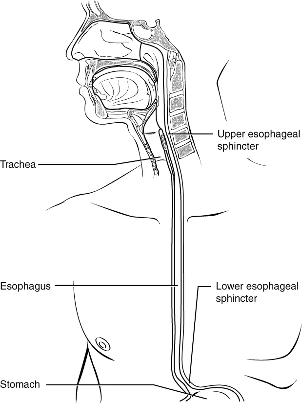
A muscular tube about 25 cm long joining the pharynx (at C6) to the stomach, running through the neck, the posterior mediastinum and the oesophageal hiatus of the diaphragm (at T10). It has three physiological narrowings: at the pharyngo-oesophageal junction, where the aortic arch and left main bronchus cross it, and at the diaphragm.
Parts
Each part has its own card; press “Show on the model” to fly to it and select it in the 3D model above.
- 01
Cervical part
Pars cervicalis oesophagi
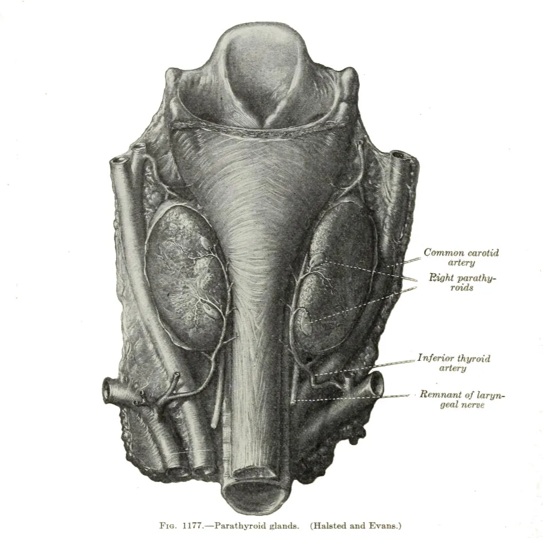
Begins at the lower border of the cricoid (C6), about 15 cm from the incisor teeth, directly behind the trachea and slightly to the left, so cervical oesophageal surgery uses a left approach. The recurrent laryngeal nerves lie in the tracheo-oesophageal grooves.
- 02
Thoracic part
Pars thoracica oesophagi
The longest part, running through the superior then posterior mediastinum, behind the left atrium and beside the thoracic aorta, with the vagi forming the oesophageal plexus round it. The aortic arch and left main bronchus indent it, forming the second physiological narrowing.
- 03
Abdominal part
Pars abdominalis oesophagi
A short 1–2 cm segment after passing the oesophageal hiatus at T10, lying in a groove on the back of the left lobe of the liver before joining the cardia at the angle of His. A hiatus hernia pushes it and part of the stomach into the thorax.
- 04
Upper and lower oesophageal sphincters
Sphincter oesophageus superior et inferior
The upper sphincter is the cricopharyngeus (striated), closed at rest to prevent air swallowing. The lower sphincter is a 3–4 cm high-pressure zone of smooth muscle, reinforced by the diaphragmatic crura, that prevents acid reflux; failure to relax causes achalasia.
- 05
Oesophageal wall
Paries oesophagi
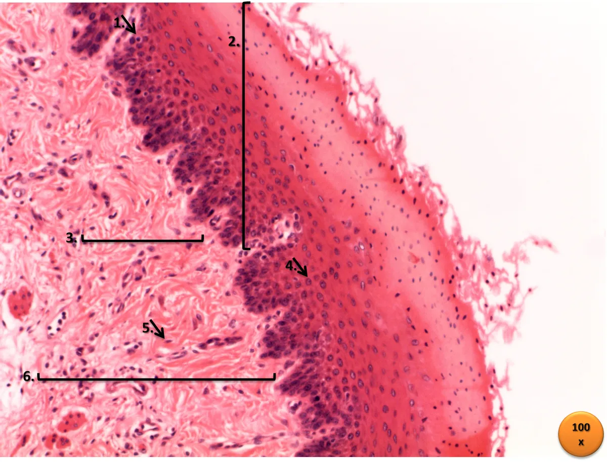
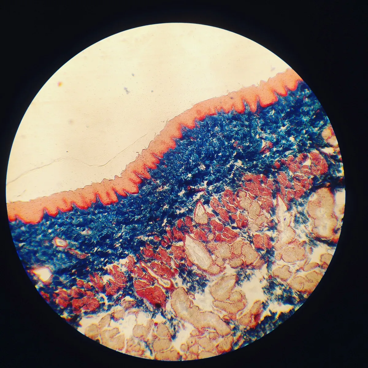
Mucosa of non-keratinised stratified squamous epithelium, submucosa with mucous glands, inner circular and outer longitudinal muscle (striated in the upper third, mixed in the middle, smooth in the lower third) and an adventitia without serosa, which eases cancer spread to neighbouring organs. Chronic reflux causes Barrett metaplasia.
- 06
Constrictions
Angustiae oesophagi
Three physiological narrowings: at its beginning behind the cricoid (about 15 cm from the incisors), where the aortic arch and left main bronchus cross (about 25 cm), and where it pierces the diaphragm (about 40 cm). Foreign bodies, caustic injuries and cancers favour these sites.
Position and relations
The oesophagus begins at the lower border of the cricoid cartilage (C6) and descends in front of the vertebral column, behind the trachea and the heart (related to the left atrium), inclining to the left and forwards to pass through the oesophageal hiatus at T10 and join the cardia at T11. Its thoracic part lies in the posterior mediastinum beside the thoracic aorta and thoracic duct; the vagal trunks form a plexus around it.
Form and structure
The oesophageal wall has no serosa (except its short abdominal part) and consists of a non-keratinised stratified squamous mucosa, a submucosa with oesophageal glands, and a two-layered muscularis (inner circular, outer longitudinal): skeletal muscle in the upper third, smooth muscle in the lower third and a mixture in between. The upper sphincter (cricopharyngeus) and the functional lower sphincter prevent reflux.
Vessels and nerves
The arteries follow its segments: the inferior thyroid artery for the cervical part, oesophageal branches of the aorta and bronchial arteries for the thoracic part, and the left gastric artery for the abdominal part. In the lower part, oesophageal veins drain both to the azygos vein (systemic) and to the left gastric vein (portal); in portal hypertension this anastomosis dilates into oesophageal varices that may rupture.
Clinical correlations
Gastro-oesophageal reflux disease arises when the anti-reflux barrier is compromised and the oesophagus clears and buffers the refluxate less well; oesophageal exposure to gastric contents is the main determinant of severity. Symptoms can still occur with a normal reflux burden if epithelial resistance is poor or visceral sensitivity is increased.[1]
Swallowing, peristalsis and the two sphincters
The adult oesophagus is 23–25 cm long and carries food one way from pharynx to stomach. The upper oesophageal sphincter (pharyngo-oesophageal sphincter), just below the back of the tongue and oropharynx, is a circular bundle that is normally contracted and closed; during swallowing it relaxes briefly to let the bolus (food, drink, mucus and saliva) through. In the pharyngeal phase, contraction of the pharyngeal wall generates the first peristaltic wave that drives the bolus through the upper sphincter and continues down the body as primary peristalsis; if that wave fails to deliver the bolus, oesophageal distension triggers secondary peristalsis until the bolus reaches the stomach. The lower oesophageal sphincter lies about 3 cm above the gastric junction and is formed by thickening and overlap of the inner circular layer; it is normally contracted to prevent reflux and opens involuntarily with peristalsis. The oesophagus also allows retrograde expulsion in vomiting, belching and the gag reflex.[2]
The upper third of the oesophagus is mainly skeletal muscle and the lower two-thirds smooth muscle; the inner circular layer is thicker than the outer longitudinal layer and generates peristalsis. The vagus provides parasympathetic motor supply to muscle and glands; the cervical and thoracic sympathetic chain (T1–T10) causes vasoconstriction, maintains tone in both sphincters and generally inhibits peristalsis; the vagus conveys pressure sensation, while the sympathetic chain carries pain. Veins drain segmentally: the cervical part to the inferior thyroid and then brachiocephalic veins, the thoracic part to the azygos and hemiazygos veins, and the distal part to the left gastric vein of the portal system. Lymph from the upper third goes to deep cervical nodes, from the middle third to superior and posterior mediastinal nodes and from the lower third to gastric and coeliac nodes. The oesophagus develops from the foregut in week 4; rare congenital anomalies include oesophageal atresia and tracheo-oesophageal fistula, stenosis, duplication and duplication cysts, rings and webs. Gastro-oesophageal reflux, often thought minor, can progress to oesophagitis, ulcers, strictures, Barrett oesophagus and adenocarcinoma.[2]
Full-text reading (StatPearls)
The StatPearls chapters on this topic, reproduced verbatim. Their CC BY-NC-ND 4.0 licence allows unmodified copies only, so they are not translated or shortened.
Anatomy, Thorax, EsophagusOmole AE, Bordoni B · StatPearls 2026 · NBK482513 · 9k characters
Loading…
Omole AE, Bordoni B. Anatomy, Thorax, Esophagus. StatPearls [Internet]. Treasure Island (FL): StatPearls Publishing; 2026. NBK482513 · PMID 29494119 · CC BY-NC-ND 4.0. Reproduced verbatim, unmodified.
Organs in this system
References
Sources for the values and descriptions on this page, indexed in PubMed (US National Library of Medicine).
- [1]Pathophysiology of Gastroesophageal Reflux DiseaseTack J et al., Gastroenterology 2018PMID 29037470
- [2]Anatomy, Thorax, EsophagusOmole AE et al., StatPearls 2026PMID 29494119
- [3]Gastrointestinal mean transit times in young and middle-aged healthy subjectsPMID 11318834Supports: Gastrointestinal transit time
- [4]Revised Estimates for the Number of Human and Bacteria Cells in the BodySender R, Fuchs S, Milo R, 2016PMID 27541692Supports: Colonic bacterial count