Skin
Model
Hover or tap to see names, click to read a description; click a name in the legend to fly to it. Procedurally built to histological proportions; colours follow illustration conventions.
Description
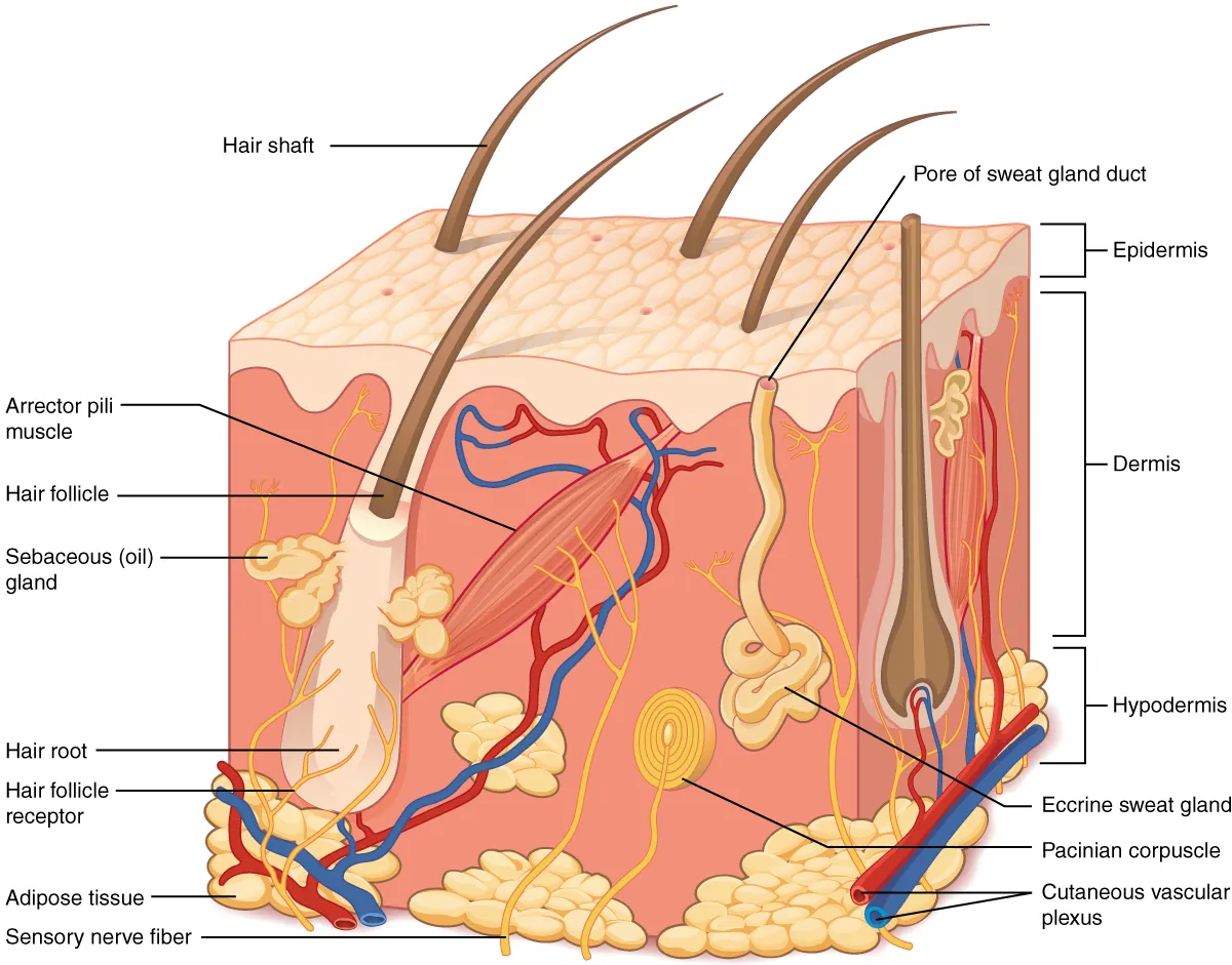
The skin consists of the epidermis, a keratinised stratified squamous epithelium continuously renewed from its basal layer, and the dermis, connective tissue with collagen, elastic fibres, vessels, nerves and appendages. Thickness varies by region and is greatest on the palms and soles. Beneath lies the fat-rich subcutaneous tissue, which cushions and insulates.
Parts
Each part has its own card; press “Show on the model” to fly to it and select it in the 3D model above.
- 01
Epidermis
Epidermis
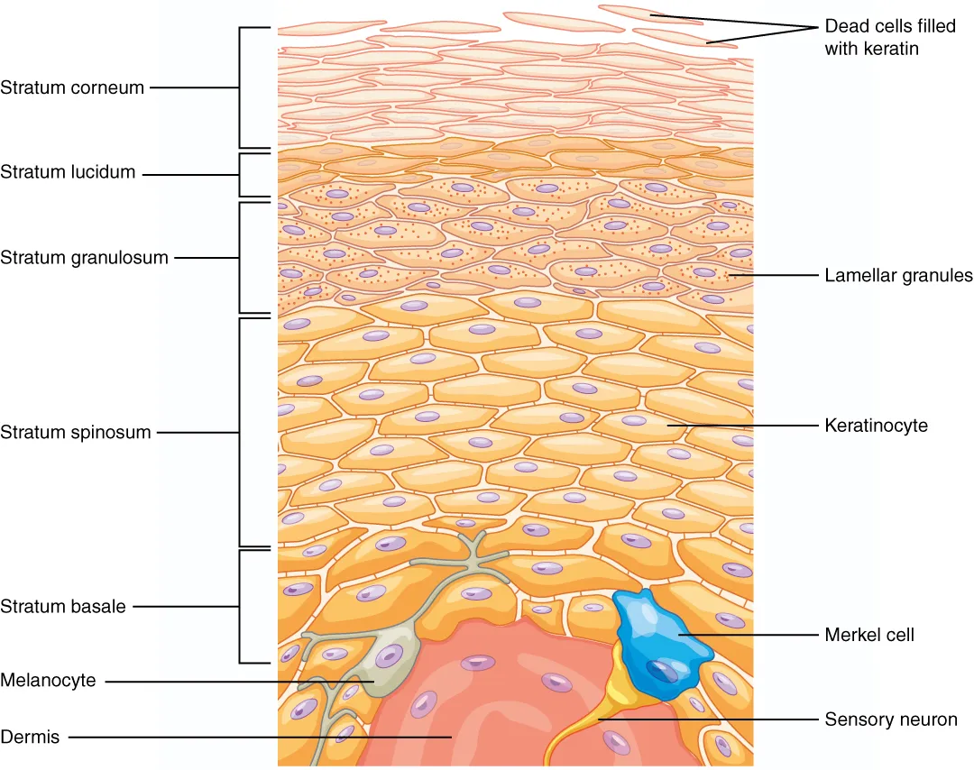
A keratinised stratified squamous epithelium without blood vessels, 0.05 mm thick on the eyelid to 1.5 mm on the sole. From deep to superficial: basal, spinous, granular, (lucid in thick skin) and cornified layers. Keratinocytes born in the basal layer take about four weeks to reach the surface and shed. It also contains melanocytes, Langerhans cells and Merkel cells.
- 02
Dermis
Dermis
Dense connective tissue of collagen and elastic fibres carrying vessels, nerves and appendages. The superficial papillary layer rises into dermal papillae with capillary loops and Meissner corpuscles; the deep reticular layer has collagen bundles aligned along skin tension lines (Langer’s lines), so incisions along them heal with finer scars.
- 03
Hypodermis (subcutaneous tissue)
Tela subcutanea
Loose connective and adipose tissue binding the skin to the deep fascia; it insulates, cushions and stores energy. Its thickness varies with region, sex and build; the scrotum, eyelids and penis have almost no fat. It is the layer used for subcutaneous injection.
- 04
Hair and hair follicles
Pili et folliculi pilorum
The hair follicle is an epidermal tube sunk into the dermis; its bulb contains matrix cells proliferating around a dermal papilla. Each follicle receives a sebaceous gland and an arrector pili muscle (smooth muscle that raises “goose bumps”). The bulge region holds stem cells that regenerate the follicle and the epidermis after injury. Palms and soles are hairless.
- 05
Nails
Unguis
A hard keratin plate on the nail bed, produced by the nail matrix under the proximal nail fold; the lunula is the visible part of the matrix. Fingernails grow about 3 mm a month, toenails about three times more slowly. Damage to the matrix leaves a permanently deformed nail.
- 06
Sebaceous and sweat glands
Glandulae cutis
Sebaceous glands release sebum by holocrine secretion into the follicle and are most numerous on the face and scalp. Eccrine sweat glands, 2–4 million of them, open directly onto the surface and control temperature; apocrine glands in the axilla, groin and areola open into follicles and become active at puberty. Ceruminous glands and the glands of Moll are modified apocrine glands.
- 07
Cutaneous receptors
Receptores cutanei
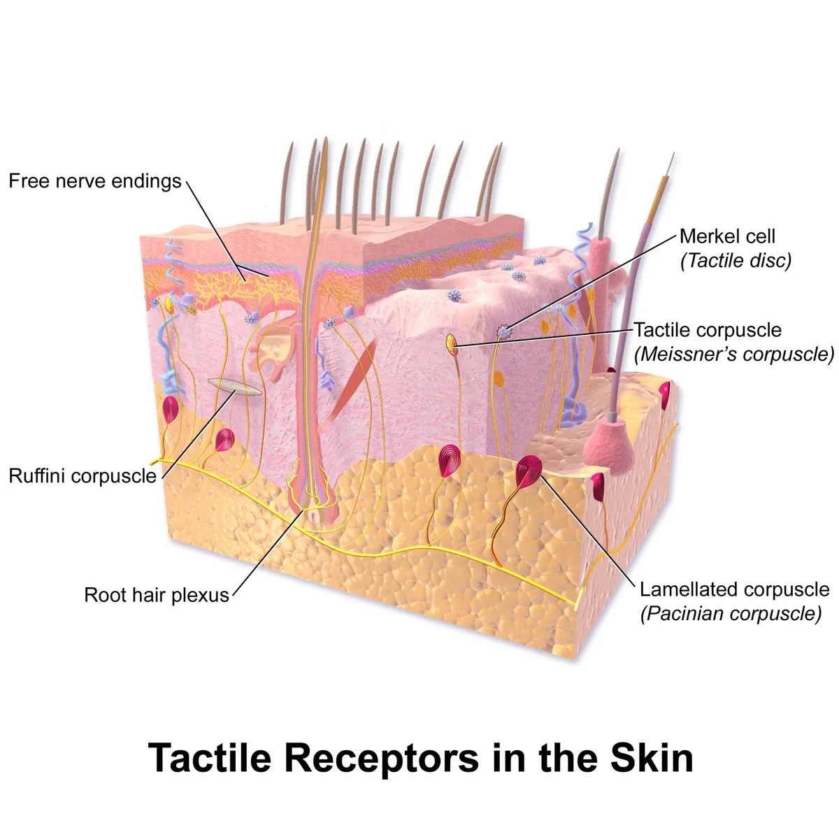
Free nerve endings (pain, temperature), Merkel discs (sustained pressure, shape), Meissner corpuscles (light touch, low-frequency vibration) in the dermal papillae, Pacinian corpuscles (high-frequency vibration) in the hypodermis and Ruffini endings (skin stretch). Their density is greatest at the fingertips and lips, where two points only 2 mm apart can be told apart.
Form and structure
From deep to superficial, the epidermis has a basal layer (stem cells, melanocytes, Merkel cells), a spinous layer, a granular layer, a clear layer (in thick skin only) and the stratum corneum. The dermis has a papillary layer, whose papillae interdigitate with epidermal ridges, and a reticular layer of thick collagen bundles; skin tension lines (Langer’s lines) follow these bundles, so incisions parallel to them heal with better scars.
Position and relations
The skin appendages include hair follicles with sebaceous glands and arrector pili muscles; eccrine sweat glands throughout the body (densest on the palms, soles and forehead) for heat loss; and apocrine glands in the axillae and genital region. The nail is a keratin plate on the nail bed, growing from the matrix beneath the proximal nail fold.
Vessels and nerves
The skin has two vascular plexuses, a deep one at the dermis–hypodermis junction and a superficial one beneath the papillae, joined to each other and equipped with arteriovenous anastomoses that adjust skin blood flow to shed or conserve heat. The epidermis is avascular and nourished by diffusion. Each spinal segment supplies a band of skin (dermatome), the basis for localising nerve lesions.
Microscopic organisation
The skin has three tiers. The epidermis is an avascular keratinised stratified squamous epithelium; from deep to superficial it comprises the basal, spinous, granular and cornified layers (plus a lucid layer on palms and soles). The dermis is connective tissue with collagen, elastin, vessels, nerves and appendages; the hypodermis (tela subcutanea) is mostly fat. Knowing their histology underpins the understanding of ageing changes and of cosmetic outcomes.[1]
Keratinocytes of the nucleated layers are joined by desmosomes and tight, gap and adherens junctions; on reaching the stratum corneum they lose their nuclei, acquire a cornified envelope and lie between lipid lamellae (ceramides, fatty acids, cholesterol). Filaggrin bundles keratin and its breakdown yields the skin’s natural moisturising factors.[2]
Clinical correlations
Atopic dermatitis is the most common chronic inflammatory skin disease. It combines barrier defects (such as filaggrin mutations), skewed immune responses and microbiome changes; this understanding has produced biologics and small molecules aimed at specific immune pathways.[3][2]
A narrow band of uninvolved papillary dermis separating the epidermis from a dermal infiltrate is called the “grenz zone”; it is a histopathological clue that distinguishes many dermal infiltrative diseases (such as lymphoma or leprosy) from lesions arising in the epidermis.[4]
Full-text reading (StatPearls)
The StatPearls chapters on this topic, reproduced verbatim. Their CC BY-NC-ND 4.0 licence allows unmodified copies only, so they are not translated or shortened.
Anatomy, Skin (Integument), EpidermisYousef H, Alhajj M, Fakoya AO, Sharma S · StatPearls 2024 · NBK470464 · 18k characters
Loading…
Yousef H, Alhajj M, Fakoya AO, Sharma S. Anatomy, Skin (Integument), Epidermis. StatPearls [Internet]. Treasure Island (FL): StatPearls Publishing; 2024. NBK470464 · PMID 29262154 · CC BY-NC-ND 4.0. Reproduced verbatim, unmodified.
Histology, Skin AppendagesYousef H, Miao JH, Alhajj M, Badri T · StatPearls 2023 · NBK482237 · 17k characters
Loading…
Yousef H, Miao JH, Alhajj M, Badri T. Histology, Skin Appendages. StatPearls [Internet]. Treasure Island (FL): StatPearls Publishing; 2023. NBK482237 · PMID 29489291 · CC BY-NC-ND 4.0. Reproduced verbatim, unmodified.
Anatomy, Skin BursaMercadante JR, Marappa-Ganeshan R · StatPearls 2022 · NBK554438 · 12k characters
Loading…
Mercadante JR, Marappa-Ganeshan R. Anatomy, Skin Bursa. StatPearls [Internet]. Treasure Island (FL): StatPearls Publishing; 2022. NBK554438 · PMID 32119325 · CC BY-NC-ND 4.0. Reproduced verbatim, unmodified.
Anatomy, Skin, Superficial Musculoaponeurotic System (SMAS) FasciaWhitney ZB, Jain M, Zito PM · StatPearls 2024 · NBK519014 · 10k characters
Loading…
Whitney ZB, Jain M, Zito PM. Anatomy, Skin, Superficial Musculoaponeurotic System (SMAS) Fascia. StatPearls [Internet]. Treasure Island (FL): StatPearls Publishing; 2024. NBK519014 · PMID 30085556 · CC BY-NC-ND 4.0. Reproduced verbatim, unmodified.
Anatomy, Skin (Integument)Lopez-Ojeda W, Pandey A, Alhajj M, Oakley AM · StatPearls 2022 · NBK441980 · 10k characters
Loading…
Lopez-Ojeda W, Pandey A, Alhajj M, Oakley AM. Anatomy, Skin (Integument). StatPearls [Internet]. Treasure Island (FL): StatPearls Publishing; 2022. NBK441980 · PMID 28723009 · CC BY-NC-ND 4.0. Reproduced verbatim, unmodified.
Anatomy, Skin, DermatomesWhitman PA, Launico MV, Adigun OO · StatPearls 2023 · NBK535401 · 9k characters
Loading…
Whitman PA, Launico MV, Adigun OO. Anatomy, Skin, Dermatomes. StatPearls [Internet]. Treasure Island (FL): StatPearls Publishing; 2023. NBK535401 · PMID 30571022 · CC BY-NC-ND 4.0. Reproduced verbatim, unmodified.
Histology, SkinAgarwal S, Krishnamurthy K · StatPearls 2023 · NBK537325 · 8k characters
Loading…
Agarwal S, Krishnamurthy K. Histology, Skin. StatPearls [Internet]. Treasure Island (FL): StatPearls Publishing; 2023. NBK537325 · PMID 30726010 · CC BY-NC-ND 4.0. Reproduced verbatim, unmodified.
Anatomy, Skin, Sudoriferous GlandPatel BC, Treister AD, McCausland C, Lio PA, Jozsa F · StatPearls 2023 · NBK513244 · 8k characters
Loading…
Patel BC, Treister AD, McCausland C, Lio PA, Jozsa F. Anatomy, Skin, Sudoriferous Gland. StatPearls [Internet]. Treasure Island (FL): StatPearls Publishing; 2023. NBK513244 · PMID 30020616 · CC BY-NC-ND 4.0. Reproduced verbatim, unmodified.
Organs in this system
References
Sources for the values and descriptions on this page, indexed in PubMed (US National Library of Medicine).
- [1]Aging skin: histology, physiology, and pathologyKhavkin J et al., Facial plastic surgery clinics of North America 2011PMID 21763983
- [2]The skin: an indispensable barrierProksch E et al., Experimental dermatology 2008PMID 19043850
- [3]Update on atopic dermatitisMeledathu S et al., The Journal of allergy and clinical immunology 2025PMID 39855361
- [4]The grenz zoneAbbas O et al., The American Journal of dermatopathology 2013PMID 23348142