Lungs
Model
Hover or tap to see names, click to read a description; click a name in the legend to fly to it. Procedurally built to histological proportions; colours follow illustration conventions.
Description
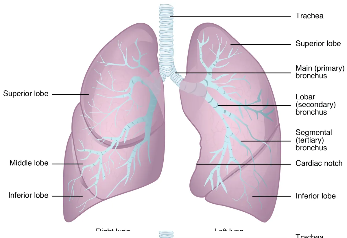
Paired spongy organs within the pleural cavities. The right lung has three lobes (superior, middle, inferior); the left lung has two lobes and a cardiac notch.
Parts
Each part has its own card; press “Show on the model” to fly to it and select it in the 3D model above.
- 01
Right upper lobe
Lobus superior pulmonis dextri
Lies above the horizontal fissure and has apical, posterior and anterior segments; its apex rises above the first rib into the root of the neck. Its bronchus branches very early, just below the tracheal bifurcation, above the pulmonary artery (the eparterial bronchus).
- 02
Right middle lobe
Lobus medius pulmonis dextri
A small wedge-shaped lobe between the horizontal and oblique fissures, with lateral and medial segments, lying against the right heart border. Its long narrow bronchus, surrounded by lymph nodes, is prone to compression, causing recurrent middle lobe collapse.
- 03
Right lower lobe
Lobus inferior pulmonis dextri
The largest lobe, below and behind the oblique fissure, with a superior segment and four basal segments (medial, anterior, lateral, posterior) resting on the right dome of the diaphragm. The superior and posterior basal segments are typical sites of aspiration pneumonia in supine patients.
- 04
Left upper lobe and lingula
Lobus superior pulmonis sinistri; Lingula
The left lung has only two lobes because the heart takes up room. The upper lobe comprises the apicoposterior and anterior segments and the lingula (superior and inferior lingular segments), the counterpart of the right middle lobe; its anterior border bears the cardiac notch.
- 05
Left lower lobe
Lobus inferior pulmonis sinistri
Lies below and behind the oblique fissure, with a superior segment and basal segments (a combined anteromedial, lateral and posterior), resting on the left dome of the diaphragm above the gastric fundus and spleen.
- 06
Fissures and pleura
Fissura obliqua et horizontalis; Pleura
The oblique fissure (both lungs) runs from about the T3 spinous process forward to the sixth costal cartilage; the horizontal fissure (right lung only) follows the fourth rib. Visceral pleura covers the lungs and dips into the fissures; parietal pleura lines the chest wall, the two separated by a pleural cavity holding a thin film of fluid.
- 07
Hilum and root of the lung
Hilum pulmonis; Radix pulmonis
Where the main bronchus, pulmonary artery, two pulmonary veins, bronchial vessels, lymphatics and nerve plexus enter and leave the lung, wrapped in a pleural sleeve that hangs down as the pulmonary ligament. Hilar nodes are a key lymphatic station in lung cancer staging.
- 08
Bronchial tree
Arbor bronchialis
Main bronchi divide into lobar and then segmental bronchi (10 on the right, 8–10 on the left) and continue through about 23 generations of branching. Bronchi have cartilage plates and glands; bronchioles (under 1 mm) lack cartilage and have smooth muscle and club cells; terminal bronchioles end the conducting zone.
- 09
Pulmonary vessels
Arteriae et venae pulmonales
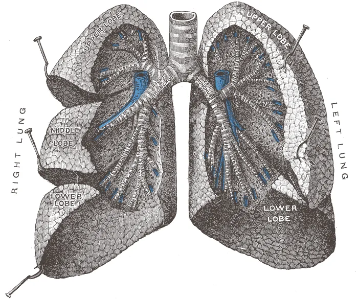
Pulmonary arteries accompany the bronchi to the centre of each lobule carrying deoxygenated blood; pulmonary veins run in the interlobular septa at the periphery and converge into two veins on each side to the left atrium. Small bronchial arteries from the aorta nourish the bronchial walls and visceral pleura.
- 10
Acinus and alveoli
Acinus pulmonis; Alveoli pulmonis
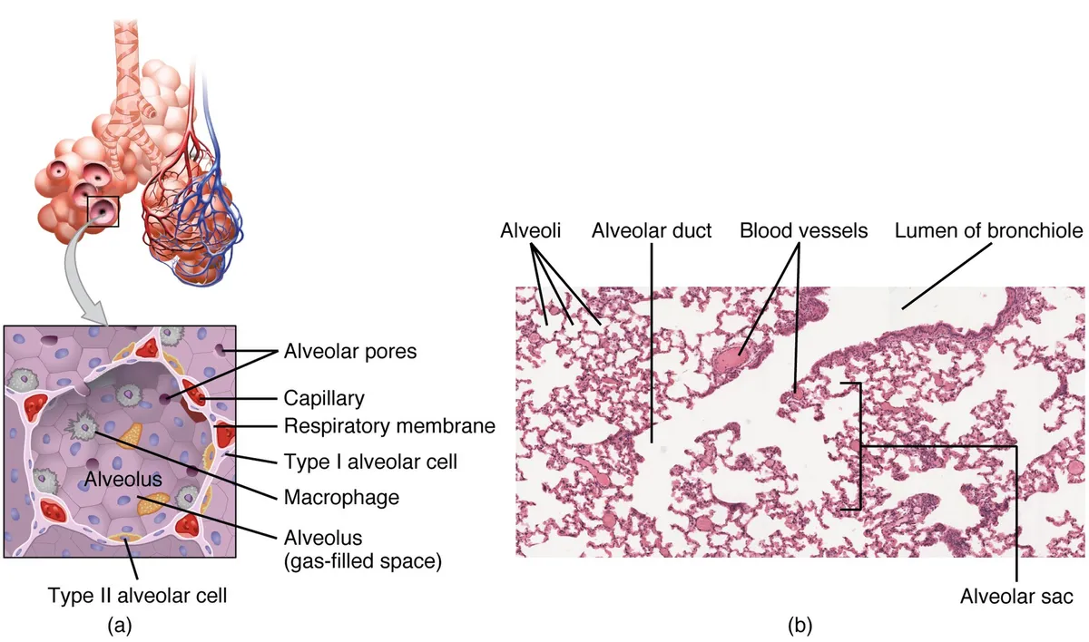
The region beyond a terminal bronchiole: respiratory bronchioles, alveolar ducts, alveolar sacs and alveoli. There are about 300–500 million alveoli with a total surface of some 70 m²; the air–blood barrier is only about 0.5 µm thick (type I pneumocyte, fused basement membrane, endothelium).
Position and relations
The two lungs occupy the thoracic cavity, each in its own pleural cavity, separated by the mediastinum. The apex rises 2–3 cm above the clavicle and the base rests on the diaphragm. The hilum on the mediastinal surface is where the main bronchus and pulmonary vessels enter and leave.
Form and structure
The right lung has three lobes (upper, middle, lower) separated by the oblique and horizontal fissures; the left lung has two lobes and a cardiac notch. Each lobe divides into bronchopulmonary segments, anatomical and surgical units with their own bronchus and pulmonary artery branch. The bronchial tree branches about 23 times, ending in some 300–500 million alveoli.[1]
Vessels and nerves
The lungs have two vascular systems: the pulmonary arteries carry deoxygenated blood from the right ventricle to the alveolar capillaries for gas exchange, while the bronchial arteries (from the aorta) nourish the bronchial walls. Pulmonary arterioles constrict when alveoli are hypoxic, matching perfusion to ventilation.[2]
Development
The lung begins to develop in the fourth week of gestation, when the tracheal bud grows out of the foregut, and continues to mature (alveolarisation) into early childhood. Survival at birth depends on adequate lung maturation in utero; abnormal development causes congenital lung malformations, and incomplete development contributes to bronchopulmonary dysplasia in preterm infants.[3]
The air–blood barrier
Stereological morphometry measures the area and thickness of the barrier between alveolar air and capillary blood and feeds them into physiological equations to estimate pulmonary diffusing capacity, which agrees with maximal oxygen consumption in experiments. The lung is a “machine” with a vast air–blood barrier built from minimal cell mass.[1]
The alveolar epithelium comprises flat type I cells covering most of the gas-exchange surface and type II cells that secrete surfactant. After alveolar injury (severe infection, inhalation injury, sepsis), type II cells proliferate and differentiate into type I cells to restore the epithelium; recovery requires an intact barrier. Defective type II cell quality control contributes to pulmonary fibrosis.[4][5]
Lung development starts at 4 weeks of gestation with the tracheal outgrowth from the foregut and continues into early childhood. Survival at birth depends on adequate development and maturation in utero; abnormal development causes congenital lung malformations, and inadequate development contributes to bronchopulmonary dysplasia in preterm infants.[3][6]
Full-text reading (StatPearls)
The StatPearls chapters on this topic, reproduced verbatim. Their CC BY-NC-ND 4.0 licence allows unmodified copies only, so they are not translated or shortened.
Anatomy, Thorax, LungsChaudhry R, Omole AE, Bordoni B · StatPearls 2024 · NBK470197 · 15k characters
Loading…
Chaudhry R, Omole AE, Bordoni B. Anatomy, Thorax, Lungs. StatPearls [Internet]. Treasure Island (FL): StatPearls Publishing; 2024. NBK470197 · PMID 29262068 · CC BY-NC-ND 4.0. Reproduced verbatim, unmodified.
Organs in this system
References
Sources for the values and descriptions on this page, indexed in PubMed (US National Library of Medicine).
- [1]Lung morphometry: the link between structure and functionWeibel ER, Cell and tissue research 2017PMID 27981379
- [2]Hypoxic pulmonary vasoconstriction: physiology and anesthetic implicationsLumb AB et al., Anesthesiology 2015PMID 25587641
- [3]Lung developmentMullassery D et al., Seminars in pediatric surgery 2015PMID 26051046
- [4]Repair and regeneration of the alveolar epithelium in lung injuryWang Y et al., FASEB journal 2024PMID 38648494
- [5]Contributions of alveolar epithelial cell quality control to pulmonary fibrosisKatzen J et al., The Journal of clinical investigation 2020PMID 32870817
- [6]Congenital Lung MalformationsPalla J et al., Pediatric annals 2019PMID 30986318
- [7]The normal human lung: ultrastructure and morphometric estimation of diffusion capacityGehr P, Bachofen M, Weibel ER, 1978PMID 644146Supports: Alveolar surface area
- [8]Physiology, LungStatPearls (NCBI Bookshelf)PMID 31424761Supports: Respiratory rate