Testes
Model
Hover or tap to see names, click to read a description; click a name in the legend to fly to it. Procedurally built to histological proportions; colours follow illustration conventions.
Description
Paired ovoid glands in the scrotum, about 4–5 cm long, covered by the tunica albuginea, with the epididymis along the posterior border. The seminiferous tubules (with Sertoli cells) produce spermatozoa; interstitial Leydig cells secrete testosterone. Scrotal temperature, a few degrees below core temperature, is needed for spermatogenesis.
Parts
Each part has its own card; press “Show on the model” to fly to it and select it in the 3D model above.
- 01
Coverings of the testis
Tunica vaginalis; Tunica albuginea
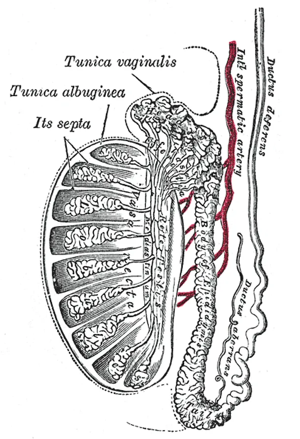
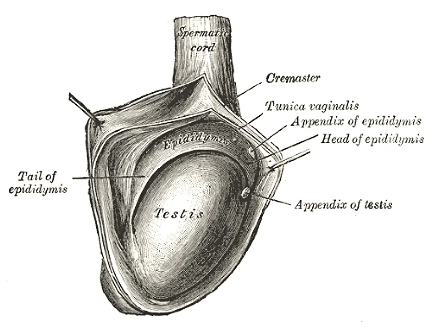
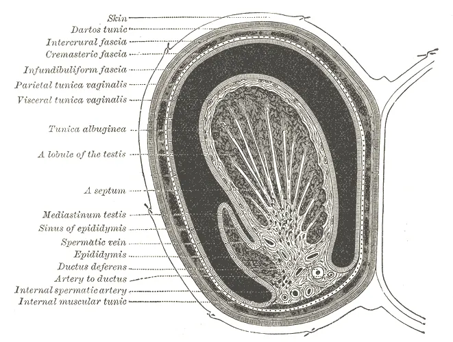
Outermost are the scrotum and the spermatic fasciae; the tunica vaginalis (two serous layers, a remnant of the processus vaginalis) covers the front and sides, and fluid in its cavity forms a hydrocele. The tunica albuginea is a thick fibrous coat, thickened posteriorly as the mediastinum testis.
- 02
Lobules and seminiferous tubules
Lobuli testis; Tubuli seminiferi contorti
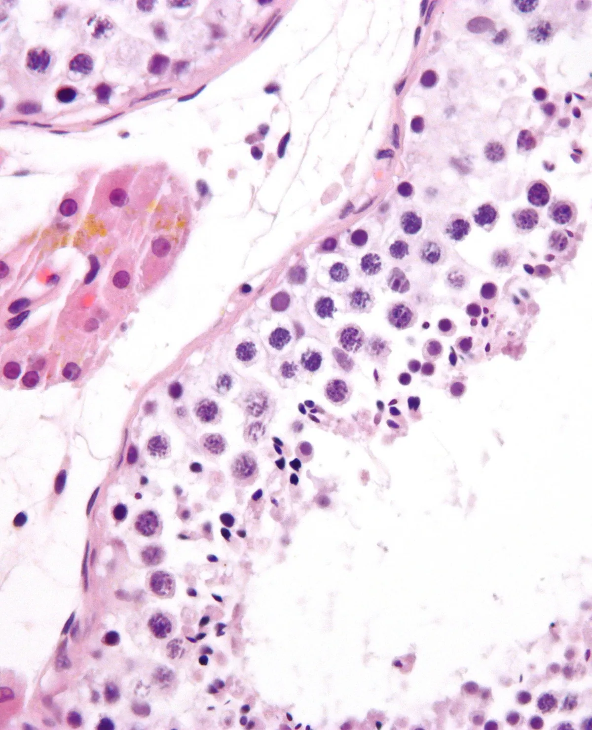
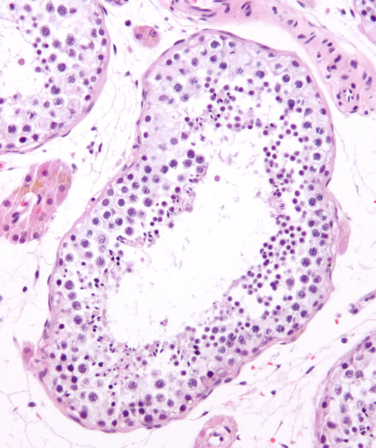
Fibrous septa divide the testis into about 250 lobules, each with 1–4 coiled seminiferous tubules (several hundred metres in all). The seminiferous epithelium holds germ cells at all stages and nourishing Sertoli cells that form the blood–testis barrier; spermatogenesis takes about 64–74 days.
- 03
Interstitial (Leydig) cells
Endocrinocyti interstitiales
Clusters in the interstitium between the tubules, close to capillaries; stimulated by LH they secrete testosterone (about 95% of male testosterone). Leydig cell tumours can cause precocious puberty or gynaecomastia.
- 04
Rete testis and efferent ductules
Rete testis; Ductuli efferentes
Straight tubules drain into an anastomosing network in the mediastinum testis; from it 12–20 efferent ductules pierce the tunica albuginea to the head of the epididymis. Their epithelium has ciliated cells and cells that reabsorb most of the testicular fluid.
- 05
Epididymis
Epididymis
A coiled duct about 6 m long folded into head, body and tail along the back of the testis; sperm take about two weeks to traverse it, acquiring motility, and are stored in the tail. Epididymitis is a common cause of scrotal pain.
- 06
Spermatic cord and ductus deferens
Funiculus spermaticus; Ductus deferens
The cord carries the ductus deferens (thick-walled, felt as a firm string), testicular artery, pampiniform venous plexus (dilated in varicocele, usually left), lymphatics and nerves through the inguinal canal. Testicular lymph drains to para-aortic nodes at L1–L2, not to inguinal nodes.
Position and relations
The two testes lie in the scrotum, the left usually hanging lower than the right. In fetal life the testis develops on the posterior abdominal wall and descends into the scrotum through the inguinal canal, drawing with it the vessels, nerves and ductus deferens that form the spermatic cord. It is invested by the tunica vaginalis (parietal and visceral layers), a remnant of the processus vaginalis.
Form and structure
The thick tunica albuginea sends septa inwards that divide the testis into about 250 lobules, each containing coiled seminiferous tubules. These drain through the straight tubules and the rete testis into the efferent ductules and the head of the epididymis. The epididymis (head, body, tail) is where spermatozoa mature and are stored before entering the ductus deferens.
Vessels and nerves
The testicular artery arises directly from the abdominal aorta at about L2. The veins form the pampiniform plexus around the artery, which cools the arterial blood; the right testicular vein drains into the inferior vena cava and the left into the left renal vein, which partly explains why varicocele is more common on the left. Testicular lymph drains to the para-aortic nodes, not to the inguinal nodes.
Physiology
Spermatogenesis has three phases: a proliferative phase that restocks the stem-cell pool, meiosis (recombination and haploidisation), and post-meiotic differentiation (spermiogenesis, with DNA hypercondensation). Spermatozoa then mature further in the epididymis, acquire seminal plasma components at ejaculation and complete capacitation in the uterine tube.[1]
Spermatogonial stem cells are the most primitive spermatogonia; by self-renewing while continuously producing differentiating daughters, they sustain lifelong sperm production. The process requires many cell types, hormones, paracrine factors and epigenetic regulators, so it is vulnerable to many causes of male infertility.[2][3]
Coverings, epididymis and descent
The testis is the male gonad, with both endocrine and exocrine roles; it is oval, about 3–5 cm long and 2–3 cm wide, lies in the scrotum separated from its partner by the scrotal septum, and feels smooth and soft through the scrotal skin. The spermatic cord suspends its upper pole, while the scrotal ligament, a remnant of the gubernaculum, anchors the lower pole to the scrotum; the left testis usually sits a little lower than the right. The two-layered tunica vaginalis covers the testis except at its posterior and superior borders, where the epididymis and spermatic cord attach; its visceral layer clings to the epididymis, testis and ductus deferens, and a small recess between the posterolateral surface of the testis and the body of the epididymis forms the sinus of the epididymis. Beneath the tunica vaginalis lies the tunica albuginea, a tough fibrous capsule. The epididymis lies along the posterior border and has a head, body and tail; it is a tightly coiled duct that would measure about 6 m if straightened, giving ample room for sperm maturation and storage. Its head, at the upper pole, receives fluid from the testicular ducts and passes sperm on to its distal part. As the testis travels from the abdomen to the scrotum it acquires the internal spermatic fascia, the cremaster muscle and fascia, and the external spermatic fascia; the cremaster raises or lowers the testis mainly in response to temperature.[4]
The testis begins as an undifferentiated gonad in the retroperitoneum. The SRY gene on the Y chromosome, the testis-determining factor, makes it differentiate into a testis; without SRY the gonad becomes an ovary. The fetal testis secretes testosterone, which allows the male genitalia to develop, and its Sertoli cells secrete Müllerian-inhibiting substance, which makes the paramesonephric ducts regress at 8–10 weeks; in the adult only the appendix testis and the prostatic utricle remain. The tunica albuginea forms by invagination as a connective tissue layer between the seminiferous tubules and the rest of the gland. In the third trimester the testis leaves the abdomen, passes through the inguinal canal and reaches the scrotum, preceded by the processus vaginalis, a peritoneal pouch. Normally this pouch undergoes apoptosis and leaves only the tunica vaginalis; if it fails to close, a communicating hydrocele and an inguinal hernia can follow. Functionless embryonic remnants include the appendix testis (cranial end of the Müllerian duct, at the upper pole in the groove between the head of the epididymis and the testis) and the appendix of the epididymis (cranial end of the mesonephric duct, variable in position but usually on the head of the epididymis); neither is constant. In about 7% of men the epididymis lies on the anterior surface of the testis, which matters at examination and surgery.[4]
Tunica vaginalis
- Embryonic origin
- Processus vaginalis
- Adult position
- Around the testis except its posterior and upper borders
Scrotal ligament
- Embryonic origin
- Gubernaculum
- Adult position
- Lower pole to the scrotum
Appendix testis
- Embryonic origin
- Cranial Müllerian duct
- Adult position
- Upper pole, groove between epididymal head and testis
Appendix of the epididymis
- Embryonic origin
- Cranial mesonephric duct
- Adult position
- Usually on the epididymal head
Prostatic utricle
- Embryonic origin
- Müllerian duct
- Adult position
- Within the prostate
| Remnant | Embryonic origin | Adult position |
|---|---|---|
| Tunica vaginalis | Processus vaginalis | Around the testis except its posterior and upper borders |
| Scrotal ligament | Gubernaculum | Lower pole to the scrotum |
| Appendix testis | Cranial Müllerian duct | Upper pole, groove between epididymal head and testis |
| Appendix of the epididymis | Cranial mesonephric duct | Usually on the epididymal head |
| Prostatic utricle | Müllerian duct | Within the prostate |
Cryptorchidism, torsion, varicocele and infertility
The testicular artery leaves the anterolateral aorta just below the renal arteries, runs retroperitoneally across the front of the ureter and enters the spermatic cord at the deep inguinal ring; the testis also receives blood from the artery to the ductus deferens and the cremasteric artery. Venous blood collects in the pampiniform plexus in front of the ductus and forms the testicular vein, which on the right joins the inferior vena cava and on the left the left renal vein. Lymph follows the arteries to the preaortic nodes. A varicocele is dilatation of these veins; it is fairly common and usually silent, but some men develop infertility and testosterone deficiency. The main mechanism is oxidative stress in the parenchyma, driven by scrotal hyperthermia, testicular hypoxia and disruption of the blood–testis barrier; ligation or percutaneous embolisation of the dilated veins can improve semen parameters, fertility and serum testosterone. Infertility affects 8–12% of couples, and a male factor is the main or a contributing cause in about half; the causes, congenital, acquired or idiopathic, all impair spermatogenesis. Semen analysis remains the cornerstone of assessment, supplemented by tests of sperm quality and function, and assisted reproduction lets many couples have biological children.[4][5][6]
Cryptorchidism (undescended testis) must be recognised early because it causes infertility and raises the risk of testicular cancer, including in the opposite testis, so regular self-examination is advised. The main treatment is orchidopexy, which should be done before 1 year of age in congenital cryptorchidism; if the testis cannot be brought into the scrotum where it can be watched, it should be removed. Every true undescended testis has an associated inguinal hernia, whereas a retractile testis does not. In premature infants descent is often incomplete and needs follow-up after birth; occasionally the testis leaves the superficial inguinal ring but strays between thigh and scrotum to lie in the perineum, an ectopic testis. Testicular torsion in young men is a surgical emergency: blood flow must be restored within 6 hours of onset and the testis fixed in the scrotum, and most urologists fix the other side at the same time. A hydrocele is serous fluid between the layers of the tunica vaginalis, caused by inflammation, trauma or, congenitally, a processus vaginalis still open to the abdomen.[4]
Full-text reading (StatPearls)
The StatPearls chapters on this topic, reproduced verbatim. Their CC BY-NC-ND 4.0 licence allows unmodified copies only, so they are not translated or shortened.
Anatomy, Abdomen and Pelvis: TestesTiwana MS, Leslie SW · StatPearls 2023 · NBK470201 · 8k characters
Loading…
Tiwana MS, Leslie SW. Anatomy, Abdomen and Pelvis: Testes. StatPearls [Internet]. Treasure Island (FL): StatPearls Publishing; 2023. NBK470201 · PMID 29261881 · CC BY-NC-ND 4.0. Reproduced verbatim, unmodified.
Organs in this system
References
Sources for the values and descriptions on this page, indexed in PubMed (US National Library of Medicine).
- [1]A concise overview of mammalian spermatogenesisSultana T et al., Systems biology in reproductive medicine 2026PMID 41511981
- [2]Spermatogonial stem cellsKubota H et al., Biology of reproduction 2018PMID 29617903
- [3]Spermatogenesis in humans and its affecting factorsNeto FT et al., Seminars in cell & developmental biology 2016PMID 27143445
- [4]Anatomy, Abdomen and Pelvis: TestesTiwana MS et al., StatPearls 2023PMID 29261881
- [5]Effect of varicoceles on spermatogenesisKang C et al., Seminars in cell & developmental biology 2022PMID 33965333
- [6]Male infertilityAgarwal A et al., Lancet 2021PMID 33308486