Perineum and pelvic floor
Model
Layers
Z-Anatomy anatomical model based on BodyParts3D, licensed CC BY-SA 4.0.
Overview
A regional model of the perineum and pelvic floor (male model): the bony pelvis with the ischial tuberosities, ischiopubic rami and pubic symphysis, levator ani (pubococcygeus, iliococcygeus, puboanalis) and coccygeus forming the pelvic diaphragm, the external anal sphincter, obturator internus, the urogenital organs (bladder, prostate, urethra, corpora cavernosa and spongiosum, testes and epididymides), the pudendal artery and nerve, the sacral plexus and the iliac and inguinal lymph nodes.
Boundaries and subdivisions
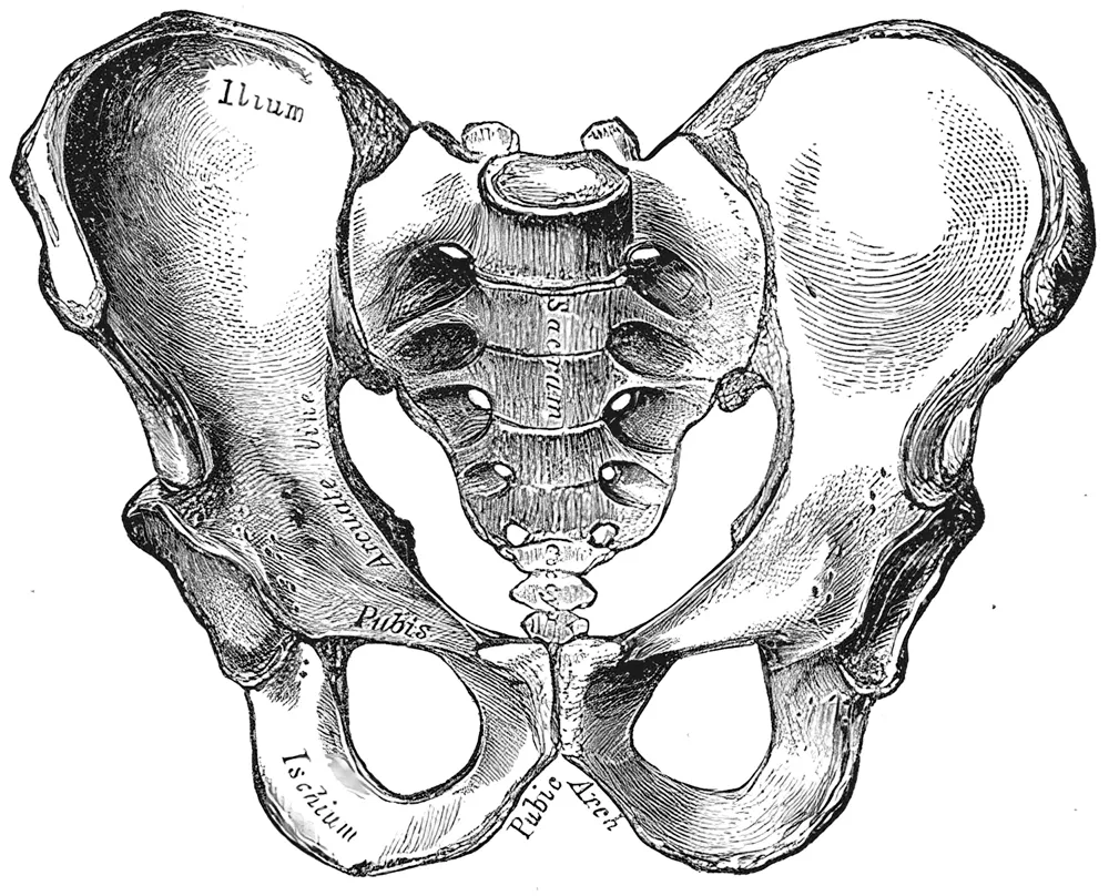
The perineum is a diamond-shaped region below the pelvic diaphragm, bounded in front by the pubic symphysis, anterolaterally by the ischiopubic rami, laterally by the ischial tuberosities, posterolaterally by the sacrotuberous ligaments and behind by the coccyx. A transverse line between the ischial tuberosities divides it into the urogenital triangle in front (urethra, root of the penis or clitoris and the vulva) and the anal triangle behind (anal canal and the two ischioanal fossae). The perineal body, between the two triangles, is the meeting point of many muscles.[1][2]
Layers from superficial to deep
The pelvic diaphragm is the muscular floor of the pelvis, formed by levator ani (pubococcygeus, puborectalis, iliococcygeus) and coccygeus, arising from the body of the pubis, the tendinous arch of the obturator fascia and the ischial spine and inserting on the coccyx and anococcygeal ligament. Below it, in the urogenital triangle, lie the perineal membrane with bulbospongiosus, ischiocavernosus and the transverse perineal muscles; in the anal triangle lie the external anal sphincter and the fat-filled ischioanal fossa, whose lateral wall is obturator internus. The 3D model is male and lacks the superficial perineal muscles: its muscle layer shows the pelvic diaphragm, the external anal sphincter and obturator internus; the thigh and gluteal muscles are left out so the perineum can be seen from below.[3][1]
Vessels, nerves and lymphatics
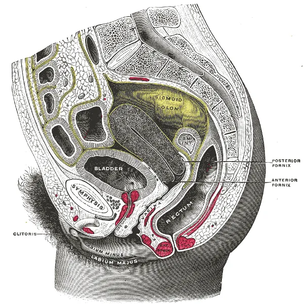
The pudendal neurovascular bundle (internal pudendal artery and vein and the pudendal nerve, S2–S4) leaves the pelvis through the greater sciatic foramen below piriformis, curves around the ischial spine and sacrospinous ligament, and enters the perineum through the lesser sciatic foramen, running in the pudendal (Alcock) canal on the lateral wall of the ischioanal fossa. Its branches are the inferior rectal, perineal and dorsal nerve and artery of the penis or clitoris. Lymph from the anal canal below the pectinate line, the perineal skin, scrotum and vulva drains to the superficial inguinal nodes; the prostate and bladder drain to the iliac nodes.[4][5][6][7]
Embryology
The muscles of the pelvic region derive from two embryonic groups: the pubocaudal group, which forms ischiococcygeus and levator ani together with the pubourethral–vesical–rectosacral ligaments, and the muscles around the cloaca, which form the sphincters and perineal muscles. Levator ani is an evolutionary remnant of the tail muscles of tailed mammals; its rudiment is visible by week 9, already split into three parts by loose mesenchyme, its funnel shape is evident by week 14, and male–female differences appear in the second trimester. In the fetus a distinct fibrous septum links the parietal and visceral walls of the ischioanal fossa and attaches to the back of the perineal membrane; in the adult these fibres spread between the fat lobules and anchor to the obturator internus fascia, gluteus maximus and sphincters, forming the framework that holds the ischioanal fat.[1][3][2]
Anatomical variants
The pudendal nerve varies widely in size, course, number of branches and structures supplied; even small cadaver series regularly find several variants, and in some people it receives contributions beyond S2–S4. The nerve to levator ani may come directly from the fourth sacral nerve or from the coccygeal plexus, and MRI in women shows morphological variants such as thinning or partial aplasia of the levator complex. Ageing changes the female pelvic floor: muscle tone falls, the genital hiatus widens, the iliococcygeus fibres point more caudally and the posterior puborectalis thins. Excess body weight distends the rectum and pushes the perineum, cervix and bladder neck downwards; in men, body weight has an even greater effect on perineal position.[7][3][1]
Clinical correlations
A pudendal nerve block is placed near the ischial spine, where the nerve curves behind the sacrospinous ligament, to relieve pain in childbirth or perineal procedures. Tearing of the perineal body during delivery can weaken the pelvic floor and contribute to pelvic organ prolapse and urinary incontinence. An ischioanal abscess can spread through the postanal space to the opposite side (horseshoe abscess). The prostate lies directly in front of the rectum and can be examined by digital rectal examination.[6][7]
Surgical and procedural considerations
Pelvic floor dysfunction can follow obstetric, urological and colorectal interventions that disrupt structural support or neural pathways. During vaginal delivery the pelvic outlet distends, stressing levator ani, the perineal body and the pudendal nerve, with possible levator avulsion, perineal tears or transient pudendal neuropraxia. The pudendal nerve is also at risk during caesarean section, radical prostatectomy, exploratory pelvic surgery and tumour resection, so its course around the ischial spine and through the pudendal canal must be known. An anal abscess is the acute phase of infection of the intersphincteric glands and an anal fistula the chronic phase of the same disease; pus may spread into the ischioanal fossa. Surgery on levator ani (for tumours, trauma or prolapse) is considered only when non-invasive management has failed.[1][7][2][3]
Key structures
Diaphragma pelvis
- Name
- Pelvic diaphragm
- Position, role
- Levator ani and coccygeus; supports the pelvic organs
M. levator ani
- Name
- Levator ani
- Position, role
- Pubococcygeus, puborectalis and iliococcygeus parts
Trigonum urogenitale
- Name
- Urogenital triangle
- Position, role
- In front of the interischial line; urethra and external genitalia
Trigonum anale
- Name
- Anal triangle
- Position, role
- Behind the interischial line; anal canal and ischioanal fossae
Fossa ischioanalis
- Name
- Ischioanal fossa
- Position, role
- Fat-filled space between levator ani and obturator internus
Canalis pudendalis
- Name
- Pudendal canal (Alcock)
- Position, role
- Split in the obturator fascia; carries the pudendal bundle
Corpus perineale
- Name
- Perineal body
- Position, role
- Fibromuscular node between the triangles where many muscles meet
| Terminologia Anatomica | Name | Position, role |
|---|---|---|
| Diaphragma pelvis | Pelvic diaphragm | Levator ani and coccygeus; supports the pelvic organs |
| M. levator ani | Levator ani | Pubococcygeus, puborectalis and iliococcygeus parts |
| Trigonum urogenitale | Urogenital triangle | In front of the interischial line; urethra and external genitalia |
| Trigonum anale | Anal triangle | Behind the interischial line; anal canal and ischioanal fossae |
| Fossa ischioanalis | Ischioanal fossa | Fat-filled space between levator ani and obturator internus |
| Canalis pudendalis | Pudendal canal (Alcock) | Split in the obturator fascia; carries the pudendal bundle |
| Corpus perineale | Perineal body | Fibromuscular node between the triangles where many muscles meet |
What each layer contains
SkinCutis5
- Anal region
- Gluteal region
- Urogenital region
- Gluteal fold
- Femoral triangle
Muscles, fasciae and tendon sheathsMusculi, fasciae et vaginae tendinum14
- (Iliopectineal bursa)
- Iliopectineal arch
- Tendinous arch of levator ani
- Sciatic bursa of obturator internus
- Subtendinous bursa of obturator internus
- Intermuscular gluteal bursae
- Inguinal ligament
- Coccygeus muscle
- Iliococcygeus muscle
- Pubo-analis muscle
- Pubococcygeus muscle
- Pyramidalis muscle
- Obturator internus
- External anal sphincter
Bones and cartilageOssa et cartilagines3
- Coccyx
- Hip bone
- Sacrum
Joints and ligamentsJuncturae et ligamenta21
- Articular capsule of hip joint
- Triradiate cartilage
- Interpubic disc
- Frenula capsulae
- Acetabular labrum
- Ligament of head of femur
- Intercornual ligament
- Ischiofemoral ligament
- Inferior pubic ligament
- Superior pubic ligament
- Pubofemoral ligament
- Posterior sacro-iliac ligament
- Sacrospinous ligament
- Sacrotuberous ligament
- Transverse acetabular ligament
- Obturator membrane
- Descending part of iliofemoral ligament
- Transverse part of iliofemoral ligament
- Pubic symphysis
- Sacrococcygeal symphysis
- Zona orbicularis
Urogenital organsOrgana urogenitalia12
- Corpus cavernosum of penis
- Corpus spongiosum of penis
- Ductus deferens
- Ejaculatory duct
- Epididymis
- Seminal gland
- Glans penis
- Prostate
- Testis
- Ureter
- Urethra
- Urinary bladder
ArteriesArteriae14
- Superior anorectal artery
- Lateral circumflex femoral artery
- Medial circumflex femoral artery
- Dorsal artery of penis
- Inferior gluteal artery
- Obturator artery
- Deep femoral artery
- Deep artery of penis
- Deep external pudendal artery
- Superficial external pudendal artery
- Internal pudendal artery
- Median sacral artery
- Right testicular artery
- Left testicular artery
VeinsVenae11
- Deep dorsal vein of penis
- Deep femoral vein
- Internal pudendal vein
- Right testicular vein
- Left testicular vein
- Lateral circumflex femoral veins
- Medial circumflex femoral veins
- Superficial dorsal veins of penis
- Inferior gluteal veins
- External pudendal veins
- Lateral sacral veins
NervesNervi9
- Cauda equina
- Posterior femoral cutaneous nerve
- Ilio-inguinal nerve
- Obturator nerve
- Pudendal nerve
- Medial crural cutaneous branches of saphenous nerve
- Femoral branch of genitofemoral nerve
- Genital branch of genitofemoral nerve
- Posterior branch of obturator nerve
LymphaticsSystema lymphoideum18
- (Intermediate deep inguinal node)
- (Proximal deep inguinal node)
- (Intermediate lacunar node)
- (Lateral lacunar node)
- (Medial lacunar node)
- Inferior epigastric nodes
- Inferior gluteal nodes
- Superior gluteal nodes
- Intermediate external iliac nodes
- Inferior superficial inguinal nodes
- Superolateral superficial inguinal nodes
- Superomedial superficial inguinal nodes
- Obturator nodes
- Pararectal nodes
- Postvesical nodes
- Prevesical nodes
- Lateral sacral nodes
- Lateral vesical nodes
Full-text reading (StatPearls)
The StatPearls chapters on this topic, reproduced verbatim. Their CC BY-NC-ND 4.0 licence allows unmodified copies only, so they are not translated or shortened.
Anatomy, Abdomen and Pelvis: Pelvic FloorBordoni B, Sugumar K, Leslie SW · StatPearls 2026 · NBK482200 · 31k characters
Loading…
Bordoni B, Sugumar K, Leslie SW. Anatomy, Abdomen and Pelvis: Pelvic Floor. StatPearls [Internet]. Treasure Island (FL): StatPearls Publishing; 2026. NBK482200 · PMID 29489277 · CC BY-NC-ND 4.0. Reproduced verbatim, unmodified.
Anatomy, Abdomen and Pelvis, Pudendal NerveKinter KJ, Newton BW · StatPearls 2023 · NBK554736 · 12k characters
Loading…
Kinter KJ, Newton BW. Anatomy, Abdomen and Pelvis, Pudendal Nerve. StatPearls [Internet]. Treasure Island (FL): StatPearls Publishing; 2023. NBK554736 · PMID 32134612 · CC BY-NC-ND 4.0. Reproduced verbatim, unmodified.
Anatomy, Abdomen and Pelvis: Ischioanal FossaSiccardi MA, Scharbach S, Bordoni B · StatPearls 2022 · NBK535387 · 11k characters
Loading…
Siccardi MA, Scharbach S, Bordoni B. Anatomy, Abdomen and Pelvis: Ischioanal Fossa. StatPearls [Internet]. Treasure Island (FL): StatPearls Publishing; 2022. NBK535387 · PMID 30571008 · CC BY-NC-ND 4.0. Reproduced verbatim, unmodified.
Anatomy, Abdomen and Pelvis: Levator Ani MuscleGowda SN, Bordoni B · StatPearls 2022 · NBK556078 · 10k characters
Loading…
Gowda SN, Bordoni B. Anatomy, Abdomen and Pelvis: Levator Ani Muscle. StatPearls [Internet]. Treasure Island (FL): StatPearls Publishing; 2022. NBK556078 · PMID 32310538 · CC BY-NC-ND 4.0. Reproduced verbatim, unmodified.
Regional anatomy
References
Sources for the values and descriptions on this page, indexed in PubMed (US National Library of Medicine).
- [1]Anatomy, Abdomen and Pelvis: Pelvic FloorBordoni B et al., StatPearls 2026PMID 29489277
- [2]Anatomy, Abdomen and Pelvis: Ischioanal FossaSiccardi MA et al., StatPearls 2022PMID 30571008
- [3]Anatomy, Abdomen and Pelvis: Levator Ani MuscleGowda SN et al., StatPearls 2022PMID 32310538
- [4]Anatomy, Bony Pelvis and Lower Limb, Gemelli MusclesLezak B et al., StatPearls 2026PMID 32491352
- [5]Anatomy, Abdomen and Pelvis: Inguinal Lymph NodeBui T et al., StatPearls 2023PMID 32491571
- [6]Anatomy, Abdomen and Pelvis, ProstateSingh O et al., StatPearls 2023PMID 31082031
- [7]Anatomy, Abdomen and Pelvis, Pudendal NerveKinter KJ et al., StatPearls 2023PMID 32134612