Spinal cord
Model
Layers
Z-Anatomy anatomical model based on BodyParts3D, licensed CC BY-SA 4.0.
Description
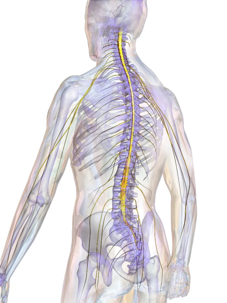
Lies within the vertebral canal, about 42–45 cm long, extending from the foramen magnum to the level of the L1–L2 vertebrae. It conducts signals between the brain and the periphery and integrates spinal reflexes.
Parts
Each part has its own card; press “Show on the model” to fly to it and select it in the 3D model above.
- 01
Grey matter
Substantia grisea medullae spinalis
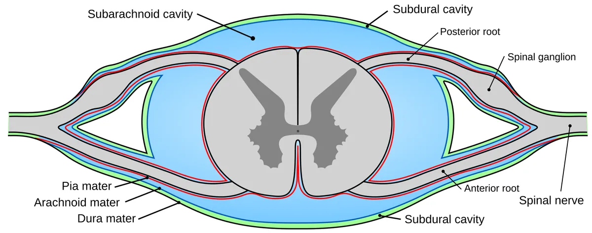
An H-shaped (butterfly) column of neuronal cell bodies around the central canal, divided into anterior and posterior horns and an intermediate zone. It is largest at the two enlargements that supply the limbs.
- 02
Anterior horn
Cornu anterius
Contains alpha and gamma motor neurons (Rexed lamina IX) arranged somatotopically: medial groups supply axial muscles, lateral groups limb muscles. Axons leave through the anterior root; damage causes flaccid weakness, wasting and fasciculations.
- 03
Posterior horn
Cornu posterius
Receives primary afferents from the posterior root; it includes the substantia gelatinosa (lamina II) that processes pain and temperature, the nucleus proprius (laminae III–IV) and the thoracic nucleus (Clarke, C8–L2) relaying proprioception to the cerebellum.
- 04
Lateral horn and intermediate zone
Cornu laterale; Substantia intermedia
From T1 to L2 the intermediolateral column holds sympathetic preganglionic neurons; from S2 to S4 a similar column holds sacral parasympathetic preganglionic neurons. Their axons leave in the anterior root and, for the sympathetic outflow, pass through white rami communicantes to the sympathetic trunk.
- 05
Central canal
Canalis centralis
A narrow ependyma-lined channel continuous with the fourth ventricle above and ending in the conus as the terminal ventricle. In adults it is often obliterated in places; dilatation (hydromyelia) or a syrinx causes cape-like loss of pain and temperature sense.
- 06
Posterior funiculus
Funiculus posterior
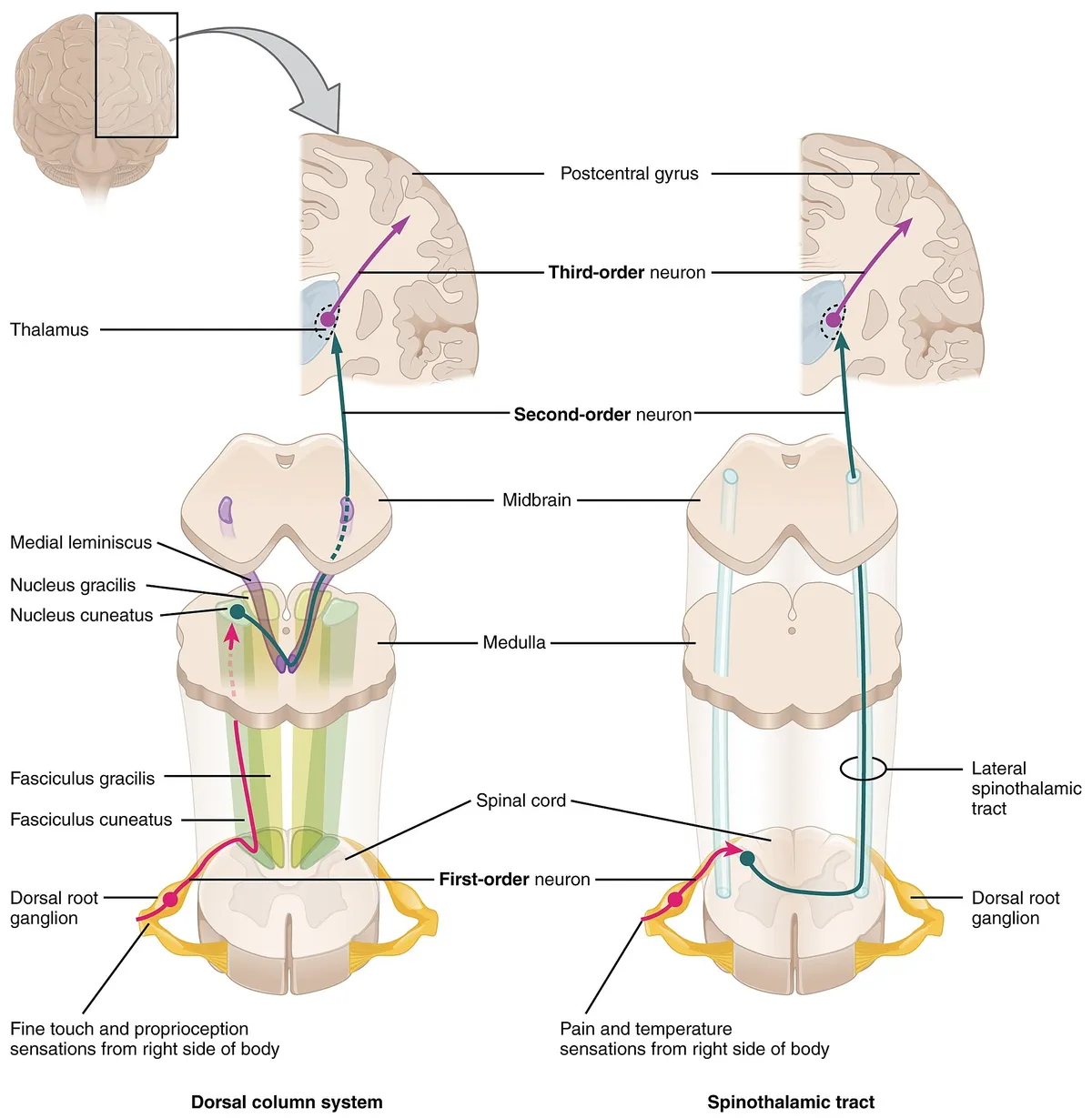
Holds the gracile fasciculus (from the lower limb) medially and the cuneate fasciculus (from the upper limb, above T6) laterally, carrying ipsilateral fine touch, vibration and conscious proprioception to the gracile and cuneate nuclei of the medulla.
- 07
Lateral funiculus
Funiculus lateralis
Contains the lateral corticospinal tract (voluntary movement, already crossed in the medulla), the rubrospinal and lateral reticulospinal tracts, the anterior and posterior spinocerebellar tracts, and the lateral spinothalamic tract carrying contralateral pain and temperature.
- 08
Anterior funiculus
Funiculus anterior
Lies between the anterior median fissure and the anterior roots; it carries the uncrossed anterior corticospinal tract, the vestibulospinal, medial reticulospinal and tectospinal tracts controlling posture and axial muscles, and the anterior spinothalamic tract for crude touch.
- 09
Enlargements and conus medullaris
Intumescentia cervicalis et lumbosacralis; Conus medullaris
The cervical (C4–T1) and lumbosacral (L2–S3) enlargements give rise to the brachial and lumbosacral plexuses. The cord tapers into the conus medullaris at L1–L2 in adults and continues as the filum terminale; the roots below form the cauda equina.
Position and relations
The spinal cord lies in the vertebral canal, from the foramen magnum (continuous with the medulla oblongata) to the level of L1–L2, ending as the conus medullaris. Below this, the lumbar and sacral roots descend as the cauda equina; lumbar puncture is therefore performed at L3–L4 or L4–L5 without touching the cord.
Form and structure
In cross-section, H-shaped grey matter lies centrally, with anterior horns (motor neurons), posterior horns (sensory neurons) and, in the thoracic and upper lumbar cord, lateral horns (sympathetic neurons). White matter surrounds it, divided into anterior, lateral and posterior funiculi carrying ascending and descending tracts. The cord has cervical and lumbar enlargements, from which the limb plexuses arise.
Vessels and nerves
The cord is supplied by one anterior spinal artery (feeding roughly the anterior two-thirds) and two posterior spinal arteries, reinforced by radicular arteries, the largest being the great radicular artery (of Adamkiewicz) in the lower thoracic or upper lumbar region. Thirty-one pairs of spinal nerves exit through the intervertebral foramina.
Conducting role
The dorsal horn is the first relay of the pain pathway: Aδ and C fibres terminate here, and the signal crosses in the anterior white commissure before ascending in the spinothalamic tract. Descending pathways from the brainstem modulate the signal right at the dorsal horn, which explains why perceived pain can be amplified or damped by the state of the central nervous system.[1]
Localising cord lesions
The spinal cord controls the voluntary muscles of the trunk and limbs and receives their sensory input; in adults it extends from the medulla oblongata to the lower border of L1. Its ascending and descending tracts are highly organised, so history and neurological examination can distinguish cord from peripheral nerve or brain disease and identify the level and part of the cord involved before MRI.[2][3]
The three key tracts are the corticospinal (motor), spinothalamic (pain, temperature) and dorsal columns (fine touch, proprioception). Because they cross at different levels, each incomplete cord syndrome has its own pattern: dorsal cord, ventral cord (often from anterior spinal artery ischaemia), central cord, Brown-Séquard (hemicord), conus medullaris and cauda equina syndromes.[4]
Transverse myelitis has many causes besides multiple sclerosis; the discovery of aquaporin-4 IgG (neuromyelitis optica) and MOG IgG has changed its diagnosis. Neuromyelitis optica typically causes recurrent optic neuritis and transverse myelitis, and its brain lesions follow the distribution of aquaporin-4 expression. Early diagnosis matters because of the high risk of relapse and severe disability.[3][5]
Development
The spinal cord arises from bipotent neuromesodermal progenitors organised within the neural tube. Most of the tube (brain and anterior cord) forms by bending and dorsal fusion of the neural groove (primary neurulation), whereas the posterior region forms by de novo lumen formation in a solid medullary cord (secondary neurulation). Neural progenitors form a pseudostratified epithelium whose division modes drive tube growth and initiate neurogenesis.[6]
Full-text reading (StatPearls)
The StatPearls chapters on this topic, reproduced verbatim. Their CC BY-NC-ND 4.0 licence allows unmodified copies only, so they are not translated or shortened.
Neuroanatomy, Spinal CordKhan YS, Lui F · StatPearls 2023 · NBK559056 · 27k characters
Loading…
Khan YS, Lui F. Neuroanatomy, Spinal Cord. StatPearls [Internet]. Treasure Island (FL): StatPearls Publishing; 2023. NBK559056 · PMID 32644482 · CC BY-NC-ND 4.0. Reproduced verbatim, unmodified.
Neuroanatomy, Spinal Cord VeinsGreen K, Reddy V, Hogg JP · StatPearls 2023 · NBK542182 · 15k characters
Loading…
Green K, Reddy V, Hogg JP. Neuroanatomy, Spinal Cord Veins. StatPearls [Internet]. Treasure Island (FL): StatPearls Publishing; 2023. NBK542182 · PMID 31194339 · CC BY-NC-ND 4.0. Reproduced verbatim, unmodified.
Anatomy, Back, Spinal Cord ArteriesKaiser JT, Reddy V, Lugo-Pico JG · StatPearls 2023 · NBK537219 · 12k characters
Loading…
Kaiser JT, Reddy V, Lugo-Pico JG. Anatomy, Back, Spinal Cord Arteries. StatPearls [Internet]. Treasure Island (FL): StatPearls Publishing; 2023. NBK537219 · PMID 30725904 · CC BY-NC-ND 4.0. Reproduced verbatim, unmodified.
Neuroanatomy, Spinal Cord ArteriesHasan S, Arain A · StatPearls 2023 · NBK539889 · 8k characters
Loading…
Hasan S, Arain A. Neuroanatomy, Spinal Cord Arteries. StatPearls [Internet]. Treasure Island (FL): StatPearls Publishing; 2023. NBK539889 · PMID 30969711 · CC BY-NC-ND 4.0. Reproduced verbatim, unmodified.
Organs in this system
References
Sources for the values and descriptions on this page, indexed in PubMed (US National Library of Medicine).
- [1]Basic anatomy and physiology of pain pathwaysBourne S et al., Neurosurgery clinics of North America 2014PMID 25240653
- [2]The spinal cord: a review of functional neuroanatomyBican O et al., Neurologic clinics 2013PMID 23186894
- [3]Spinal Cord Anatomy and LocalizationHardy TA, Continuum 2021PMID 33522735
- [4]Incomplete Cord Syndromes: Clinical and Imaging ReviewKunam VK et al., Radiographics 2018PMID 29995620
- [5]Diagnosing neuromyelitis opticaMakhani N et al., Neuroimaging clinics of North America 2013PMID 23608690
- [6]Early spinal cord development: from neural tube formation to neurogenesisSaade M et al., Nature reviews. Neuroscience 2025PMID 39915695
- [7]The human brain in numbers: a linearly scaled-up primate brainHerculano-Houzel S, 2009PMID 19915731Supports: Neuron count
- [8]Determinants of conduction velocity in myelinated nerve fibersWaxman SG, 1980PMID 6245357Supports: Conduction velocity
- [9]Quantitative imaging of energy expenditure in human brainPMID 22487547Supports: Cerebral energy expenditure