Inguinal region and femoral triangle
Model
Layers
Z-Anatomy anatomical model based on BodyParts3D, licensed CC BY-SA 4.0.
Overview
A regional model of the (right) inguinal region and femoral triangle: the inguinal ligament, the abdominal wall layers that form the inguinal canal (external oblique aponeurosis, internal oblique, transversus abdominis, transversalis fascia), the spermatic cord with the ductus deferens, the femoral triangle bounded by sartorius and adductor longus with iliopsoas and pectineus in its floor, the femoral artery, vein and nerve, the great saphenous vein and the inguinal lymph nodes.
Boundaries and subdivisions
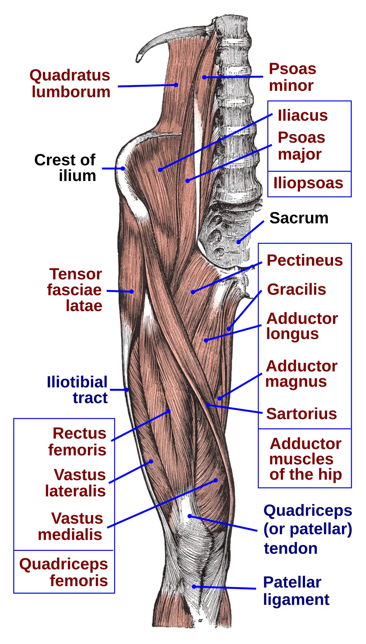
The inguinal ligament, the rolled-under lower border of the external oblique aponeurosis, runs from the anterior superior iliac spine to the pubic tubercle and marks the boundary between abdominal wall and thigh. Above it lies the inguinal canal, about 4 cm long, running obliquely downwards and medially from the deep inguinal ring (an opening in the transversalis fascia, just lateral to the inferior epigastric artery) to the superficial ring (a slit in the external oblique aponeurosis above and lateral to the pubic tubercle). Below the ligament lies the femoral triangle, bounded by the inguinal ligament above, the medial border of sartorius laterally and the medial border of adductor longus medially; its floor is iliopsoas and pectineus, its roof the fascia lata.[1][2][3]
Layers from superficial to deep
The anterior wall of the inguinal canal is the external oblique aponeurosis (with internal oblique fibres in its lateral third); the posterior wall is transversalis fascia, reinforced medially by the conjoint tendon; the floor is the inguinal and lacunar ligaments; the roof is the arching fibres of internal oblique and transversus abdominis. In the male the canal carries the spermatic cord (ductus deferens, testicular artery, pampiniform venous plexus, nerves and lymphatics) wrapped in three fascial coverings borrowed from the abdominal wall; in the female it carries the round ligament of the uterus. In both sexes the ilio-inguinal nerve runs through it.[4][5]
Vessels, nerves and lymphatics
Within the femoral triangle, from lateral to medial, lie the femoral nerve, femoral artery, femoral vein and femoral canal (the “NAVL” mnemonic). The artery, vein and canal lie inside the femoral sheath, the nerve outside it; the femoral nerve branches about 2 cm below the inguinal ligament, medial to sartorius and lateral to the femoral artery. The femoral canal, most medial, contains fat and the deep inguinal nodes (Cloquet’s node). The superficial inguinal nodes lie below the inguinal ligament and receive lymph from the lower limb, the skin below the umbilicus, the anal canal below the pectinate line, the scrotum and the vulva, draining in turn to the deep inguinal and external iliac nodes.[6][7][8]
Embryology
During embryonic life the testis descends from the posterior abdominal wall into the scrotum, guided by the gubernaculum, which tethers its lower pole to the developing scrotal swelling. A peritoneal pouch, the processus vaginalis, follows the testis and normally obliterates once descent is complete. If it fails to close or closes late, peritoneal fluid can track down and form a hydrocele, and a patent processus vaginalis predisposes to indirect inguinal hernia; incomplete descent produces cryptorchidism at various levels. The femoral sheath is formed in front by an extension of transversalis fascia and behind by the iliopsoas fascia, so it develops with the mesodermal layers of the abdominal wall. The muscles and fascia of the femoral triangle derive from mesoderm within the lower limb bud, which appears at about week 4.[2][8][3]
Anatomical variants
The femoral triangle is highly conserved; it changes only with the congenital absence of a bordering muscle or rare malformations of its contents, and its overall size simply follows body size. The more important variation lies in the nerves: the ilioinguinal nerve, which runs through the inguinal canal, arises from L1 alone in only 65% of people (otherwise from T12–L1, L1–L2 or L2–L3), and the femoral nerve occasionally splits in the abdomen and rejoins just above the inguinal canal. These differences partly explain failed groin blocks and persistent pain after hernia repair.[3][9][10]
Clinical correlations
An indirect inguinal hernia enters the deep ring lateral to the inferior epigastric artery, follows the canal and may descend into the scrotum; a direct hernia bulges through the posterior wall in the inguinal (Hesselbach) triangle, medial to that artery. A femoral hernia passes through the femoral canal, below and lateral to the pubic tubercle; it is commoner in women and prone to strangulation. The femoral artery at the midpoint of the inguinal ligament is used for pulse palpation and arterial catheterisation, and the femoral vein just medial to it for central venous access. Enlarged inguinal nodes call for examination of the whole drainage area, including the anal canal and external genitalia.[7]
Surgical and procedural considerations
Inguinal hernia is a very common surgical disease; repair reduces the contents and sac, closes the defect and reinforces the posterior wall of the canal. Many techniques have been used, and today most repairs use mesh, open or laparoscopic. The femoral canal, the medial compartment of the femoral sheath, is where abdominal contents protrude as a femoral hernia; in both sexes, elective repair now favours mesh (open or laparoscopic) over suture alone, and a strangulated femoral hernia is an emergency. Operations around the femoral triangle must protect the femoral artery, vein and nerve, which are vital to the limb, and the inguinal nodes in the triangle are examined to assess disease spread. An adductor canal block places local anaesthetic around the saphenous nerve and the sensory branches to the knee within the canal, giving analgesia for knee, ankle and foot surgery while preserving quadriceps strength.[2][8][3][11]
Key structures
Lig. inguinale
- Name
- Inguinal ligament
- Position, role
- ASIS → pubic tubercle; floor of the inguinal canal
Anulus inguinalis profundus
- Name
- Deep inguinal ring
- Position, role
- Opening in transversalis fascia lateral to the inferior epigastric artery
Anulus inguinalis superficialis
- Name
- Superficial inguinal ring
- Position, role
- Slit in the external oblique aponeurosis above the pubic tubercle
Funiculus spermaticus
- Name
- Spermatic cord
- Position, role
- Ductus deferens, testicular artery, pampiniform plexus
Trigonum femorale
- Name
- Femoral triangle (Scarpa)
- Position, role
- Inguinal ligament, sartorius, adductor longus; femoral nerve, artery and vein
Vagina femoralis
- Name
- Femoral sheath
- Position, role
- Three compartments: artery, vein, femoral canal
Trigonum inguinale
- Name
- Inguinal triangle (Hesselbach)
- Position, role
- Weak area of the posterior wall; site of direct hernia
| Terminologia Anatomica | Name | Position, role |
|---|---|---|
| Lig. inguinale | Inguinal ligament | ASIS → pubic tubercle; floor of the inguinal canal |
| Anulus inguinalis profundus | Deep inguinal ring | Opening in transversalis fascia lateral to the inferior epigastric artery |
| Anulus inguinalis superficialis | Superficial inguinal ring | Slit in the external oblique aponeurosis above the pubic tubercle |
| Funiculus spermaticus | Spermatic cord | Ductus deferens, testicular artery, pampiniform plexus |
| Trigonum femorale | Femoral triangle (Scarpa) | Inguinal ligament, sartorius, adductor longus; femoral nerve, artery and vein |
| Vagina femoralis | Femoral sheath | Three compartments: artery, vein, femoral canal |
| Trigonum inguinale | Inguinal triangle (Hesselbach) | Weak area of the posterior wall; site of direct hernia |
What each layer contains
SkinCutis6
- Anterior region of thigh
- Hip region
- Hypogastric region
- Inguinal region
- Posterior region of thigh
- Femoral triangle
Muscles, fasciae and tendon sheathsMusculi, fasciae et vaginae tendinum45
- (Adductor minimus)
- (Iliopectineal bursa)
- Adductor brevis
- Adductor longus
- Adductor magnus
- Iliopectineal arch
- Tendinous arch of levator ani
- Sciatic bursa of gluteus maximus muscle
- Sciatic bursa of obturator internus
- Bursa of piriformis muscle
- Subcutaneous trochanteric bursa
- Subtendinous bursa of iliacus muscle
- Subtendinous bursa of obturator internus
- Superior bursa of biceps femoris muscle
- Trochanteric bursa of gluteus maximus muscle
- Trochanteric bursa of gluteus minimus muscle
- Intermuscular gluteal bursae
- Piriformis fascia
- Inguinal ligament
- Inferior gemellus muscle
- Superior gemellus muscle
- Gluteus medius muscle
- Gluteus minimus muscle
- Gracilis muscle
- Iliacus muscle
- Iliococcygeus muscle
- External abdominal oblique muscle
- Internal abdominal oblique muscle
- Pectineus muscle
- Piriformis muscle
- Pubo-analis muscle
- Pubococcygeus muscle
- Pyramidalis muscle
- Quadratus femoris muscle
- Rectus abdominis muscle
- Transversus abdominis muscle
- Vastus intermedius muscle
- Vastus lateralis muscle
- Vastus medialis muscle
- Obturator externus
- Obturator internus
- Psoas major
- Medial femoral intermuscular septum
- Tensor fasciae latae
- Trochanteric bursa of gluteus medius muscle
Bones and cartilageOssa et cartilagines4
- Hip bone
- Femur
- Sacrum
- Vertebra L5
Joints and ligamentsJuncturae et ligamenta19
- Articular capsule of hip joint
- Triradiate cartilage
- Interpubic disc
- Intervertebral disc L5-S1
- Frenula capsulae
- Acetabular labrum
- Ligament of head of femur
- Ischiofemoral ligament
- Inferior pubic ligament
- Superior pubic ligament
- Pubofemoral ligament
- Anterior sacro-iliac ligament
- Transverse acetabular ligament
- Obturator membrane
- Nucleus pulposus L5-S1
- Descending part of iliofemoral ligament
- Transverse part of iliofemoral ligament
- Pubic symphysis
- Zona orbicularis
ArteriesArteriae28
- Superior anorectal artery
- Appendicular artery
- Lateral circumflex femoral artery
- Medial circumflex femoral artery
- Dorsal artery of penis
- Inferior epigastric artery
- Superficial epigastric artery
- Femoral artery
- Inferior gluteal artery
- Superior gluteal artery
- External iliac artery
- Internal iliac artery
- Iliolumbar artery
- Obturator artery
- Deep femoral artery
- Deep artery of penis
- Deep external pudendal artery
- Superficial external pudendal artery
- Internal pudendal artery
- Median sacral artery
- Right testicular artery
- Perforating femoral arteries
- Anterior division of internal iliac artery
- Posterior division of internal iliac artery
- Descending branch of lateral circumflex femoral artery
- Iliacus branch of iliolumbar artery
- Lumbar branch of iliolumbar artery
- Spinal branch of iliolumbar artery
VeinsVenae19
- Deep dorsal vein of penis
- Inferior epigastric vein
- Superficial epigastric vein
- Femoral vein
- External iliac vein
- Internal iliac vein
- Iliolumbar vein
- Deep femoral vein
- Internal pudendal vein
- Right testicular vein
- Left testicular vein
- Lateral circumflex femoral veins
- Medial circumflex femoral veins
- Superficial dorsal veins of penis
- Inferior gluteal veins
- Superior gluteal veins
- Perforating veins
- External pudendal veins
- Lateral sacral veins
NervesNervi16
- Lateral femoral cutaneous nerve
- Femoral nerve
- Superior gluteal nerve
- Ilio-inguinal nerve
- Sciatic nerve
- Nerve to piriformis muscle
- Nerve to quadratus femoris muscle
- Obturator nerve
- Pudendal nerve
- Anterior root of posterior femoral cutaneous nerve
- Anterior cutaneous branches of femoral nerve
- Medial crural cutaneous branches of saphenous nerve
- Anterior branch of obturator nerve
- Femoral branch of genitofemoral nerve
- Genital branch of genitofemoral nerve
- Posterior branch of obturator nerve
LymphaticsSystema lymphoideum23
- (Intermediate deep inguinal node)
- (Proximal deep inguinal node)
- (Intermediate lacunar node)
- (Lateral lacunar node)
- (Medial lacunar node)
- Appendicular nodes
- Inferior epigastric nodes
- Inferior gluteal nodes
- Superior gluteal nodes
- Intermediate external iliac nodes
- Medial external iliac nodes
- Inferior superficial inguinal nodes
- Superolateral superficial inguinal nodes
- Superomedial superficial inguinal nodes
- Juxta-intestinal mesenteric nodes
- Obturator nodes
- Pararectal nodes
- Postvesical nodes
- Precaecal nodes
- Prevesical nodes
- Retrocaecal nodes
- Median sacral nodes
- Lateral vesical nodes
Urogenital organsOrgana urogenitalia12
- Corpus cavernosum of penis
- Corpus spongiosum of penis
- Ductus deferens
- Ejaculatory duct
- Epididymis
- Seminal gland
- Glans penis
- Prostate
- Testis
- Ureter
- Urethra
- Urinary bladder
Full-text reading (StatPearls)
The StatPearls chapters on this topic, reproduced verbatim. Their CC BY-NC-ND 4.0 licence allows unmodified copies only, so they are not translated or shortened.
Anatomy, Abdomen and Pelvis: Inguinal Region (Inguinal Canal)Tuma F, Lopez RA, Varacallo MA · StatPearls 2023 · NBK470204 · 12k characters
Loading…
Tuma F, Lopez RA, Varacallo MA. Anatomy, Abdomen and Pelvis: Inguinal Region (Inguinal Canal). StatPearls [Internet]. Treasure Island (FL): StatPearls Publishing; 2023. NBK470204 · PMID 29261933 · CC BY-NC-ND 4.0. Reproduced verbatim, unmodified.
Anatomy, Abdomen and Pelvis: Femoral TriangleBasinger H, Hogg JP · StatPearls 2023 · NBK541140 · 12k characters
Loading…
Basinger H, Hogg JP. Anatomy, Abdomen and Pelvis: Femoral Triangle. StatPearls [Internet]. Treasure Island (FL): StatPearls Publishing; 2023. NBK541140 · PMID 31082184 · CC BY-NC-ND 4.0. Reproduced verbatim, unmodified.
Anatomy, Abdomen and Pelvis: Femoral SheathMahabadi N, Lew V, Kang M · StatPearls 2023 · NBK482388 · 10k characters
Loading…
Mahabadi N, Lew V, Kang M. Anatomy, Abdomen and Pelvis: Femoral Sheath. StatPearls [Internet]. Treasure Island (FL): StatPearls Publishing; 2023. NBK482388 · PMID 29494010 · CC BY-NC-ND 4.0. Reproduced verbatim, unmodified.
Anatomy, Abdomen and Pelvis: Adductor Canal (Subsartorial Canal, Hunter Canal)Migirov A, Arbor TC, Vilella RC · StatPearls 2024 · NBK556046 · 8k characters
Loading…
Migirov A, Arbor TC, Vilella RC. Anatomy, Abdomen and Pelvis: Adductor Canal (Subsartorial Canal, Hunter Canal). StatPearls [Internet]. Treasure Island (FL): StatPearls Publishing; 2024. NBK556046 · PMID 32310506 · CC BY-NC-ND 4.0. Reproduced verbatim, unmodified.
Regional anatomy
References
Sources for the values and descriptions on this page, indexed in PubMed (US National Library of Medicine).
- [1]Anatomy, Bony Pelvis and Lower Limb: Iliopsoas MuscleBordoni B et al., StatPearls 2026PMID 30285403
- [2]Anatomy, Abdomen and Pelvis: Inguinal Region (Inguinal Canal)Tuma F et al., StatPearls 2023PMID 29261933
- [3]Anatomy, Abdomen and Pelvis: Femoral TriangleBasinger H et al., StatPearls 2023PMID 31082184
- [4]Anatomy, Abdomen and Pelvis: Uterus Round LigamentChaudhry SR et al., StatPearls 2023PMID 29763145
- [5]Anatomy, Abdomen and Pelvis: TestesTiwana MS et al., StatPearls 2023PMID 29261881
- [6]Anatomy, Bony Pelvis and Lower Limb: Thigh Quadriceps MuscleBordoni B et al., StatPearls 2023PMID 30020706
- [7]Anatomy, Abdomen and Pelvis: Inguinal Lymph NodeBui T et al., StatPearls 2023PMID 32491571
- [8]Anatomy, Abdomen and Pelvis: Femoral SheathMahabadi N et al., StatPearls 2023PMID 29494010
- [9]Anatomy, Abdomen and Pelvis, Posterior Abdominal Wall NervesNunn JF et al., StatPearls 2022PMID 32491537
- [10]Anatomy, Bony Pelvis and Lower Limb: Thigh Femoral NerveRefai NA et al., StatPearls 2023PMID 32310525
- [11]Anatomy, Abdomen and Pelvis: Adductor Canal (Subsartorial Canal, Hunter Canal)Migirov A et al., StatPearls 2024PMID 32310506