Digestive system
Model
Z-Anatomy anatomical model based on BodyParts3D, licensed CC BY-SA 4.0.
Overview
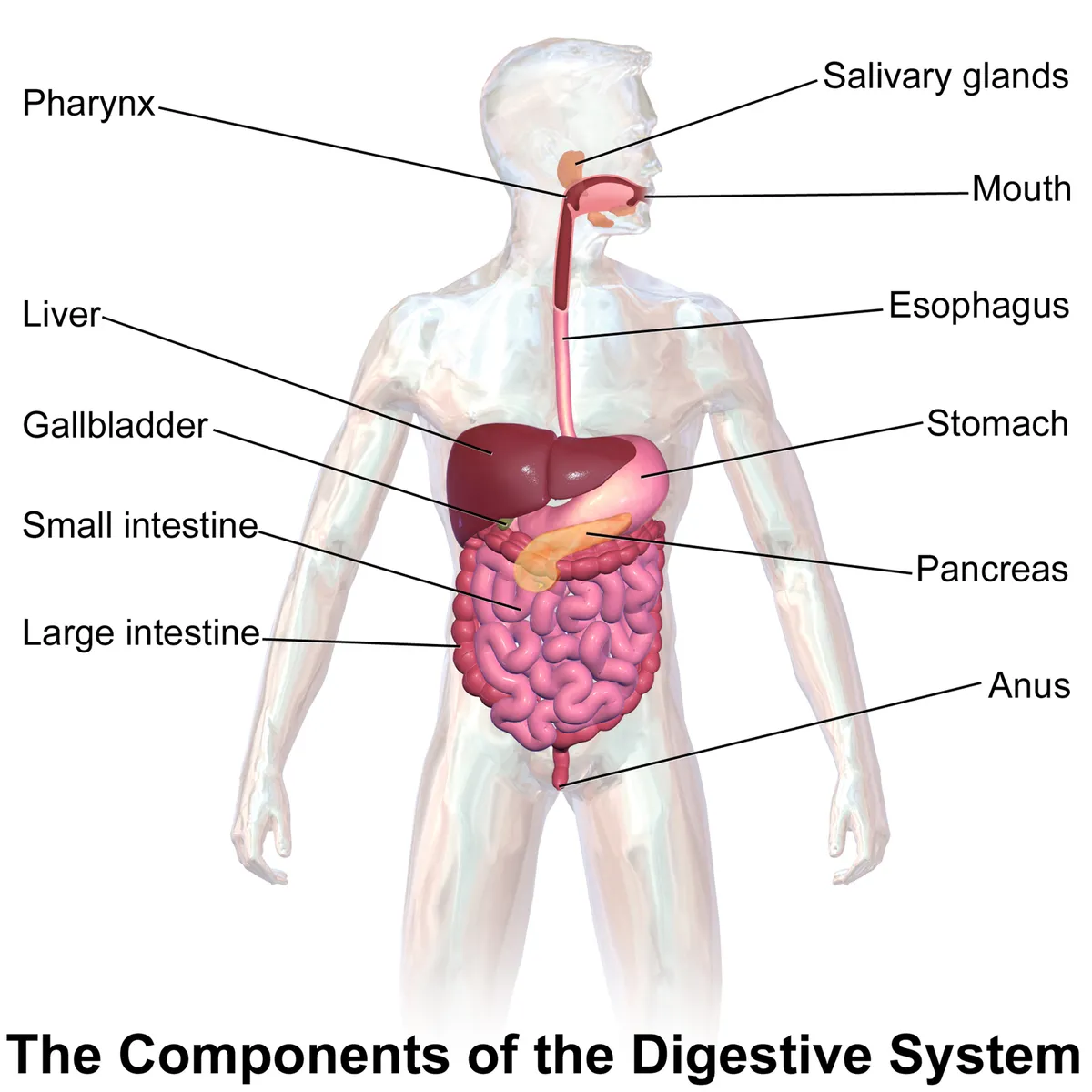
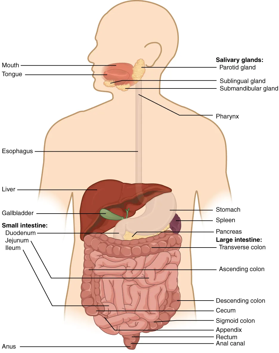
The digestive system comprises the alimentary canal (mouth, pharynx, oesophagus, stomach, small and large intestines, anal canal) and the accessory glands (salivary glands, liver, pancreas). It carries out mechanical and chemical digestion, absorption of nutrients and elimination of faeces.
Functions
- 01Mechanical digestion: mastication, mixing and propulsion
- 02Enzymatic (chemical) digestion
- 03Absorption of nutrients, water and electrolytes
- 04Elimination of faeces
The liver, the largest gland
The liver accounts for about 2.5% of body weight and handles nutrient metabolism, detoxification and energy storage. It is also an immune organ: blood from the gut reaches it first through the portal vein, so it hosts many resident macrophages (Kupffer cells) and other immune cells that clear microbes coming from the bowel, and it produces acute-phase proteins during inflammation.[1][2]
Clinical correlations
Liver fibrosis is the common pathway of many chronic liver diseases (viral hepatitis, obesity-related fatty liver disease). The key event is activation of hepatic stellate cells, which then become the main source of extracellular matrix; studies show that the process is partly reversible once the cause is removed.[3]
General plan of the gut wall
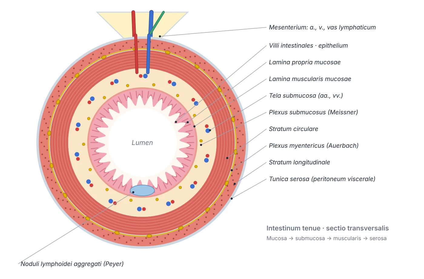
From the oesophagus to the rectum the gut wall keeps a four-layer plan. The mucosa comprises the epithelium, a lamina propria holding immune cells, blood vessels and lymphatics, and the muscularis mucosae. The submucosa is dense connective tissue carrying vessels, lymphatics and the submucosal (Meissner) plexus. The muscularis externa has an inner circular and an outer longitudinal layer, with the myenteric (Auerbach) plexus between them. Outermost is a serosa (visceral peritoneum) in intraperitoneal segments, or an adventitia in the oesophagus and retroperitoneal segments. Each segment modifies this plan: the stomach has mucosal folds (rugae) that let it distend and gastric glands whose parietal cells secrete HCl and chief cells pepsinogen; the small intestine has circular folds (valves of Kerckring) and villi; submucosal Brunner glands occur only in the duodenum; and Peyer patches are far more numerous in the ileum than in the jejunum.[4][5]
The muscle coat is adapted to each segment’s task. The upper third of the oesophagus is skeletal muscle and the lower two-thirds smooth muscle; the inner circular layer is thicker than the longitudinal and thickens further at the lower end to form the lower oesophageal sphincter, about 3 cm above the gastric junction. The upper sphincter, formed by cricopharyngeus with fibres of the pharyngeal constrictors, stays closed except during swallowing. Primary peristalsis continues the pharyngeal phase of swallowing; if a bolus remains, distension of the wall triggers secondary peristalsis. In the stomach the upper part (cardia, fundus, body) relaxes to store food, while the lower part generates mixing waves about every 20 seconds, each pushing a small amount of chyme through the pylorus.[6][4]
Foregut, midgut and hindgut
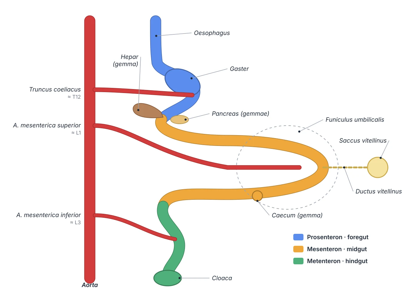
The primitive gut tube forms in week 4 as cephalocaudal and lateral folding of the embryo encloses part of the endoderm-lined yolk sac. Endoderm gives the epithelial lining and the parenchyma of the glands; the surrounding splanchnic mesoderm gives muscle, connective tissue and the wall layers; a dorsal mesentery suspends the tube from the posterior body wall. The tube has three parts, each with its own artery: the foregut is supplied by the coeliac trunk, the midgut by the superior mesenteric artery and the hindgut by the inferior mesenteric artery; the duodenum derives from both foregut and midgut. The stomach rotates 90° clockwise about its long axis, so the left vagus comes to lie anteriorly and the right vagus posteriorly; a further rotation about the anteroposterior axis pulls the pylorus up to the right and the cardia down to the left.[5][4]
The midgut grows so fast that in week 6 the primary intestinal loop herniates physiologically through the umbilicus, joined to the yolk sac by the vitelline duct, with the superior mesenteric artery as the loop’s axis. The cranial limb forms the distal duodenum, jejunum and upper ileum; the caudal limb forms the lower ileum, caecum, appendix, ascending colon and right two-thirds of the transverse colon. The loop turns 90° anticlockwise as it herniates and a further 180° as it returns to the abdomen in week 10, 270° in all; its lumen is temporarily occluded and recanalised by weeks 8–10. The hindgut forms the left third of the transverse colon down to the upper anal canal; the urorectal septum divides the cloaca into the urogenital sinus in front and the rectum behind. The anal canal below the pectinate line comes from the proctodeum (ectoderm), which is why vessels, lymphatics and nerves differ above and below that line.[5][7]
Foregut
- Derivatives
- Oesophagus, stomach, duodenum to the major papilla, liver, gallbladder, pancreas
- Artery
- Coeliac trunk
- Sympathetic supply
- Greater splanchnic nerve (T5–T9) → coeliac ganglia
Midgut
- Derivatives
- Duodenum beyond the papilla, jejunum, ileum, caecum, appendix, ascending colon, right 2/3 of transverse colon
- Artery
- Superior mesenteric artery
- Sympathetic supply
- Lesser splanchnic nerve (T10–T11) → superior mesenteric ganglia
Hindgut
- Derivatives
- Left 1/3 of transverse colon, descending and sigmoid colon, rectum, anal canal above the pectinate line
- Artery
- Inferior mesenteric artery
- Sympathetic supply
- Inferior mesenteric plexus
| Segment | Derivatives | Artery | Sympathetic supply |
|---|---|---|---|
| Foregut | Oesophagus, stomach, duodenum to the major papilla, liver, gallbladder, pancreas | Coeliac trunk | Greater splanchnic nerve (T5–T9) → coeliac ganglia |
| Midgut | Duodenum beyond the papilla, jejunum, ileum, caecum, appendix, ascending colon, right 2/3 of transverse colon | Superior mesenteric artery | Lesser splanchnic nerve (T10–T11) → superior mesenteric ganglia |
| Hindgut | Left 1/3 of transverse colon, descending and sigmoid colon, rectum, anal canal above the pectinate line | Inferior mesenteric artery | Inferior mesenteric plexus |
Arteries, portal venous system and lymphatics
The coeliac trunk leaves the abdominal aorta at T12 and divides into the left gastric, common hepatic and splenic arteries. The stomach is ringed by two arcades: along the lesser curvature the left gastric artery joins the right gastric (from the proper hepatic artery); along the greater curvature the right gastro-omental (from the gastroduodenal) joins the left gastro-omental (from the splenic), and 3–5 short gastric arteries supply the fundus. In the duodenum the superior pancreaticoduodenal artery (coeliac territory) anastomoses with the inferior pancreaticoduodenal (superior mesenteric territory), exactly at the foregut–midgut boundary. Jejunum and ileum receive jejunal and ileal branches that form arcades in the mesentery and end as vasa recta. The colon receives the ileocolic, right colic and middle colic arteries (superior mesenteric) and the left colic and sigmoid arteries (inferior mesenteric), linked by the marginal artery of Drummond along the whole colonic frame. The rectum has three sources: the superior rectal (end of the inferior mesenteric), the middle rectal (internal iliac) and the inferior rectal (internal pudendal).[4][5][7]
The veins follow the arteries, but almost all blood from the stomach to the upper rectum, together with that of the spleen, pancreas and gallbladder, reaches the portal vein: the inferior mesenteric vein drains into the splenic vein, which joins the superior mesenteric vein behind the neck of the pancreas. The portal vein runs in the hepatoduodenal ligament behind the proper hepatic artery and the common bile duct, divides into right and left branches at the porta hepatis and opens into the sinusoids, where nutrient-rich portal blood mixes with oxygen-rich arterial blood. The liver receives about 25% of cardiac output, roughly 75% of it through the portal vein. When flow through the liver is obstructed (cirrhosis, thrombosis), portal pressure rises, causing oesophageal varices, ascites and hepatorenal syndrome.[8][7]
Lymphatics follow the arteries. In the small intestine lymph starts in the mucosa, passes nodes beside the gut, along the arcades and around the root of the superior mesenteric artery, then reaches the cisterna chyli and thoracic duct, which opens at the junction of the left internal jugular and subclavian veins. This route carries most absorbed lipid, is part of mucosal immunity and is also a path of metastasis: an enlarged left supraclavicular (Virchow) node can signal a gastrointestinal cancer. Gastric lymph drains in four zones (gastric, suprapyloric, pancreaticosplenic and subpyloric nodes) before converging on the coeliac nodes, the basis of lymphadenectomy in gastric cancer surgery.[5][4]
Enteric nervous system and autonomic supply
The gut has its own intrinsic nervous system, the enteric nervous system, often called the “brain of the gut”. The myenteric (Auerbach) plexus lies between the two muscle layers and governs motility; the submucosal (Meissner) plexus regulates secretion and local blood flow. The outer longitudinal layer shortens and lengthens the tube and the thicker inner circular layer constricts its lumen; the myenteric plexus coordinates them into peristaltic waves. Autonomic nerves only modulate this intrinsic activity: parasympathetic (vagal) fibres increase secretion and motility, sympathetic fibres reduce both and constrict vessels.[4][5]
Sympathetic supply is organised in three tiers matching the three embryonic segments. Preganglionic fibres of the greater splanchnic nerve (T5–T9) synapse in the coeliac ganglia, whose postganglionic fibres follow the coeliac branches to foregut organs; the lesser splanchnic nerve (T10–T11) synapses in the superior mesenteric ganglia for the midgut; the hindgut is supplied by the inferior mesenteric plexus. On the stomach the anterior and posterior vagal trunks run along the lesser curvature and give gastric branches. Visceral pain travels back with the sympathetic fibres to the spinal cord, which is why pain from the small bowel is vague, poorly localised and felt in the middle of the abdomen rather than over the organ itself.[5][7][4]
Peritoneum: intraperitoneal and retroperitoneal organs
The peritoneum is a two-layered serous membrane: the parietal layer (from somatic mesoderm) lines the abdominal wall and the visceral layer (from splanchnic mesoderm) covers the organs. Intraperitoneal organs, which have a mesentery and are mobile, are the stomach, spleen, liver, first and fourth parts of the duodenum, jejunum, ileum, transverse colon and sigmoid colon. Retroperitoneal organs lie behind the posterior parietal peritoneum and are fixed: the abdominal oesophagus, second and third parts of the duodenum, ascending and descending colon, pancreas, kidneys, ureters, suprarenal glands and aorta. The ventral mesentery becomes the lesser omentum (hepatogastric and hepatoduodenal ligaments, the latter carrying the portal triad); the dorsal mesentery gives the greater omentum, the gastrosplenic ligament and the transverse and sigmoid mesocolons.[9]
The epiploic foramen (of Winslow) links the greater and lesser sacs; it is bounded by the hepatoduodenal ligament in front, the inferior vena cava behind, the duodenum below and the caudate lobe above, and a surgeon passes a finger through it to clamp the hepatoduodenal ligament in liver haemorrhage or to enter the lesser sac. Normally the peritoneal cavity holds less than 100 mL of fluid. Peritonitis usually follows perforation of a hollow viscus or contamination by blood, gastric or pancreatic juice, and presents with rigidity and rebound tenderness; cirrhotic ascites is linked to portal hypertension. The large semipermeable surface of the peritoneum is used for peritoneal dialysis and ventriculoperitoneal shunting; the cavity is also a site of malignant spread (peritoneal carcinomatosis) and of pseudomyxoma peritonei.[9]
Principal organs
Humans have two dentitions: 20 primary teeth gradually replaced by 32 permanent teeth, arranged in two dental arches on the maxillae and the mandible. Each tooth has an enamel-covered crown and a cementum-covered root held in its socket by the periodontal ligament; inside, dentine surrounds the pulp. The mandible moves at the temporomandibular joints under four muscles of mastication supplied by the mandibular nerve (V3); the teeth receive sensation from the superior and inferior alveolar nerves and blood from the maxillary artery.
TongueLinguaA mass of striated muscle covered by mucosa on the floor of the mouth, with a body and a root separated by the V-shaped terminal sulcus. It takes part in chewing, swallowing and speech and carries the taste papillae.
Salivary glandsGlandulae salivariae majoresThree pairs of major salivary glands (parotid, submandibular, sublingual) and hundreds of minor glands secrete about 0.5–1.5 litres of saliva a day, which moistens food, starts starch digestion with amylase and protects the teeth and mucosa.
PharynxPharynxA musculomembranous tube about 12–15 cm long from the skull base to the lower border of the cricoid cartilage (C6), lying behind the nose, mouth and larynx and divided into the nasopharynx, oropharynx and laryngopharynx. It is the crossroads of the airway and the food passage: during swallowing the constrictors drive the bolus into the oesophagus while the larynx is raised and closed.
OesophagusOesophagusA muscular tube about 25 cm long joining the pharynx (at C6) to the stomach, running through the neck, the posterior mediastinum and the oesophageal hiatus of the diaphragm (at T10). It has three physiological narrowings: at the pharyngo-oesophageal junction, where the aortic arch and left main bronchus cross it, and at the diaphragm.
StomachGasterThe dilated segment of the alimentary canal in the epigastric and left hypochondriac regions, with an adult capacity of about 1–1.5 L. The gastric mucosa secretes hydrochloric acid and pepsinogen.
LiverHeparThe largest solid organ (about 1.4–1.5 kg), situated in the right hypochondrium. It secretes bile, metabolises carbohydrates, lipids and proteins, synthesises plasma proteins and detoxifies xenobiotics.
GallbladderVesica biliarisA pear-shaped sac 7–10 cm long lying in the gallbladder fossa on the visceral surface of the liver and holding about 30–50 mL of bile. Bile is concentrated and stored here, then delivered to the duodenum through the cystic and common bile ducts when cholecystokinin makes the gallbladder contract after a meal.
DuodenumDuodenumThe first part of the small intestine, about 25 cm long, curving in a C around the head of the pancreas and mostly retroperitoneal. It has four parts, superior, descending, horizontal and ascending; the bile and pancreatic ducts open on the major duodenal papilla in the descending part. The duodenum neutralises gastric acid and is where pancreatic enzymes and bile begin digestion.
Vermiform appendixAppendix vermiformisA blind tube averaging 6–10 cm, arising from the posteromedial wall of the caecum where the three taeniae coli converge, about 2 cm below the ileocaecal junction. The position of its tip varies widely (retrocaecal, pelvic and others). Its wall is rich in lymphoid tissue; appendicitis is the commonest abdominal surgical emergency.
IntestinesIntestinumThe small intestine (duodenum, jejunum, ileum), about 6–7 m long, absorbs most nutrients; the large intestine (caecum, colon, rectum), about 1.5 m long, absorbs water and electrolytes.
Reference values
- Whole-gut transit time is typically 24 to 72 hours.
- The colon harbours roughly 4 × 10¹³ bacteria, which make up the gut microbiota.
References
Sources for the values and descriptions on this page, indexed in PubMed (US National Library of Medicine).
- [1]Clinical and surgical anatomy of the liver: a review for cliniciansJuza RM et al., Clinical anatomy 2014PMID 24453062
- [2]Liver macrophages: development, dynamics, and functionsNusse Y et al., Cellular & molecular immunology 2025PMID 40461700
- [3]Pathogenesis of liver fibrosisHernandez-Gea V et al., Annual review of pathology 2011PMID 21073339
- [4]Anatomy, Abdomen and Pelvis: StomachChaudhry SR et al., StatPearls 2024PMID 29493959
- [5]Anatomy, Abdomen and Pelvis, Small IntestineCollins JT et al., StatPearls 2025PMID 29083773
- [6]Anatomy, Thorax, EsophagusOmole AE et al., StatPearls 2026PMID 29494119
- [7]Anatomy, Abdomen and Pelvis: Large IntestineOmole AE et al., StatPearls 2025PMID 29261962
- [8]Anatomy, Abdomen and Pelvis, Portal Venous System (Hepatic Portal System)Harkins JM et al., StatPearls 2023PMID 32119476
- [9]Anatomy, Abdomen and Pelvis, PeritoneumKalra A et al., StatPearls 2023PMID 30521209
- [10]Gastrointestinal mean transit times in young and middle-aged healthy subjectsPMID 11318834Supports: Gastrointestinal transit time
- [11]Revised Estimates for the Number of Human and Bacteria Cells in the BodySender R, Fuchs S, Milo R, 2016PMID 27541692Supports: Colonic bacterial count