The human body, from the whole to the single cell
An open anatomy atlas for medical students: true models you can dissect layer by layer, sections through the organs, real histology slides and articles cited from PubMed, with Terminologia Anatomica names in eight languages.
1,004
models
2,647
anatomical structures
365
cited PubMed sources
8
languages
3D library
Every model in one place
Layered regional atlases, whole systems, organs, inside-organ scenes and cells, opened full-frame with a toolbar: section planes, standard views, auto-rotate, screenshots and shareable view links.
- 13 Regional
- 11 Whole system
- 64 Organ
- 864 Specimens
- 36 Inside organs
- 16 Cells and tissues
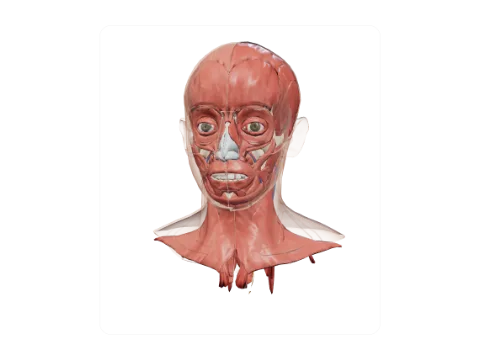 Head and neck
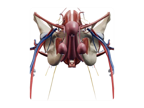 Perineum and pelvic floor
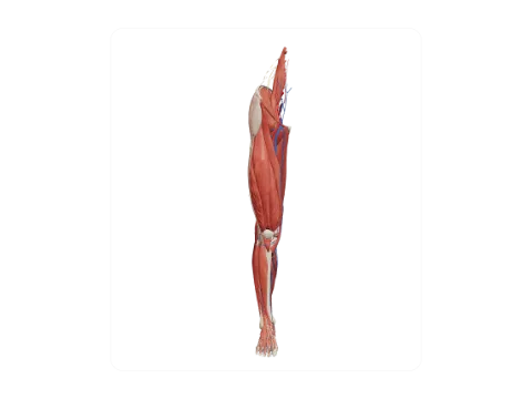 Lower limb
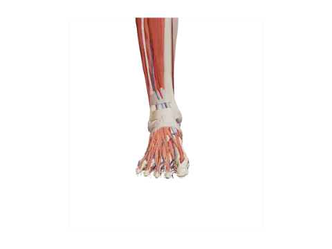 Ankle and foot
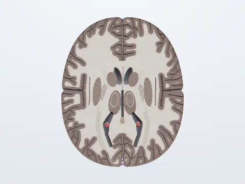 Brain, axial section
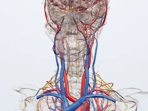 Cranial nerves
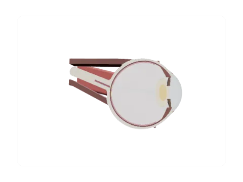 Eyeball, horizontal section
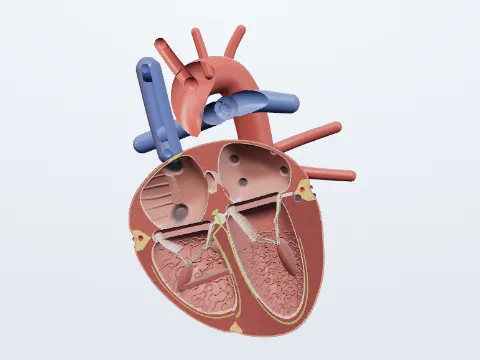 Heart
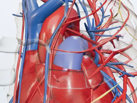 Pulmonary trunk
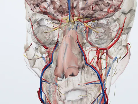 Nose and nasal cavity
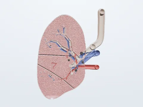 Right lung, coronal section
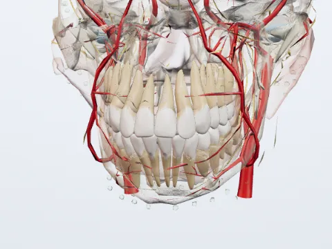 Teeth and masticatory apparatus
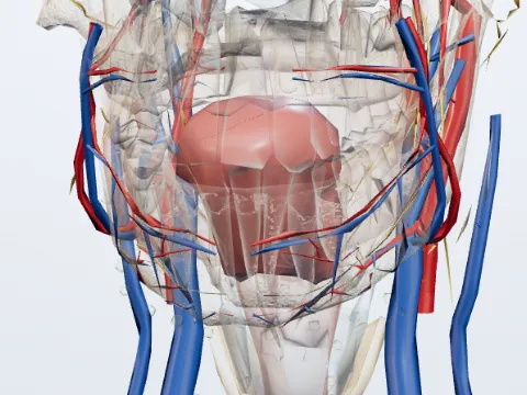 Tongue
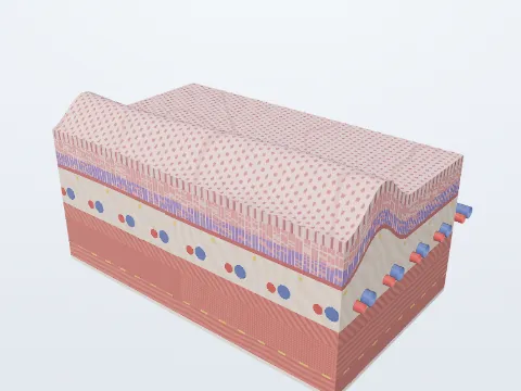 Stomach
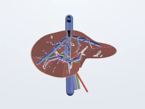 Liver, coronal section
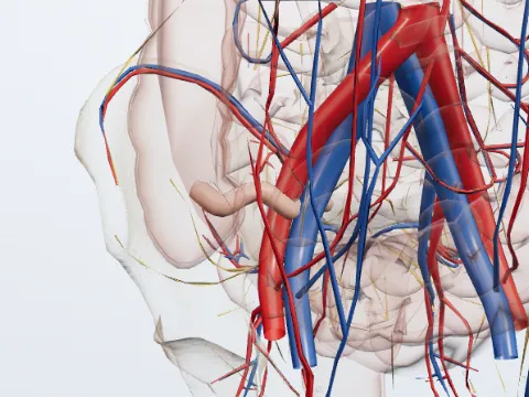 Vermiform appendix
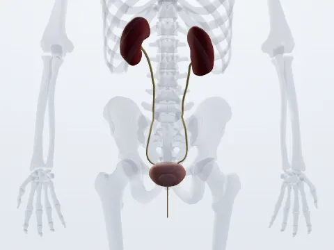 Urinary system
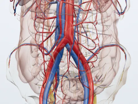 Ureter
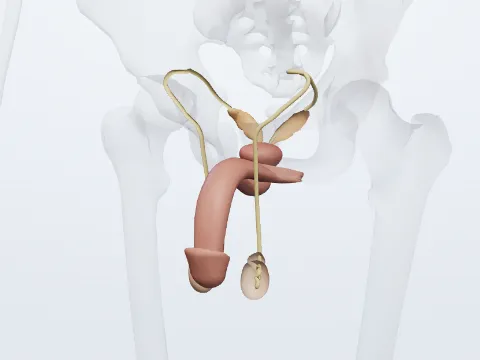 Reproductive system
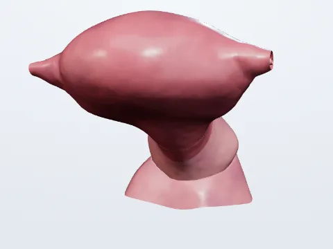 Uterus
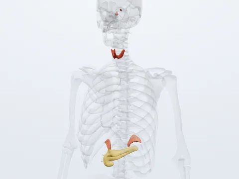 Endocrine system
 Pituitary gland
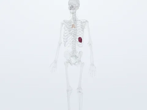 Lymphatic system
 Lymph nodes
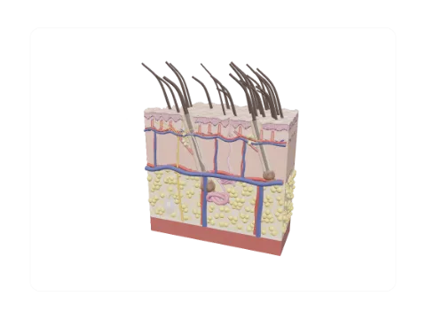 Male skin block (terminal hair): layers and appendages
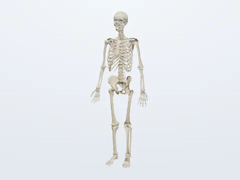 Skeletal system
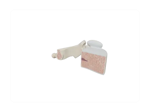 Cervical vertebra (C5)
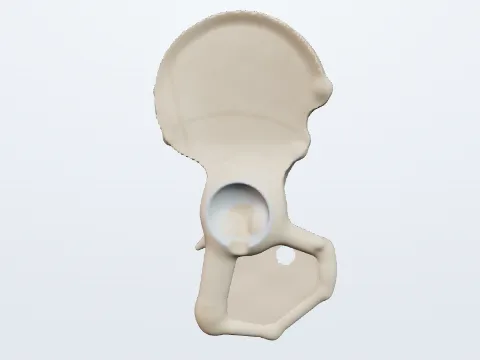 Hip bone: parts
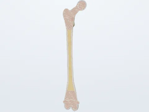 Femur: parts and section
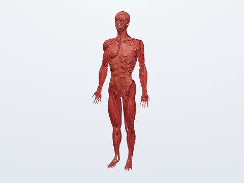 Muscular system
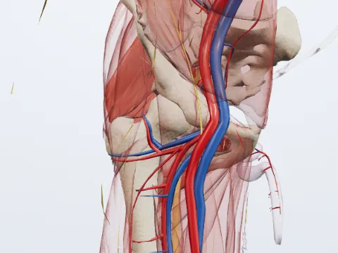 Gluteus maximus
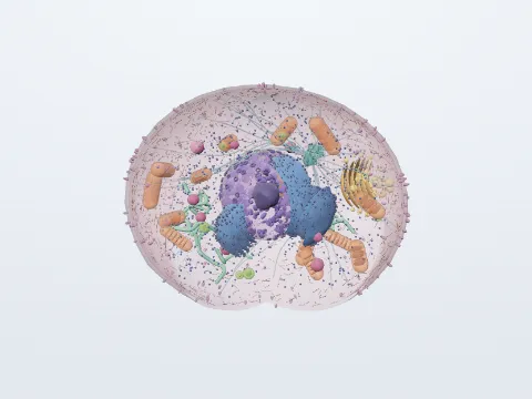 Animal cell
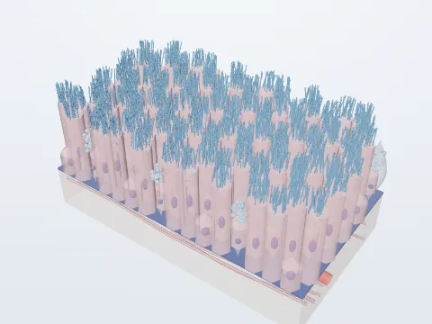 Pseudostratified ciliated
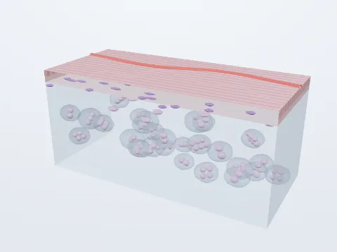 Hyaline cartilage
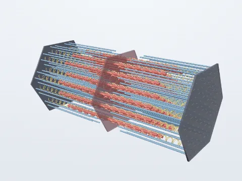 Sarcomere
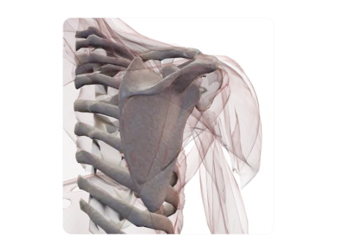 Scapula
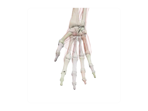 Bones of the hand
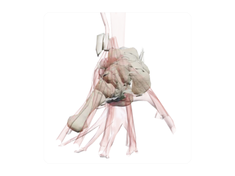 Calcaneus
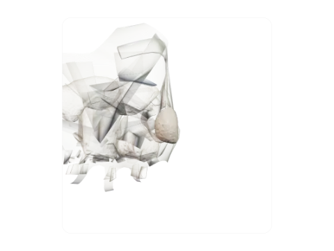 Pisiform
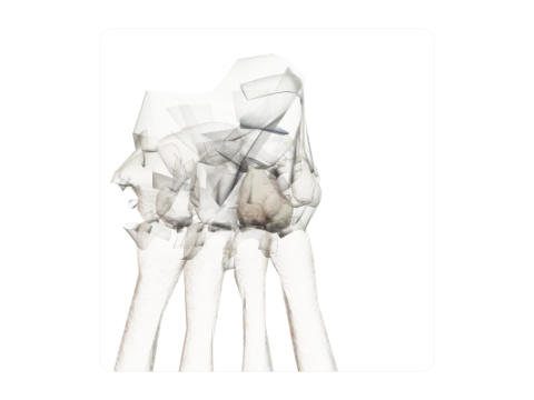 Hamate
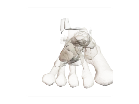 Cuboid
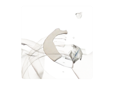 First rib
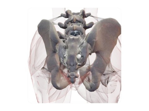 Sacrum and coccyx
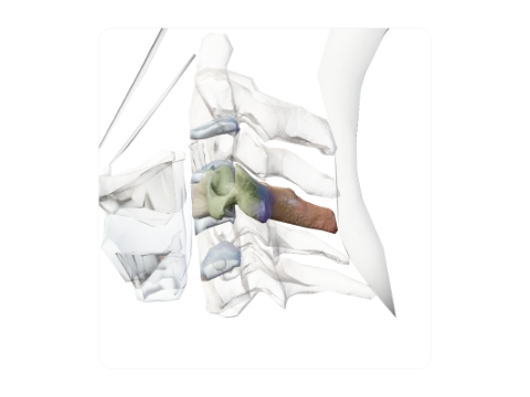 Fourth cervical vertebra (C4)
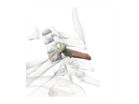 First thoracic vertebra (T1)
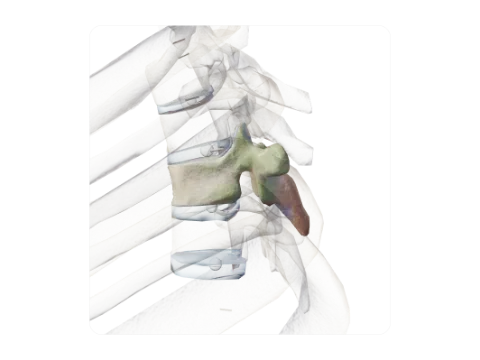 Fifth thoracic vertebra (T5)
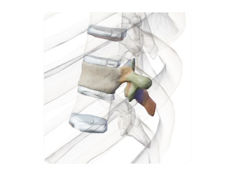 Ninth thoracic vertebra (T9)
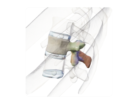 First lumbar vertebra (L1)
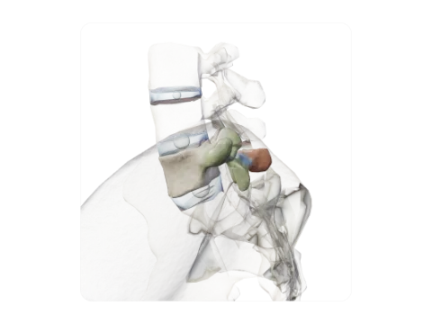 Fifth lumbar vertebra (L5)
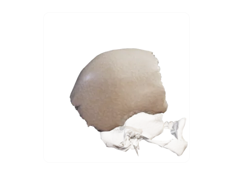 Parietal bone
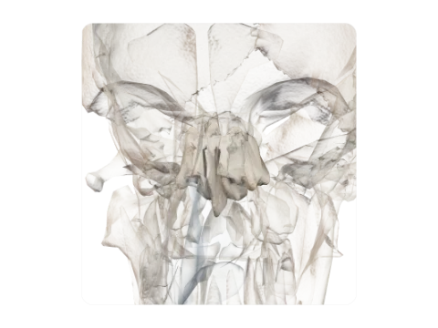 Ethmoid bone
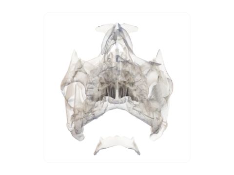 Palatine bones
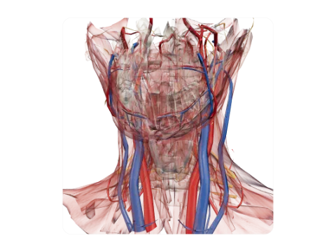 Hyoid bone
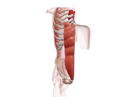 Rectus abdominis
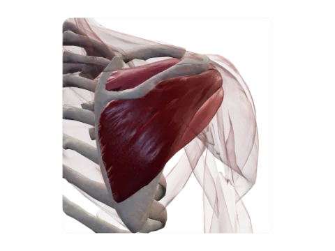 Rotator cuff
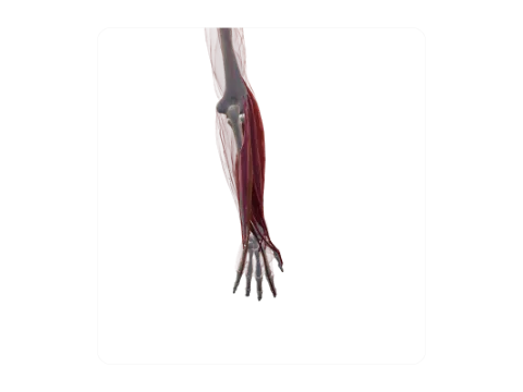 Forearm extensor muscles
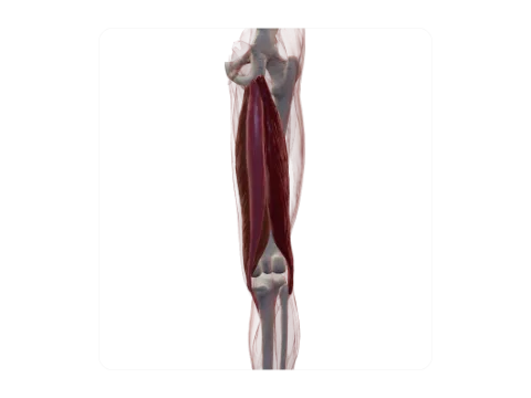 Hamstrings
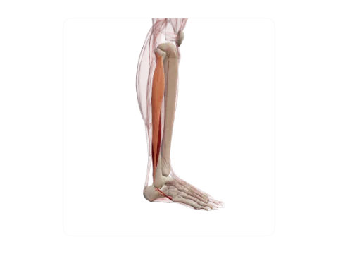 Fibularis longus and brevis
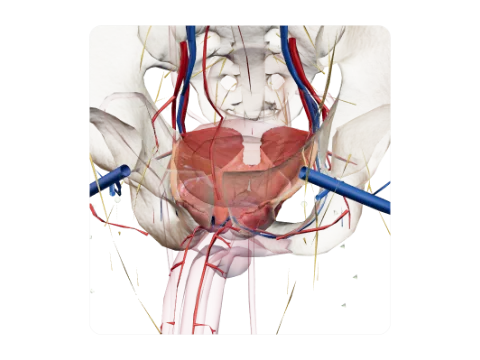 Pelvic floor (levator ani and coccygeus)
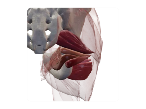 Deep lateral rotators of the hip
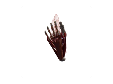 Intrinsic muscles of the sole
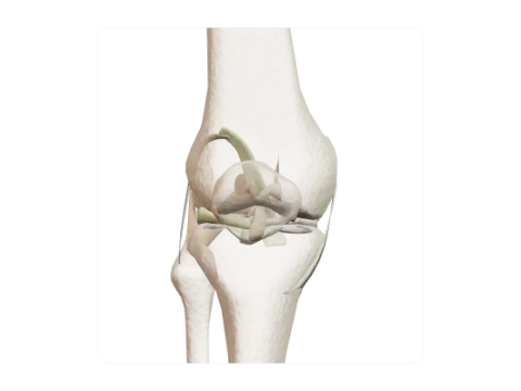 Knee joint
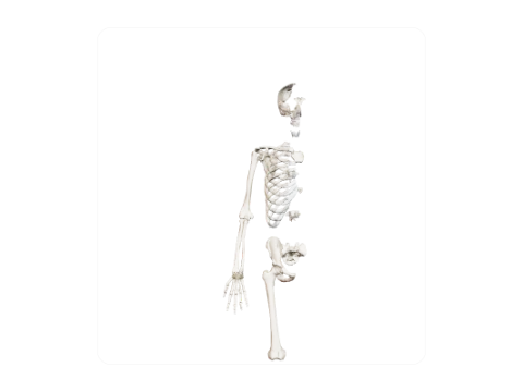 Wrist joint
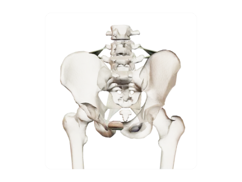 Ligaments of the pelvis
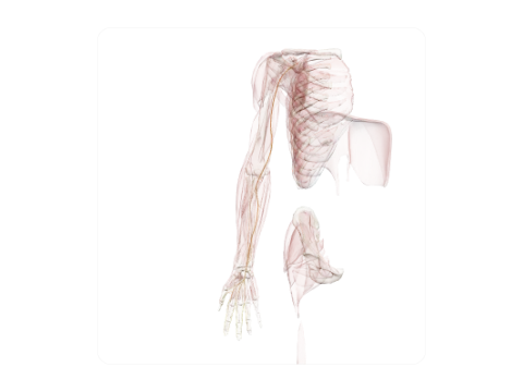 Median nerve
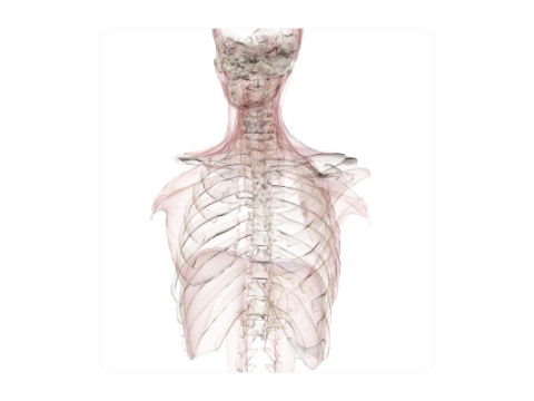 Vagus nerve (X)
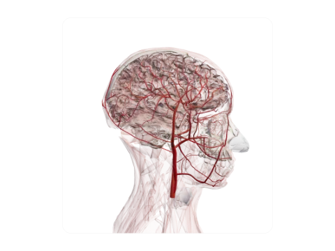 Carotid arteries and branches
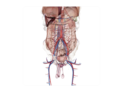 Renal vessels
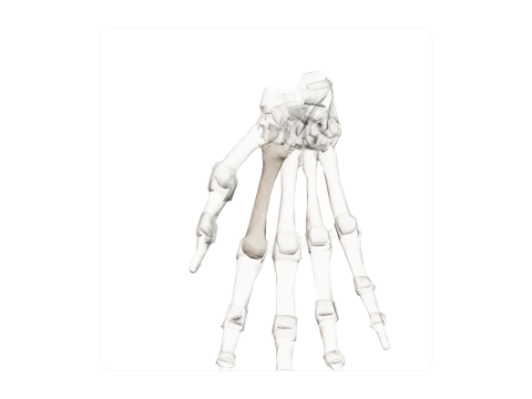 Second metacarpal
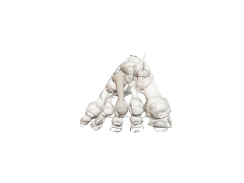 Third metatarsal
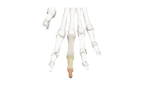 Phalanges of the middle finger
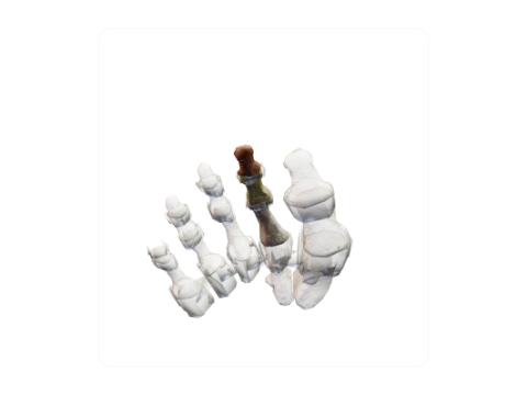 Phalanges of the second toe
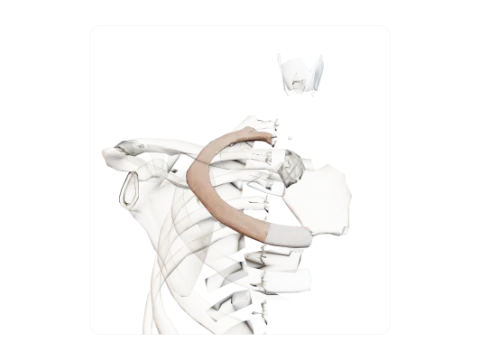 Second rib
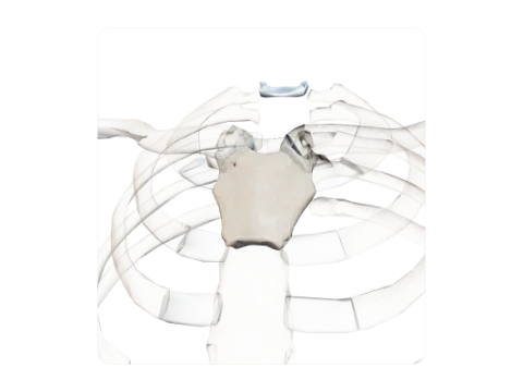 Manubrium of the sternum
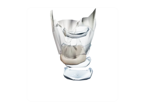 Cricoid cartilage
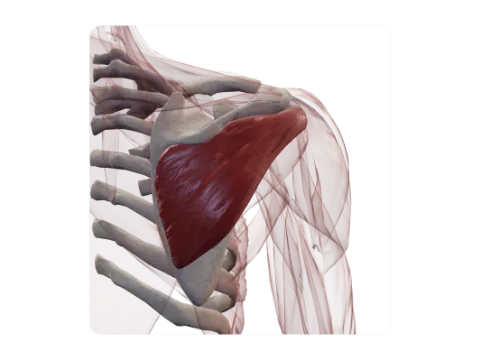 Infraspinatus
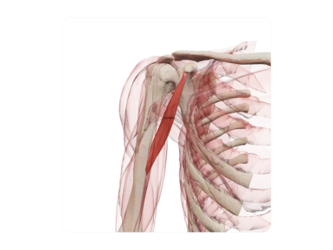 Coracobrachialis
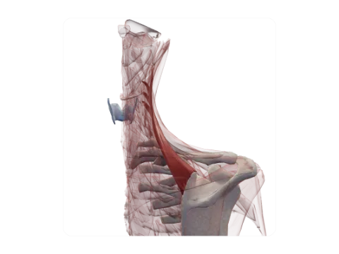 Levator scapulae
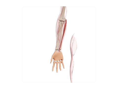 Palmaris longus and palmar aponeurosis
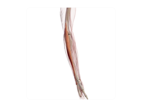 Extensor carpi radialis longus and brevis
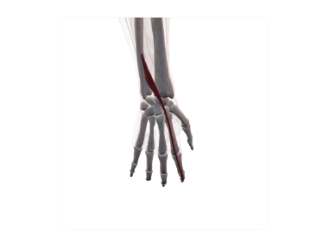 Extensor indicis
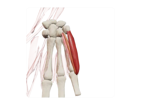 Hypothenar muscles
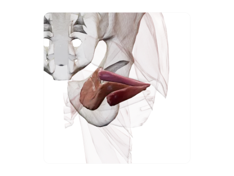 Obturator internus and gemelli
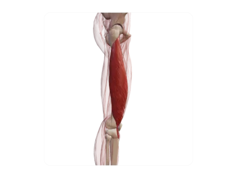 Vastus lateralis
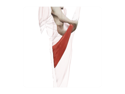 Adductor brevis
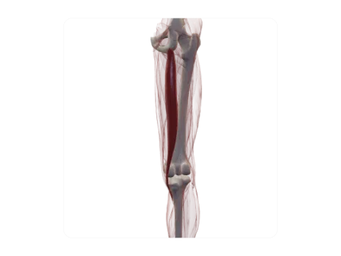 Semitendinosus
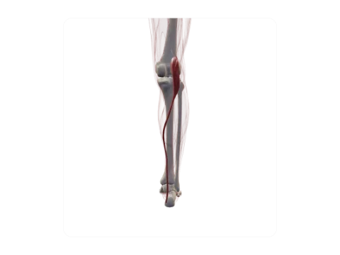 Plantaris
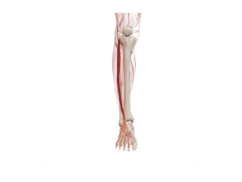 Extensor digitorum longus and fibularis tertius
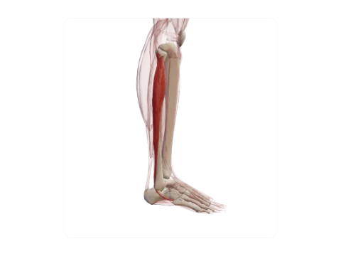 Fibularis longus
 Flexor digitorum brevis and plantar aponeurosis
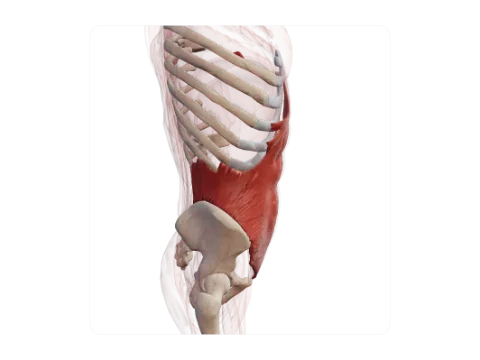 Internal oblique
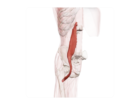 Psoas major
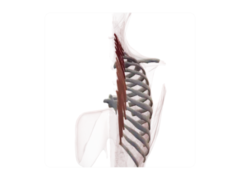 Semispinalis
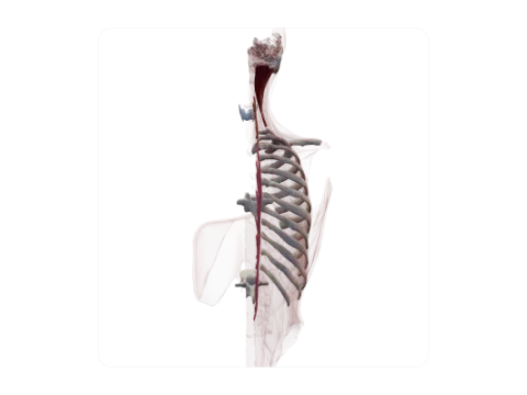 Spinalis
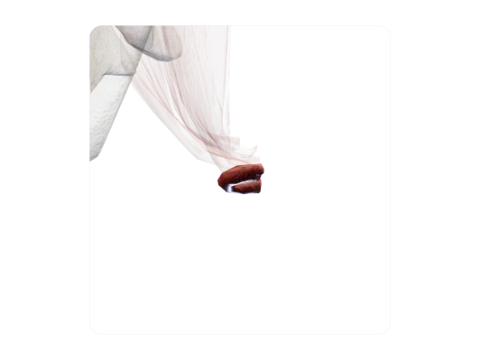 External anal sphincter
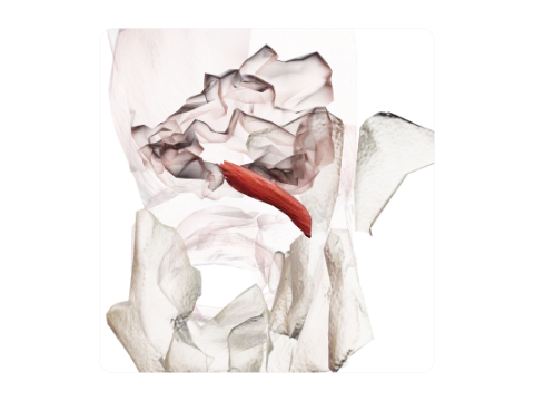 Corrugator supercilii
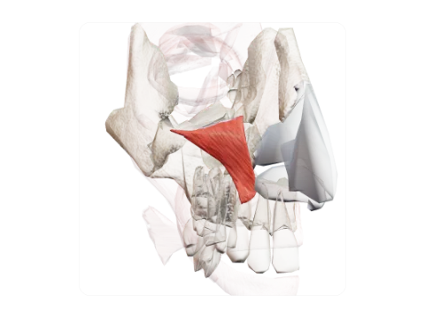 Levator labii superioris
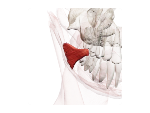 Risorius
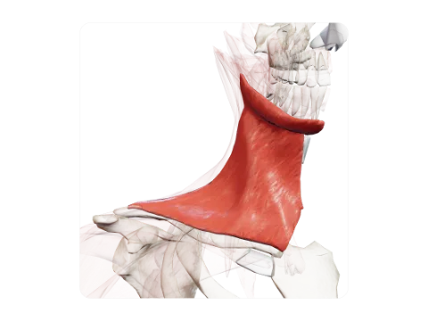 Platysma
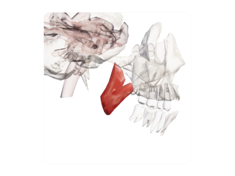 Medial pterygoid
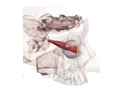 Lateral rectus
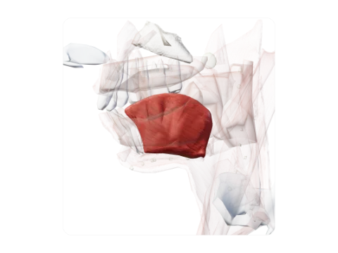 Genioglossus
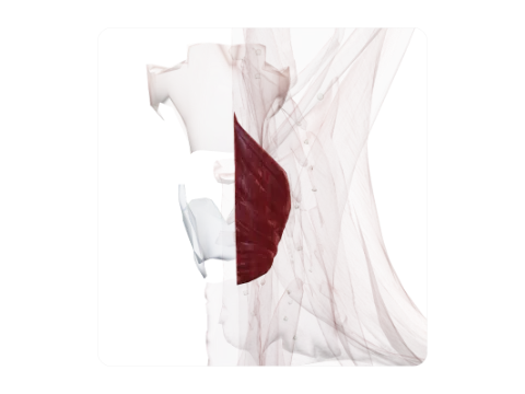 Inferior pharyngeal constrictor
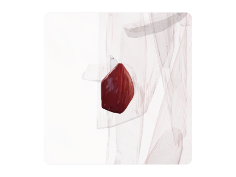 Posterior cricoarytenoid
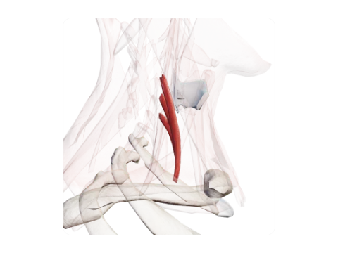 Scalenus anterior
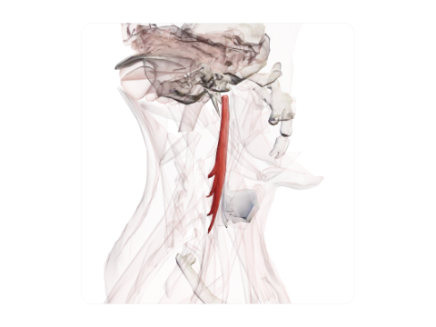 Longus capitis
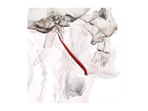 Stylohyoid
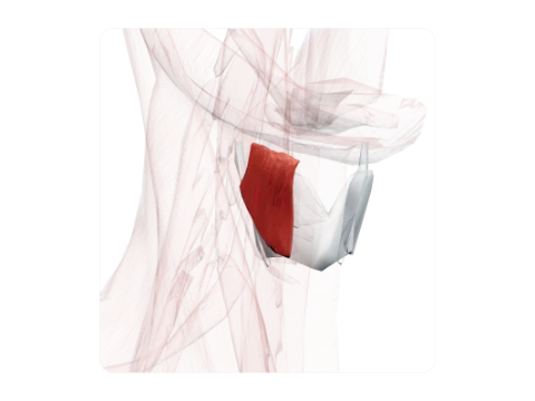 Thyrohyoid
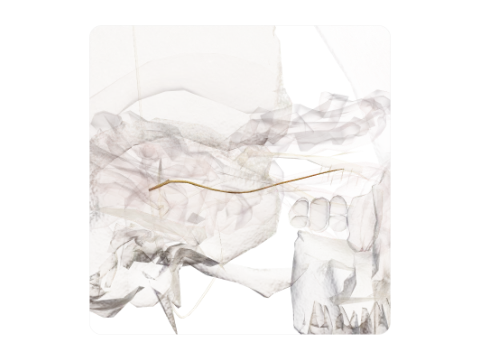 Trochlear nerve (IV)
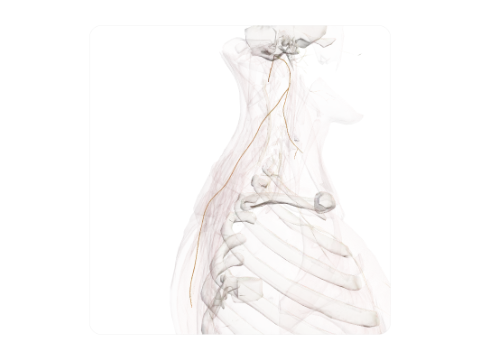 Accessory nerve (XI)
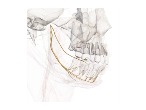 Mandibular nerve (V3) and its branches
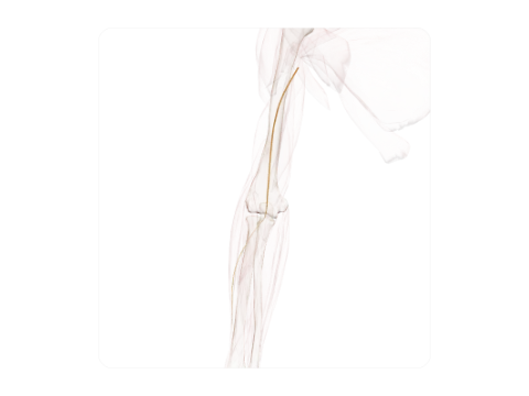 Musculocutaneous nerve
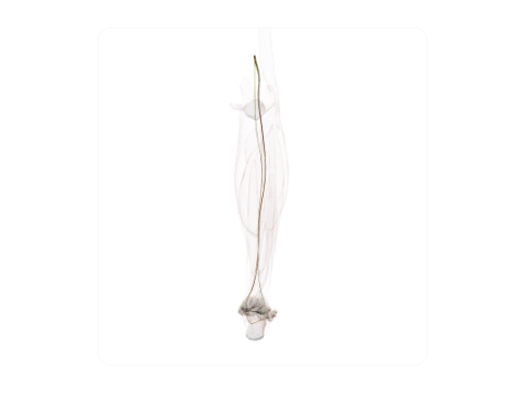 Tibial nerve
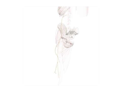 Lateral femoral cutaneous nerve
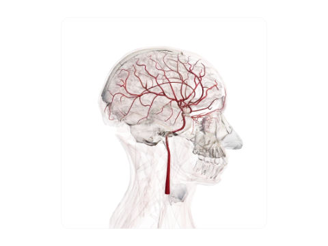 Internal carotid artery
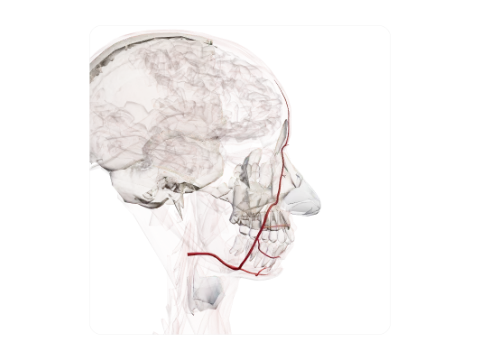 Facial artery
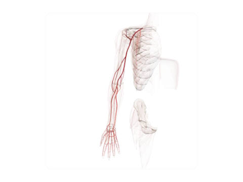 Axillary artery
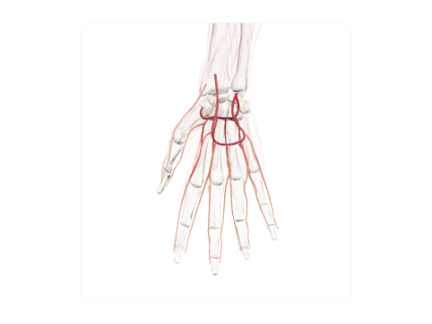 Superficial and deep palmar arches
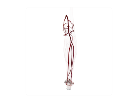 Popliteal artery
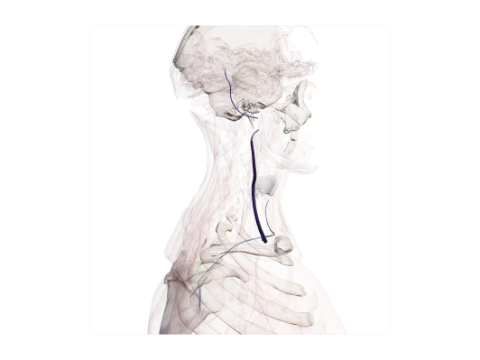 External jugular vein
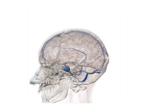 Dural venous sinuses
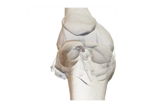 Lateral meniscus
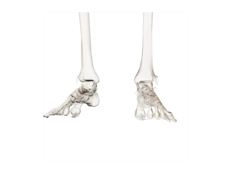 Deltoid (medial collateral) ligament of the ankle
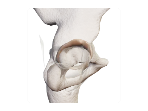 Acetabular labrum
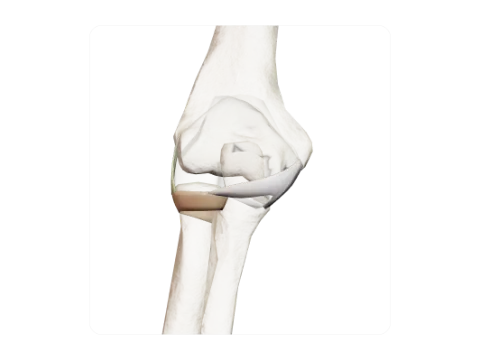 Elbow collateral and annular ligaments
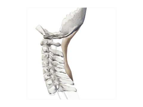 Nuchal ligament
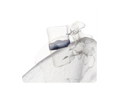 Lumbar intervertebral disc L4–L5
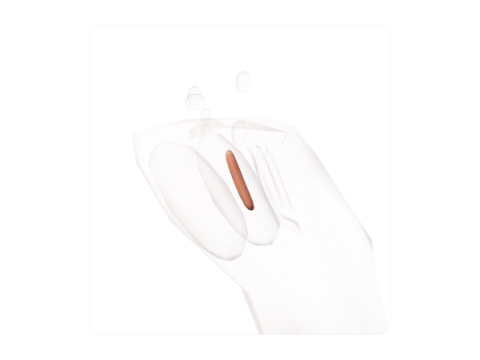 Nucleus ambiguus
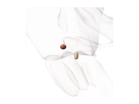 Anterior and posterior cochlear nuclei
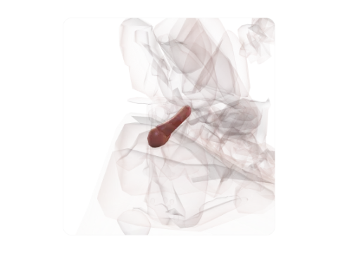 Inferior colliculus
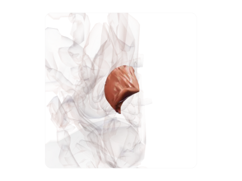 Crus cerebri (base of the cerebral peduncle)
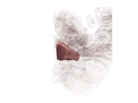 Anterior quadrangular lobule
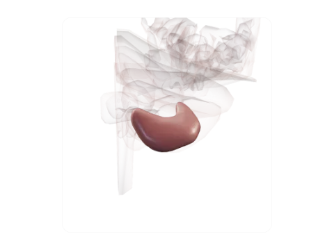 Gracile (paramedian) lobule
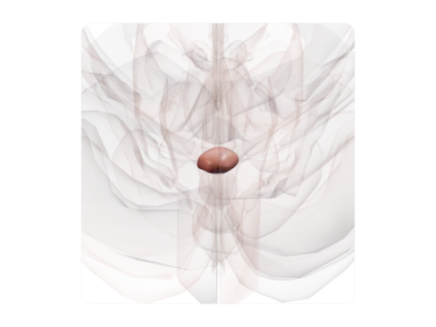 Nodule of vermis
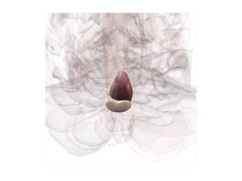 Declive and folium of vermis
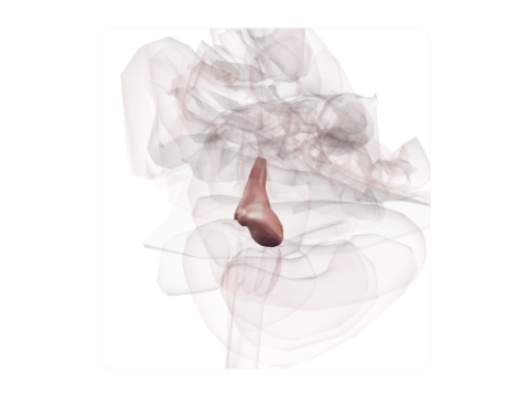 Superior cerebellar peduncle
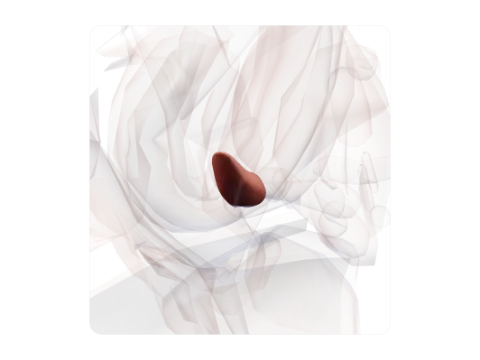 Lateral geniculate body
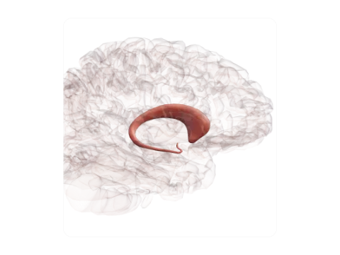 Caudate nucleus
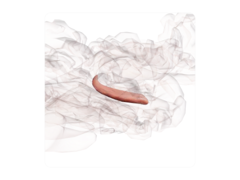 Hippocampus
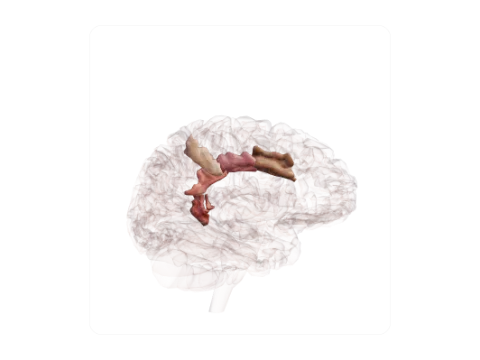 Cingulate gyrus
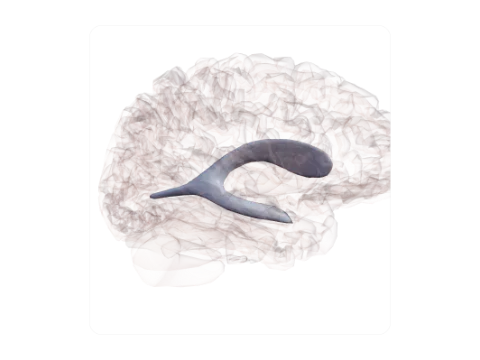 Lateral ventricle
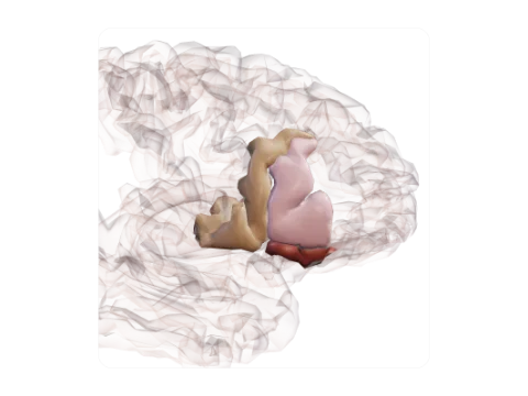 Inferior frontal gyrus (Broca area)
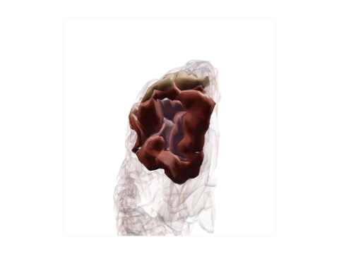 Orbital gyri
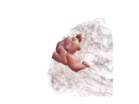 Angular gyrus
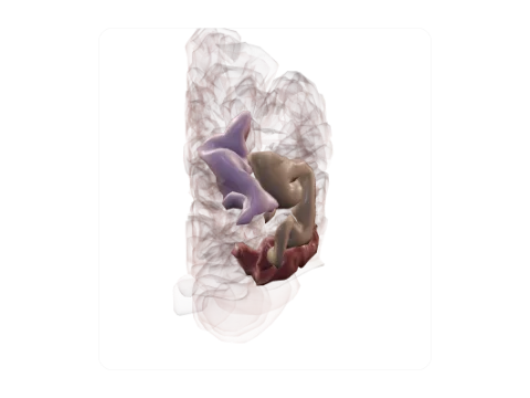 Lateral occipital gyri
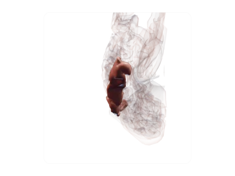 Inferior temporal gyrus
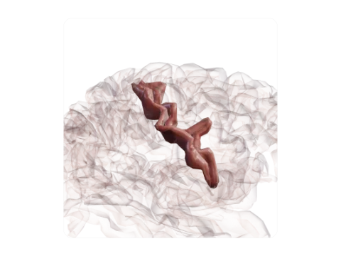 Central sulcus (Rolando)
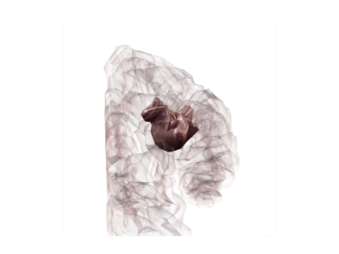 Intraparietal sulcus
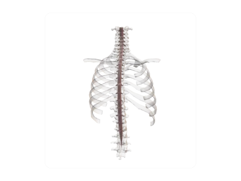 Spinal dura mater
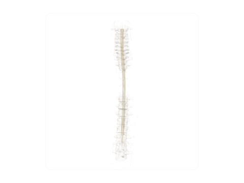 Tectospinal tract
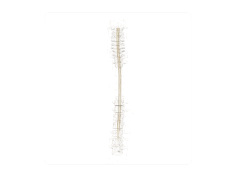 Lateral (medullary) reticulospinal tract
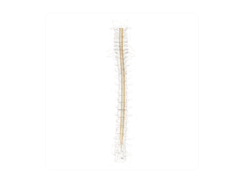 Anterior spinocerebellar tract (Gowers)
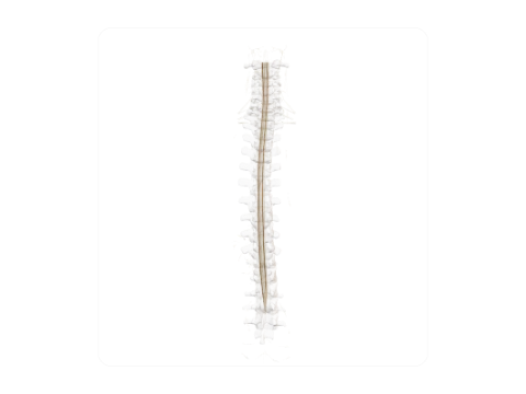 Posterolateral tract (Lissauer)
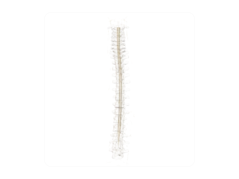 Nucleus proprius of posterior horn
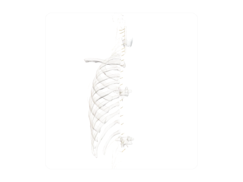 Anterior (ventral) root of spinal nerve
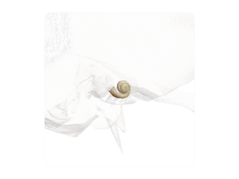 Scala vestibuli and scala tympani
 Vestibule of inner ear
 Iris
 Retina
 Nasolacrimal duct
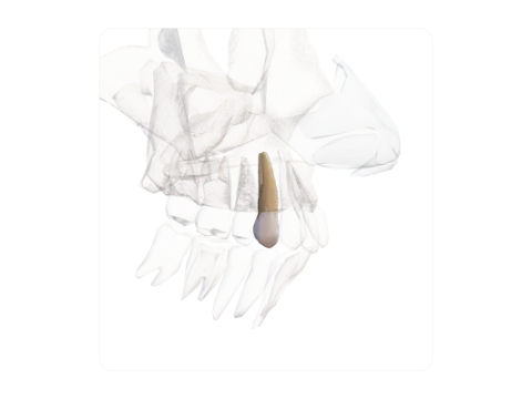 Maxillary first premolar
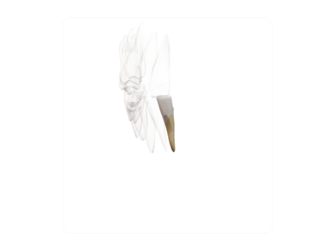 Mandibular central incisor
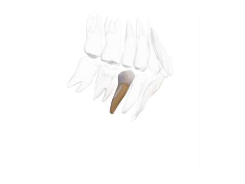 Mandibular second premolar
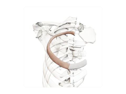 Fourth rib
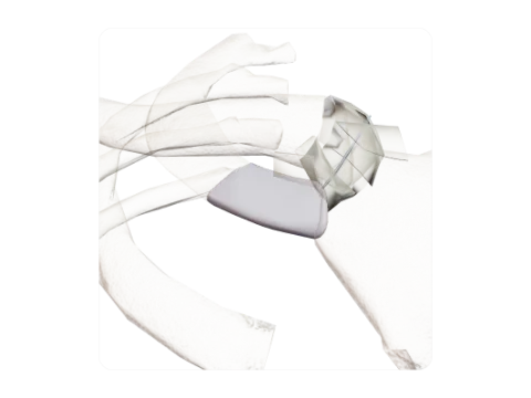 First costal cartilage
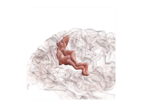 Postcentral sulcus
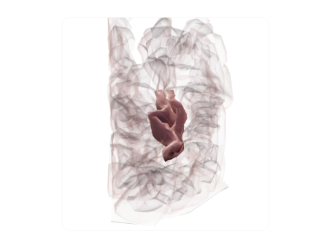 Transverse occipital sulcus
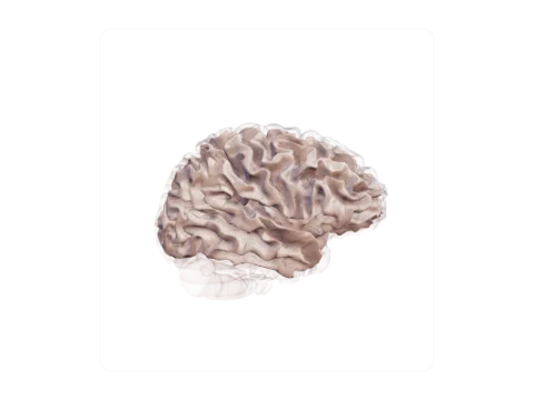 Cerebral white matter (centrum semiovale)
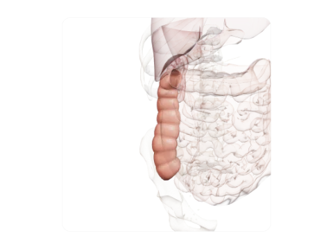 Ascending colon
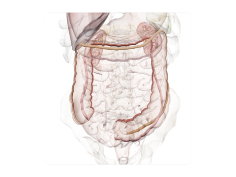 Taeniae coli
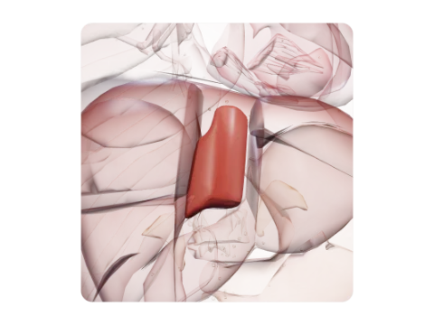 Liver segment I (caudate lobe)
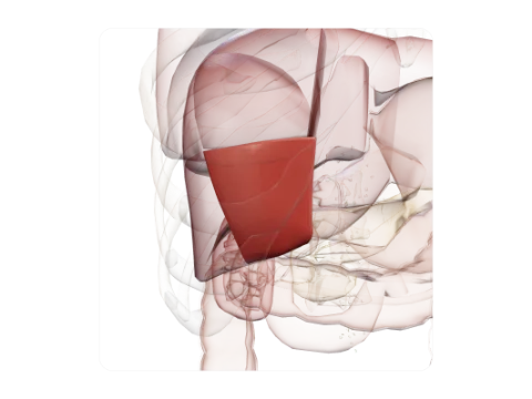 Liver segment V
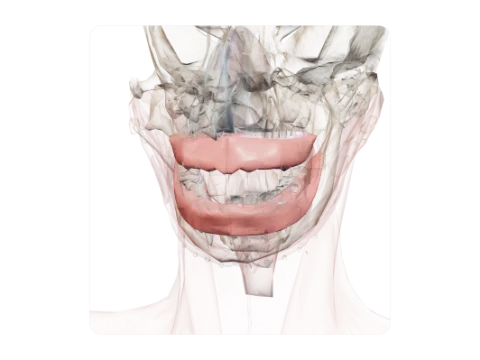 Gingiva (gums)
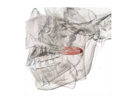 Soft palate and uvula
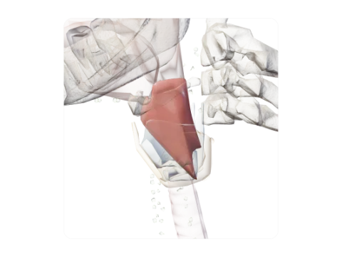 Laryngopharynx (hypopharynx)
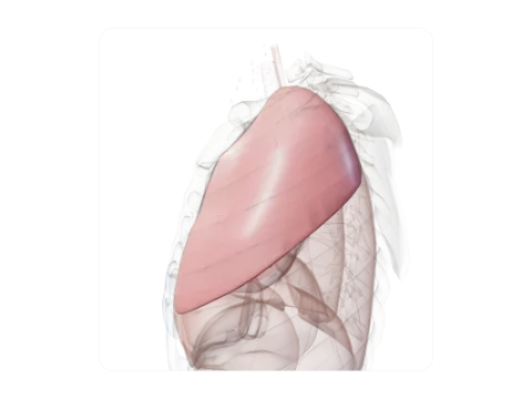 Superior lobe of left lung (with lingula)
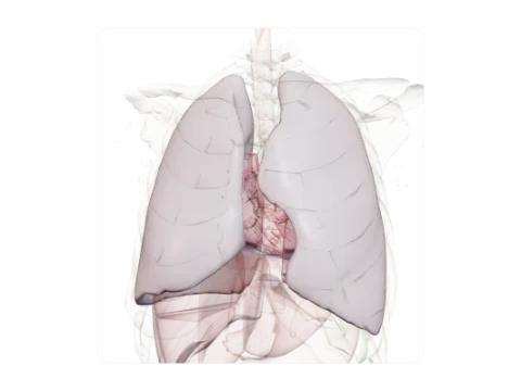 Pleura
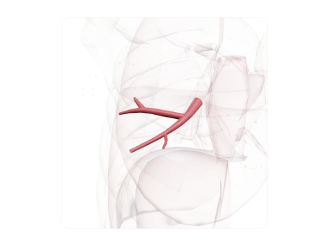 Lateral segmental bronchus of middle lobe (B4)
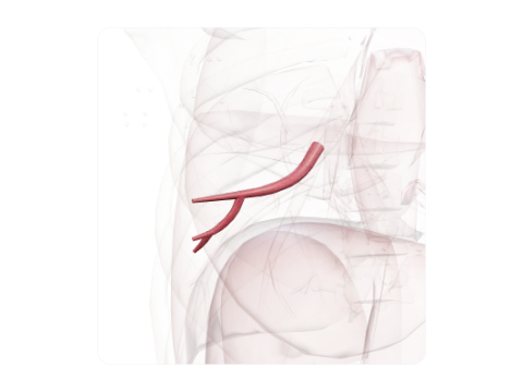 Anterior basal segmental bronchus, right (B8)
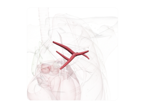 Anterior segmental bronchus, left (B3)
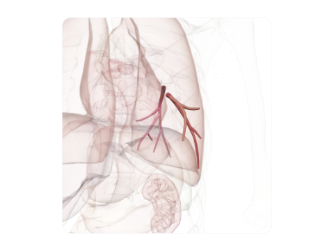 Anteromedial basal segmental bronchus, left (B7+8)
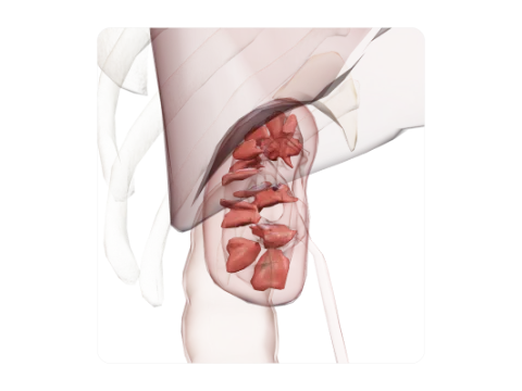 Renal pyramids (medulla)
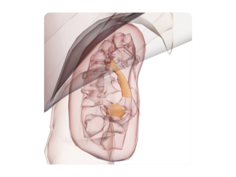 Major calices and renal pelvis
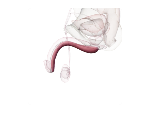 Corpus spongiosum of penis
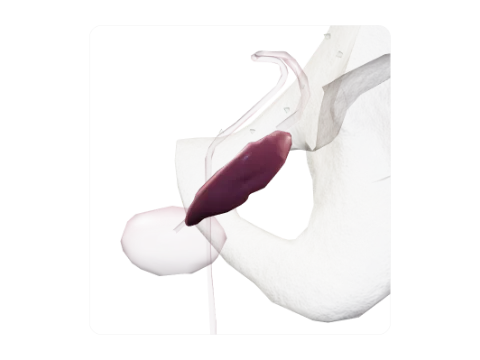 Seminal vesicle
 Superior parathyroid gland
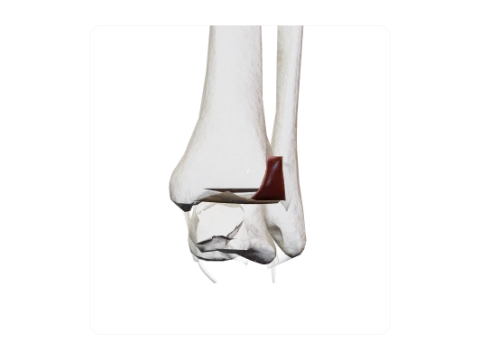 Posterior and transverse tibiofibular ligaments
 Bifurcate ligament
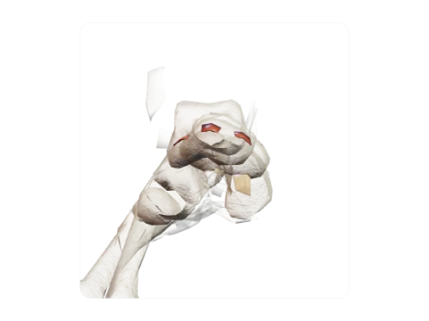 Cuneonavicular ligaments
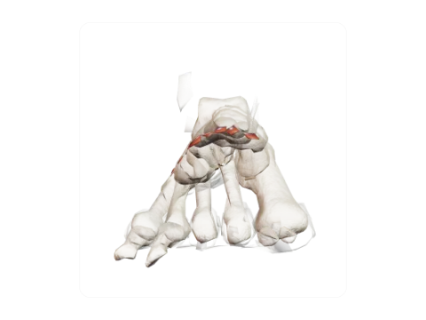 Dorsal and plantar tarsometatarsal ligaments
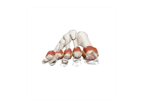 Metatarsophalangeal joints
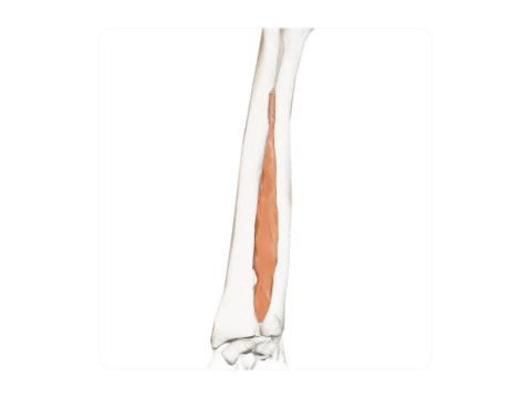 Interosseous membrane of forearm
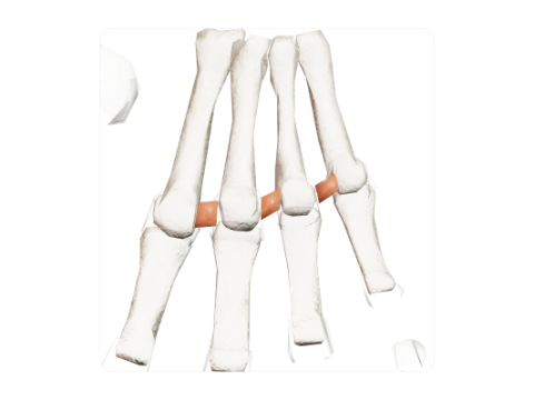 Deep transverse metacarpal ligament
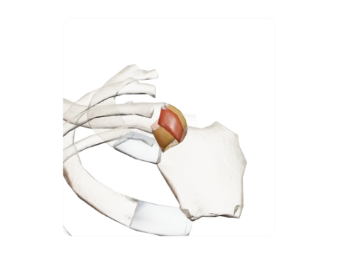 Sternoclavicular joint
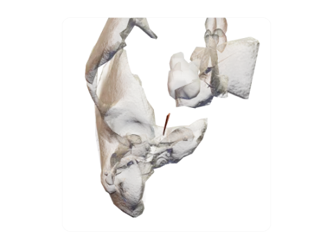 Pterygospinous ligament
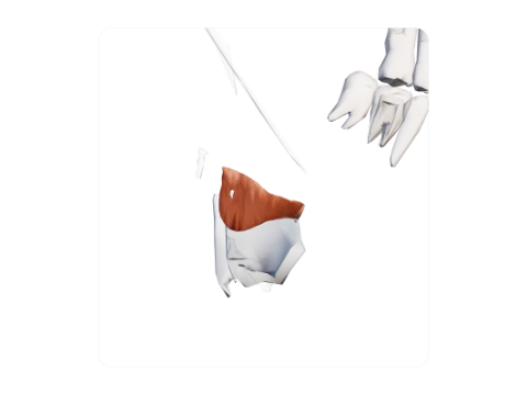 Quadrangular membrane
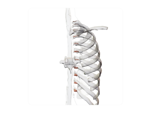 Costotransverse ligaments
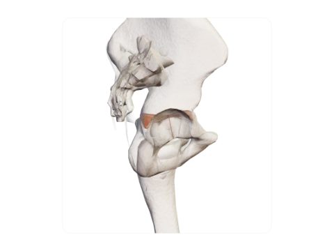 Triradiate cartilage
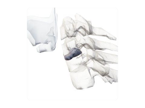 Intervertebral disc C6–C7
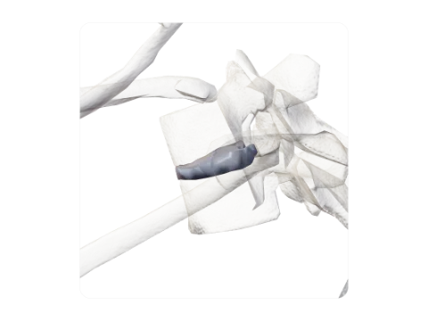 Intervertebral disc T3–T4
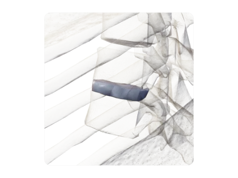 Intervertebral disc T8–T9
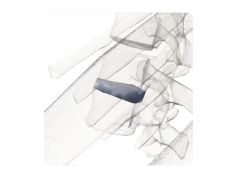 Intervertebral disc T12–L1
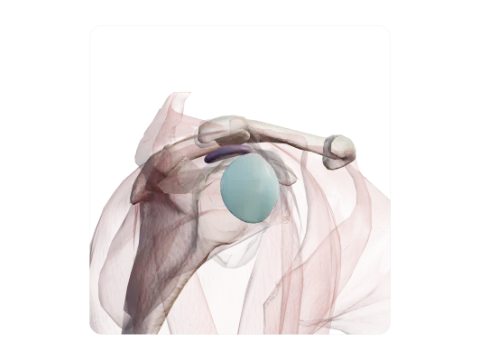 Subacromial–subdeltoid bursa
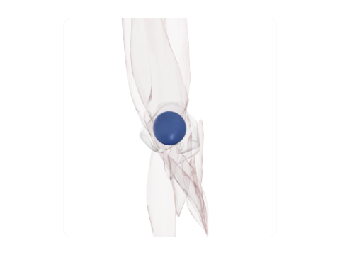 Subtendinous bursa of triceps brachii
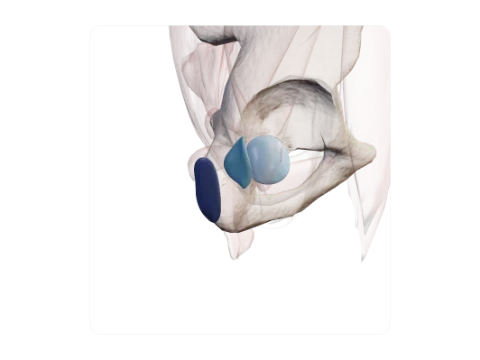 Trochanteric bursae
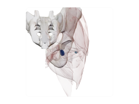 Bursae of obturator internus
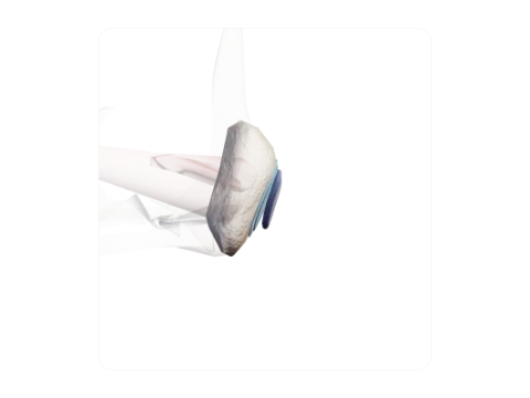 Prepatellar bursae
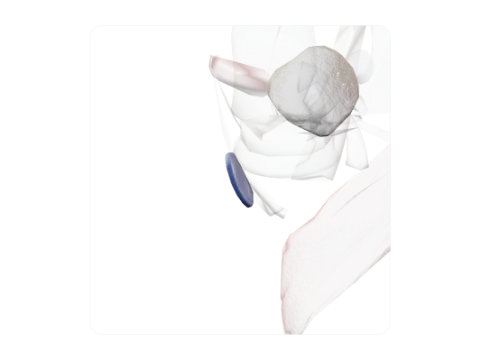 Semimembranosus bursa
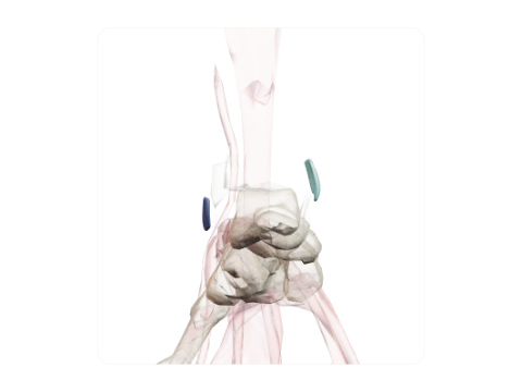 Subcutaneous malleolar bursae
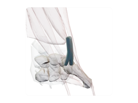 Tendon sheath of extensores carpi radiales
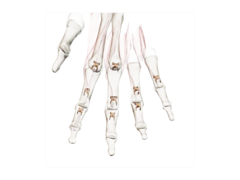 Fibrous sheaths of the fingers (pulleys)
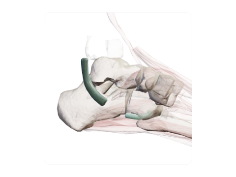 Tendon sheaths of fibularis muscles
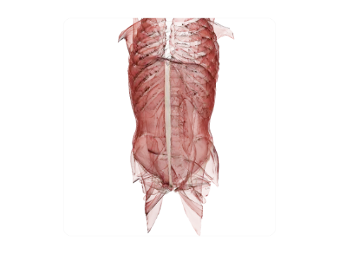 Linea alba
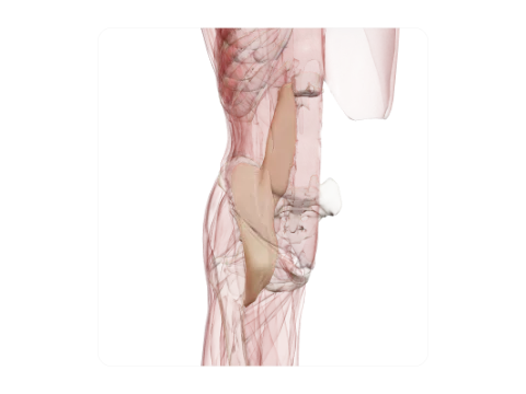 Iliopsoas fascia
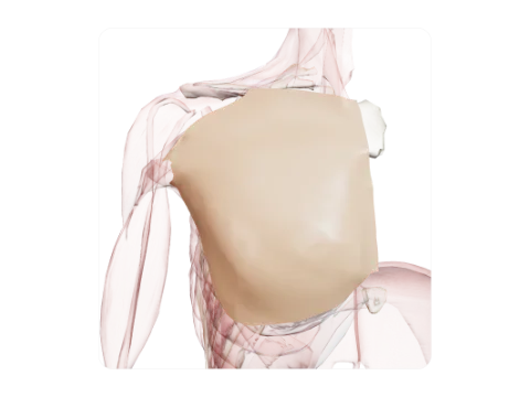 Pectoral fascia
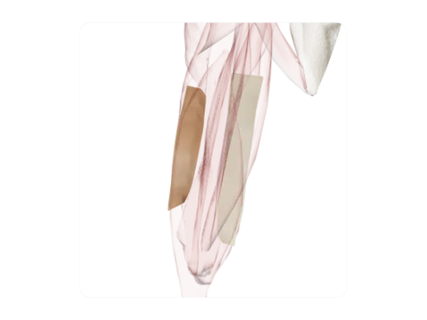 Intermuscular septa of the arm
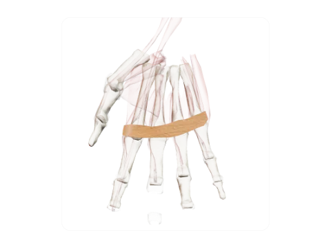 Superficial transverse metacarpal ligament
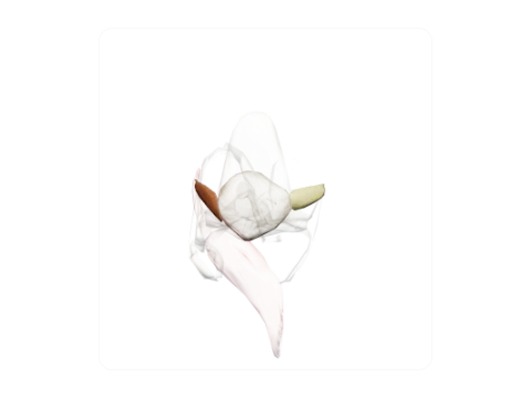 Patellar retinacula
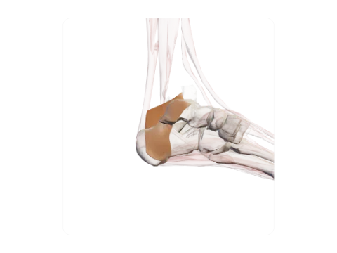 Flexor retinaculum of the ankle
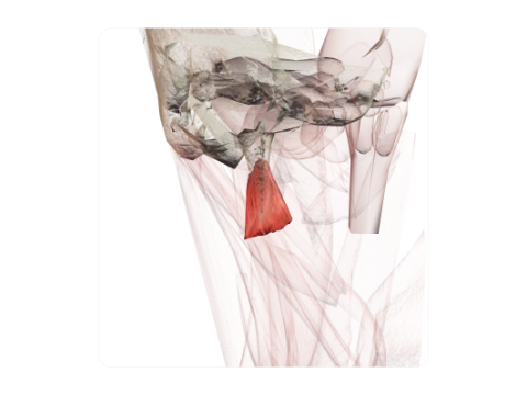 Rectus capitis lateralis
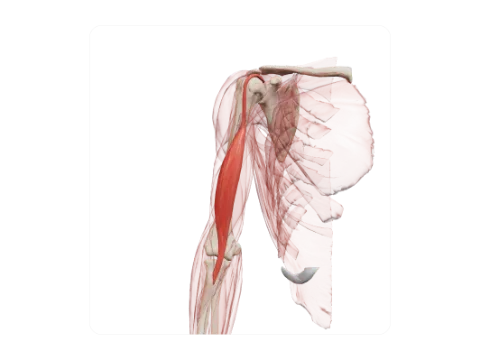 Long head of biceps brachii
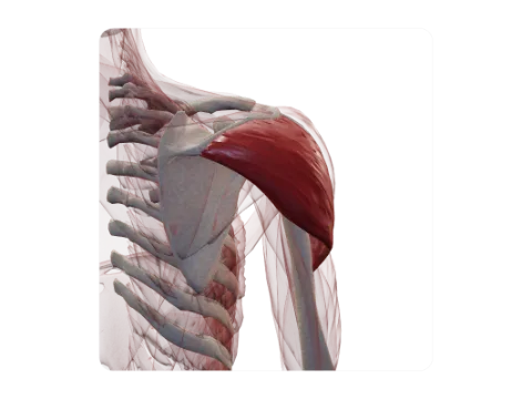 Spinal part of deltoid
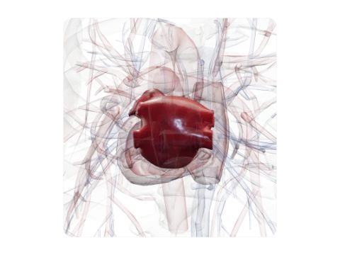 Left atrium
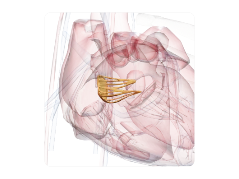 Septal leaflet of the tricuspid valve
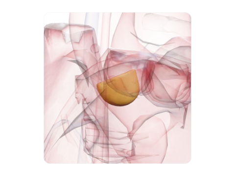 Right coronary cusp
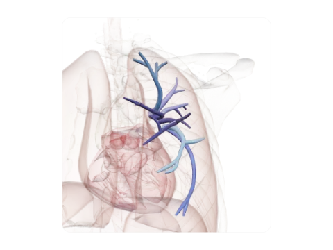 Segmental arteries of the left upper lobe
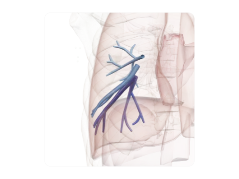 Segmental arteries of the right lower lobe
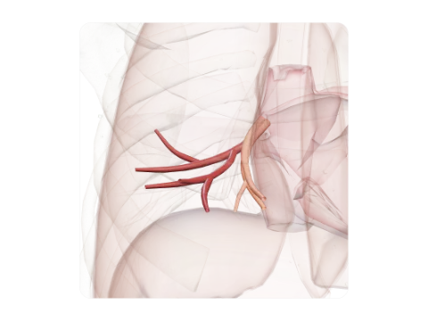 Veins of the right middle lobe
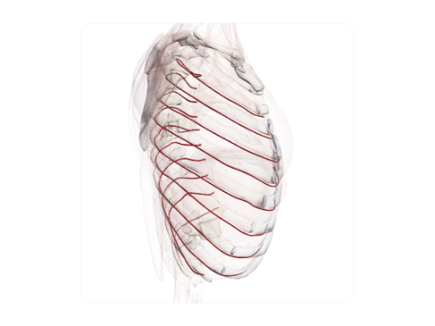 Posterior intercostal arteries
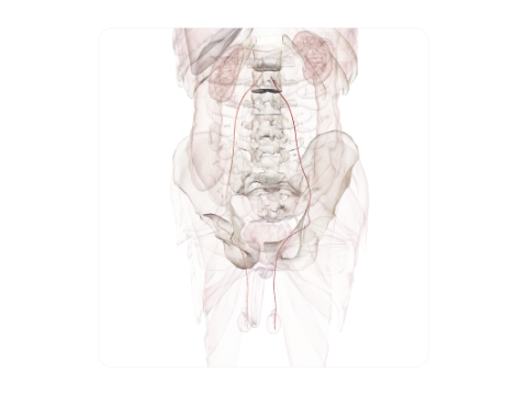 Testicular arteries
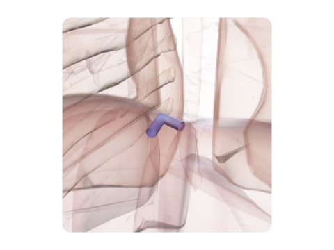 Hepatic veins
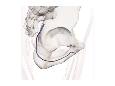 Internal pudendal vein
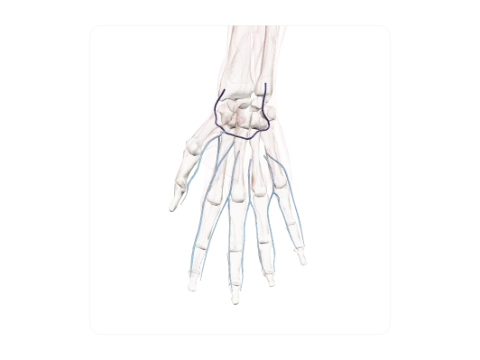 Deep venous palmar arch and palmar digital veins
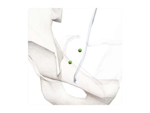 Inferior epigastric nodes
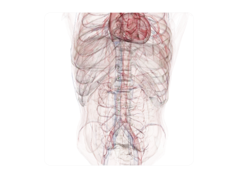 Caval nodes
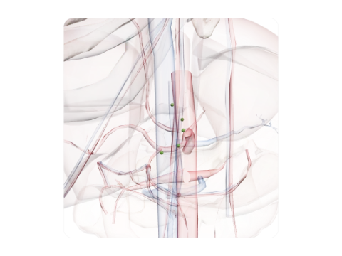 Right gastric nodes
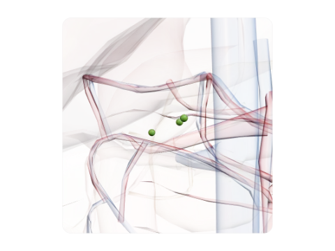 Superior pancreaticoduodenal nodes
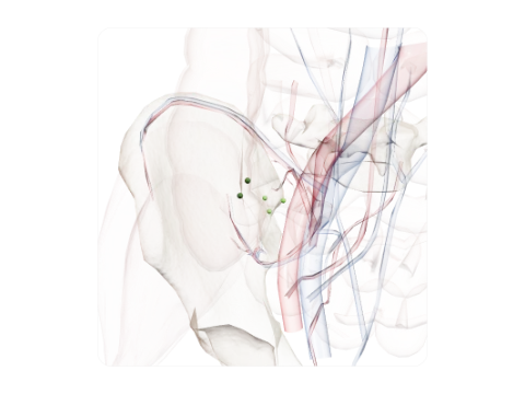 Precaecal and retrocaecal nodes
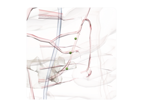 Left colic nodes
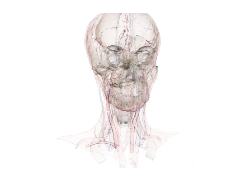 Facial nodes
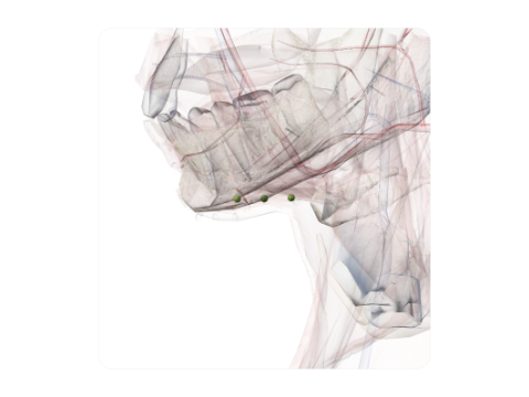 Submandibular nodes
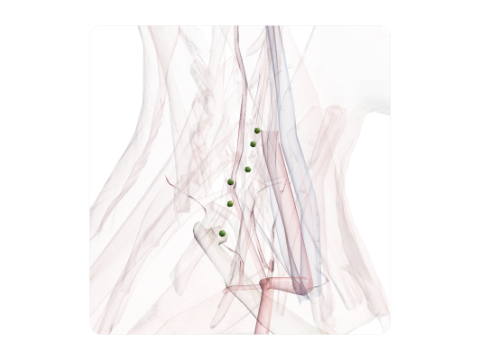 Superficial lateral cervical nodes
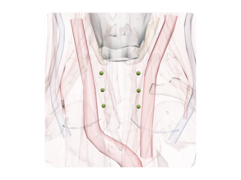 Cervical paratracheal nodes
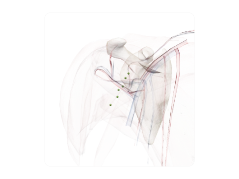 Posterior axillary nodes
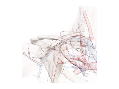 Infraclavicular nodes
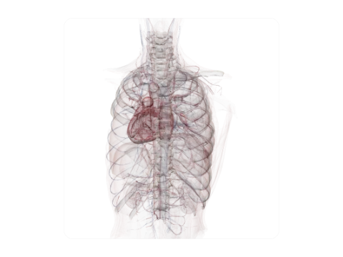 Intercostal nodes
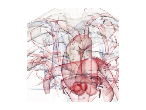 Inferior tracheobronchial (subcarinal) nodes
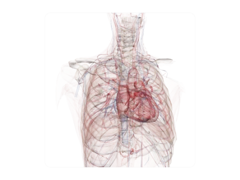 Pericardial nodes
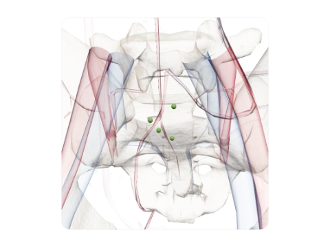 Median sacral nodes
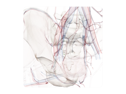 Lateral sacral nodes
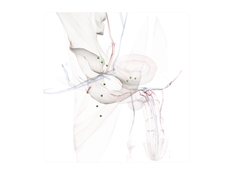 Superficial inguinal nodes
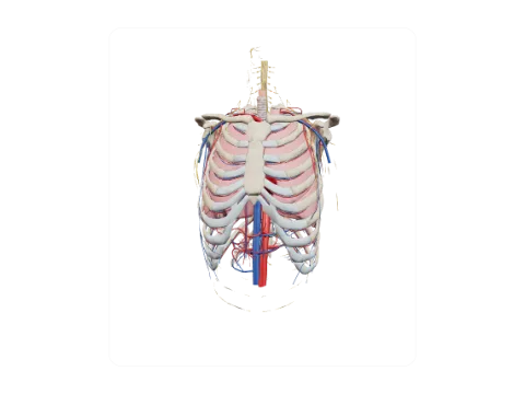 Thorax
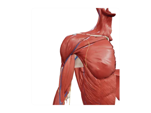 Shoulder and axilla
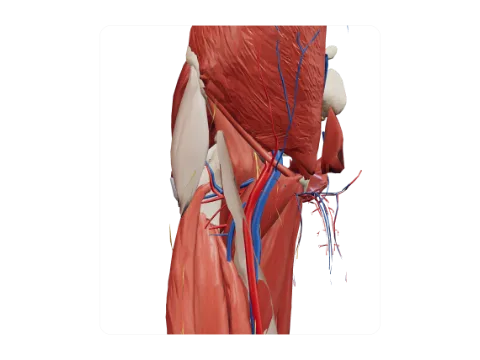 Inguinal region and femoral triangle
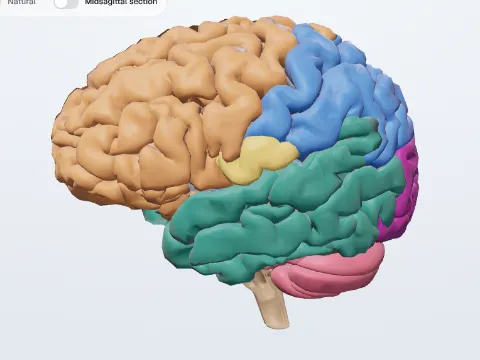 Brain
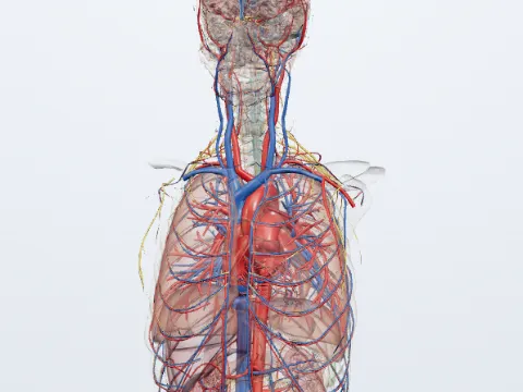 Meninges and ventricles
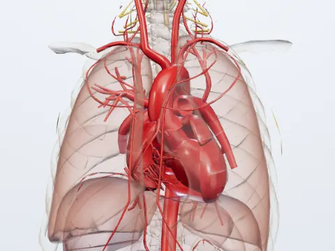 Autonomic nervous system
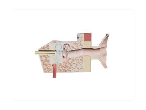 Ear, coronal section through the canal, middle ear and cochlea
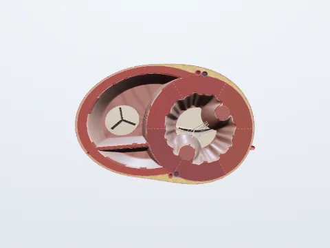 Heart, short-axis section
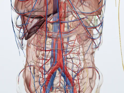 Hepatic portal vein
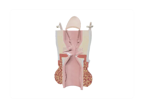 Larynx, coronal section through the vocal folds
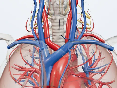 Trachea
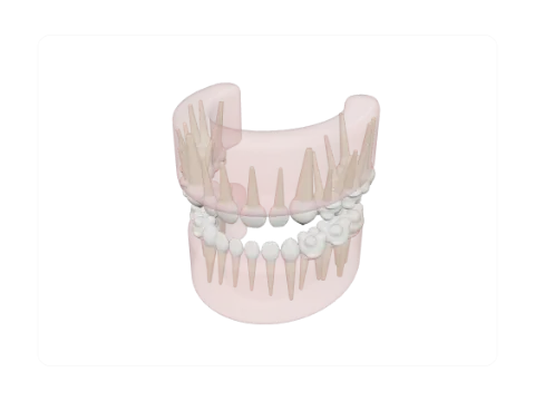 Primary dentition (20 teeth) with the first permanent molars
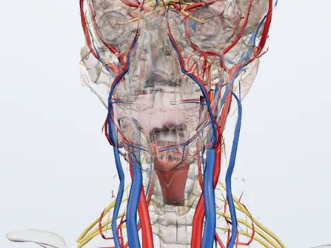 Pharynx
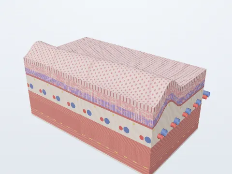 Layers of the stomach wall
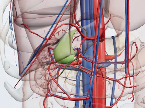 Gallbladder
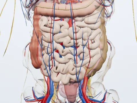 Intestines
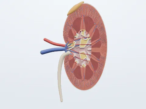 Kidney, coronal section
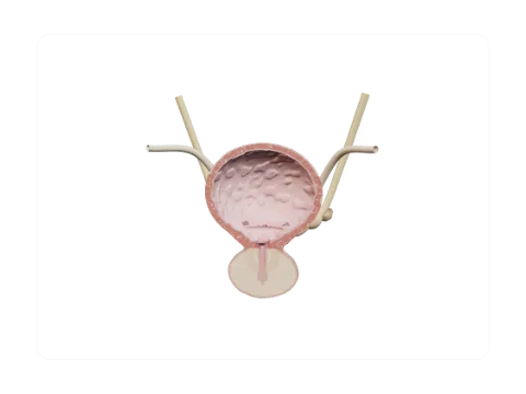 Bladder and prostate, coronal section
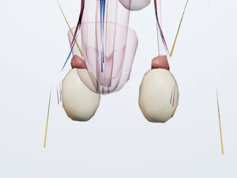 Testes
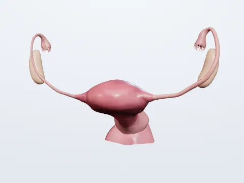 Uterine tubes
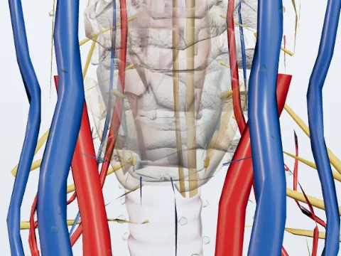 Parathyroid glands
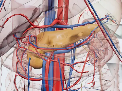 Pancreas
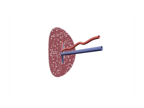 Spleen, section through the hilum
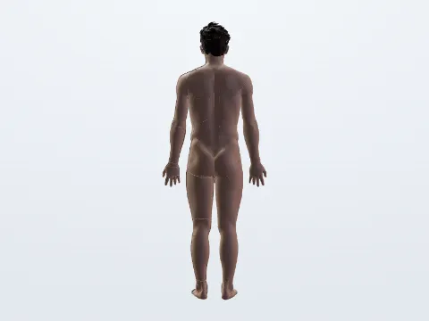 Integumentary system
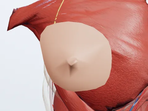 Breast (mammary gland)
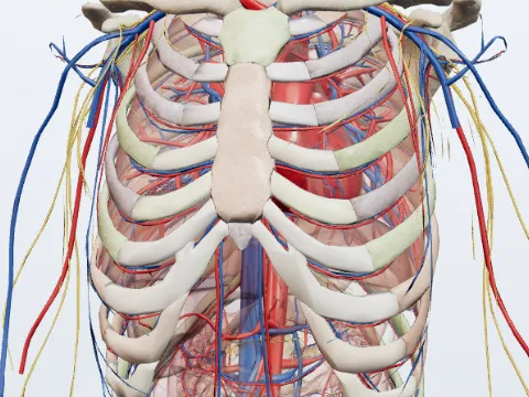 Thoracic cage
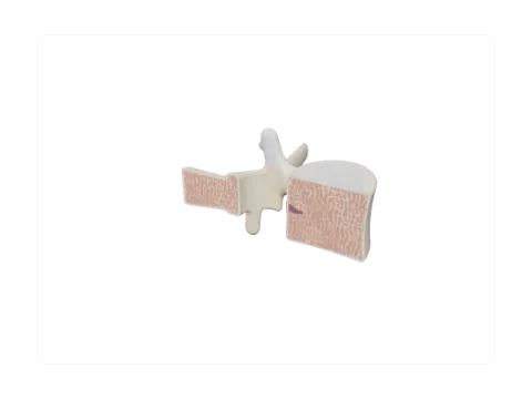 Lumbar vertebra (L3)
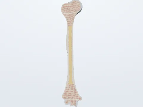 Humerus: parts and section
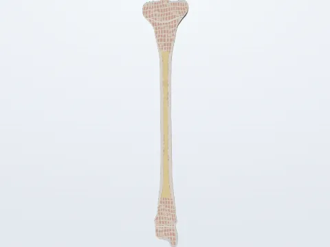 Tibia: parts and section
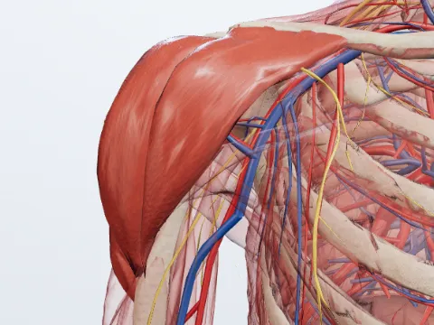 Deltoid
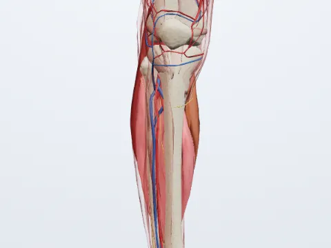 Triceps surae
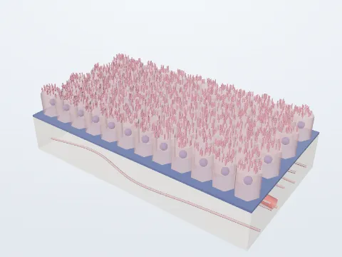 Simple cuboidal
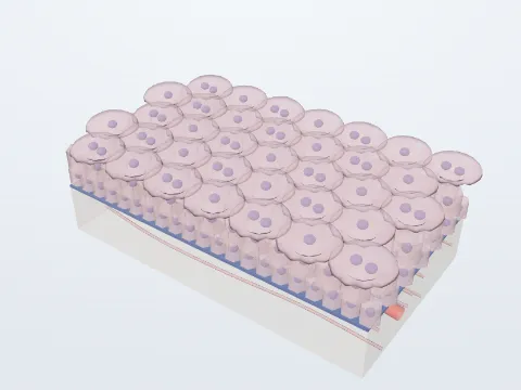 Transitional (urothelium)
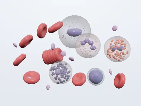 Blood cells
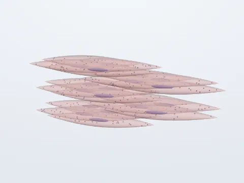 Smooth muscle
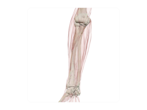 Radius
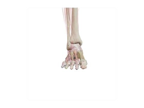 Bones of the foot
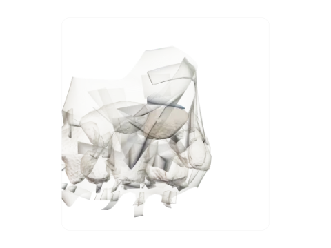 Lunate
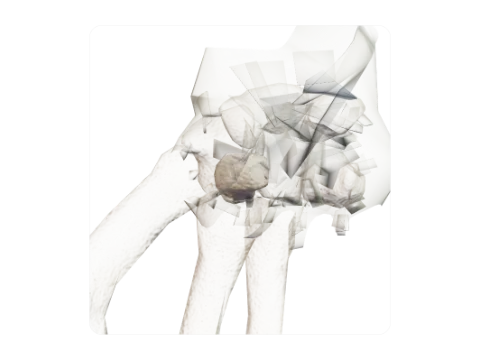 Trapezoid
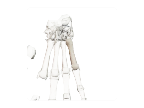 Fifth metacarpal
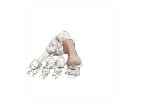 First metatarsal and sesamoids
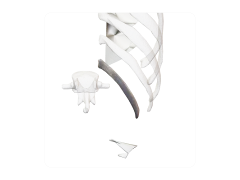 Twelfth rib
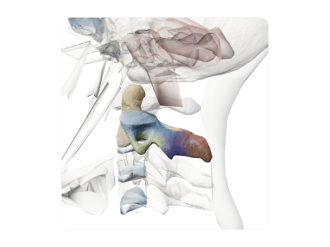 Axis (C2)
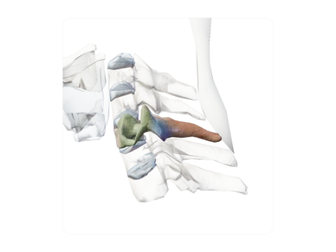 Sixth cervical vertebra (C6)
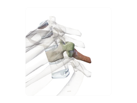 Third thoracic vertebra (T3)
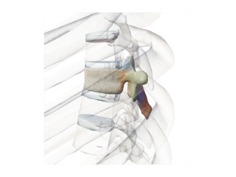 Seventh thoracic vertebra (T7)
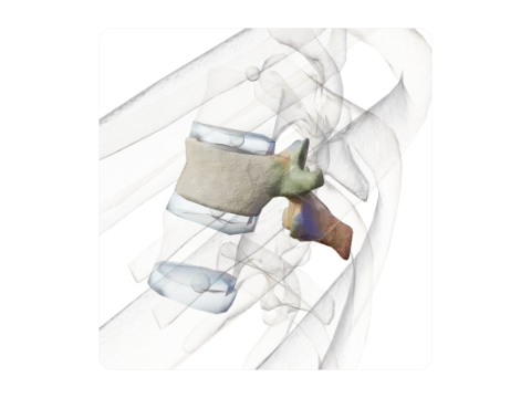 Eleventh thoracic vertebra (T11)
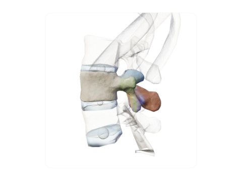 Third lumbar vertebra (L3)
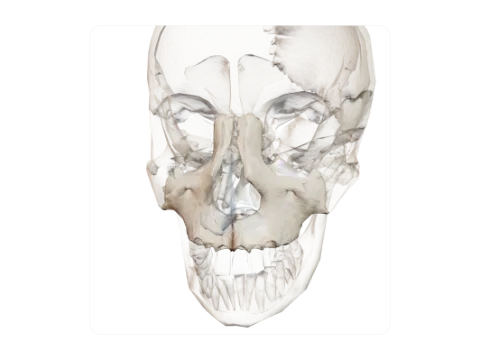 Maxilla
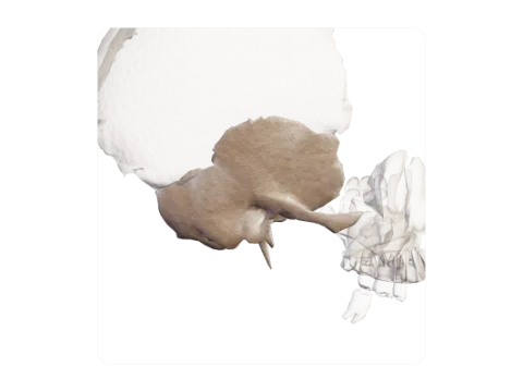 Temporal bone
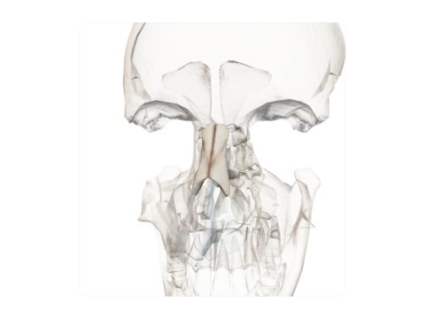 Nasal bones
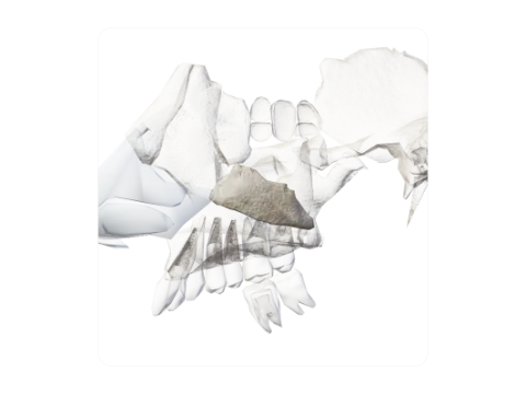 Inferior nasal concha
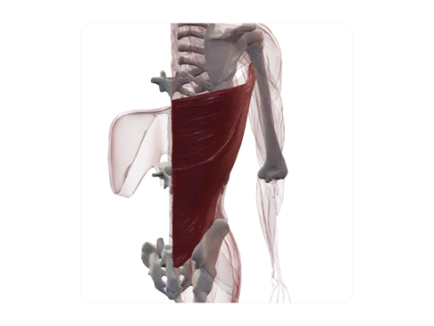 Latissimus dorsi
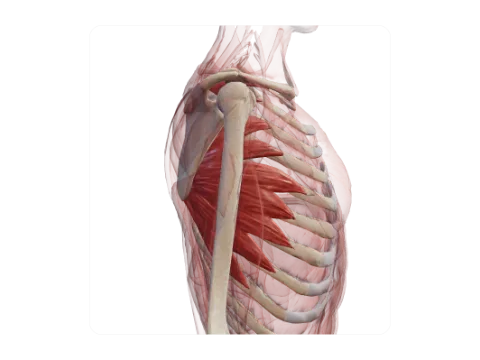 Serratus anterior
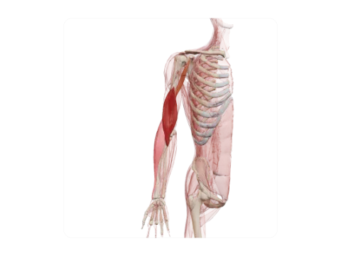 Brachialis, brachioradialis and coracobrachialis
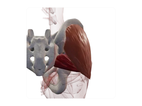 Gluteus medius, gluteus minimus and piriformis
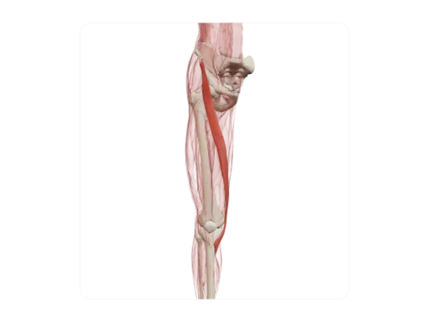 Sartorius
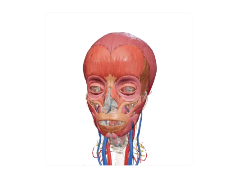 Muscles of the head
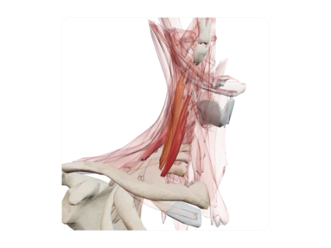 Scalene muscles
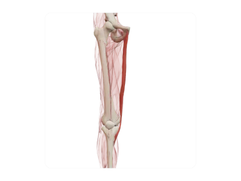 Gracilis
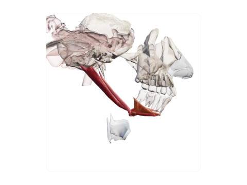 Suprahyoid muscles
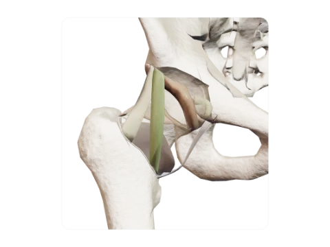 Hip joint
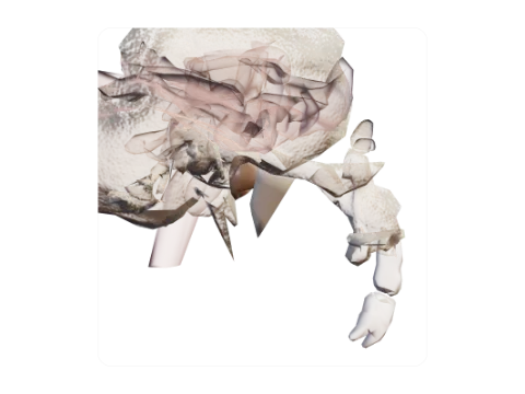 Temporomandibular joint
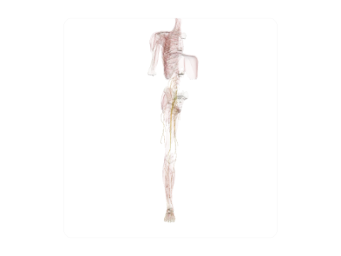 Lumbosacral plexus
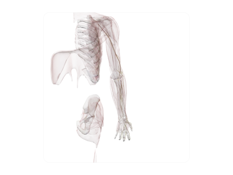 Radial nerve
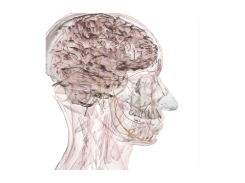 Trigeminal nerve (V)
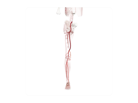 Arteries of the lower limb
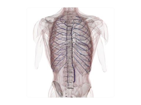 Azygos venous system
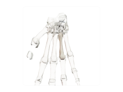 Fourth metacarpal
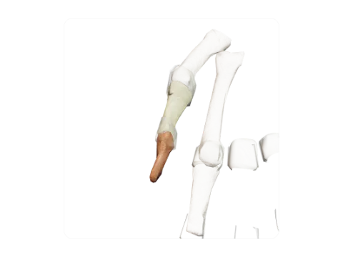 Phalanges of the thumb
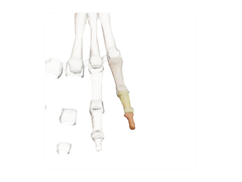 Phalanges of the little finger
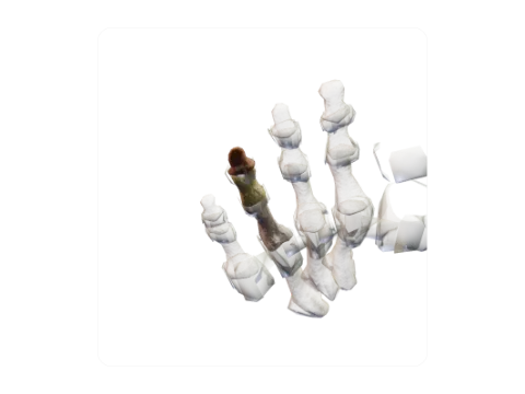 Phalanges of the fourth toe
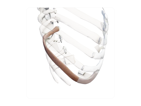 Tenth rib
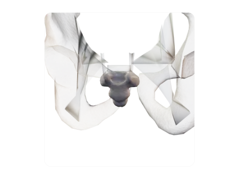 Coccyx
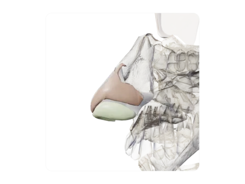 Nasal cartilages
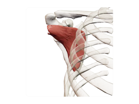 Subscapularis
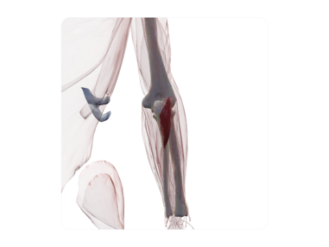 Anconeus
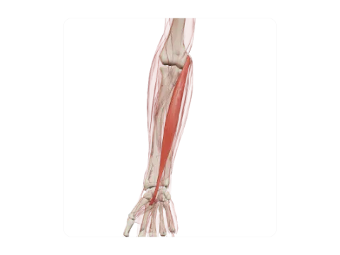 Flexor carpi radialis
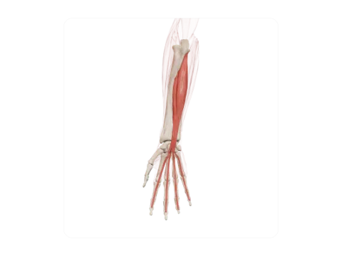 Flexor digitorum profundus
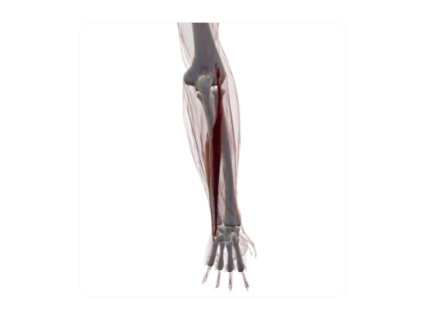 Extensor carpi ulnaris
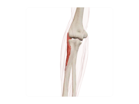 Supinator
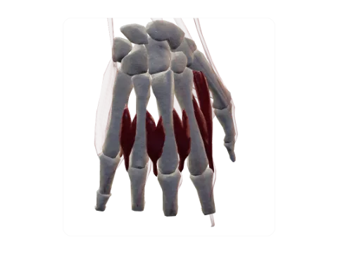 Interossei of the hand
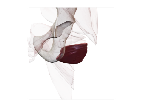 Quadratus femoris
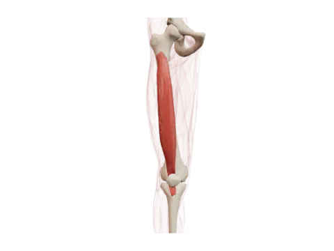 Vastus intermedius
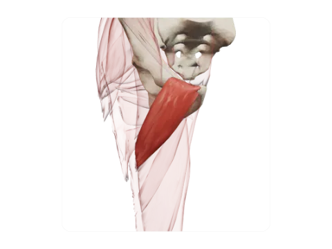 Pectineus
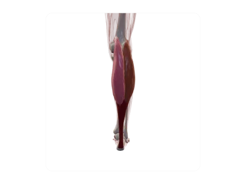 Gastrocnemius and calcaneal tendon
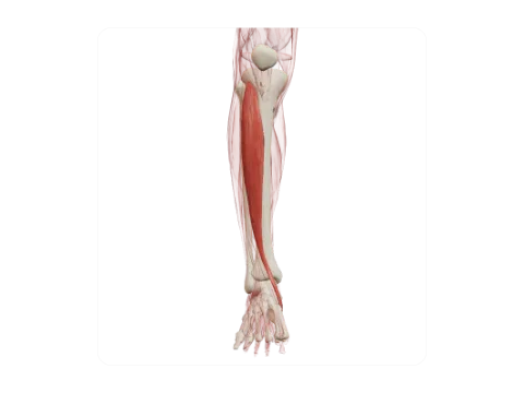 Tibialis anterior
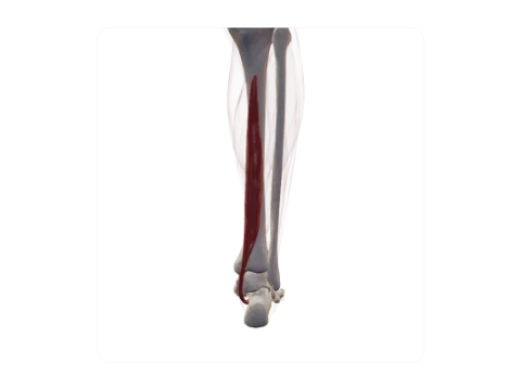 Flexor digitorum longus
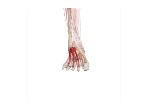 Muscles of the dorsum of the foot
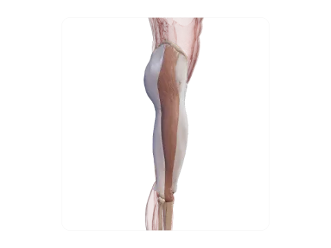 Fascia lata and iliotibial tract
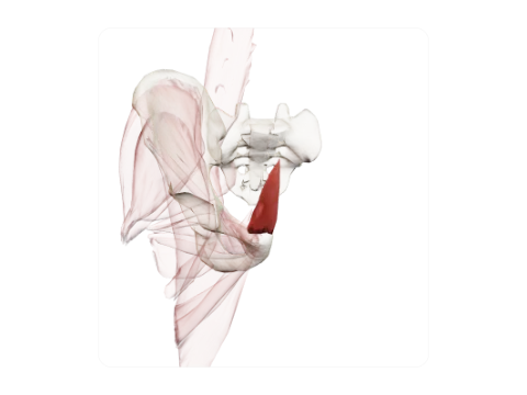 Pyramidalis
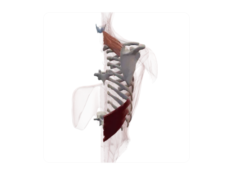 Serratus posterior superior and inferior
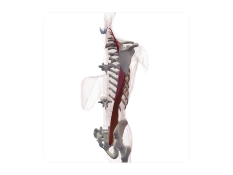 Iliocostalis
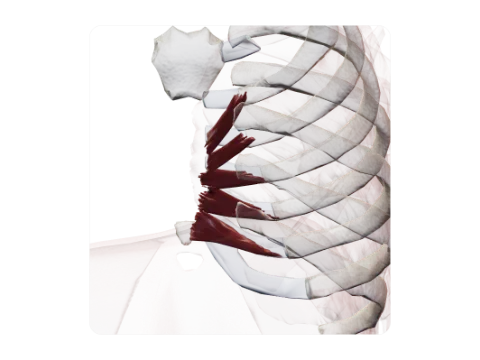 Transversus thoracis
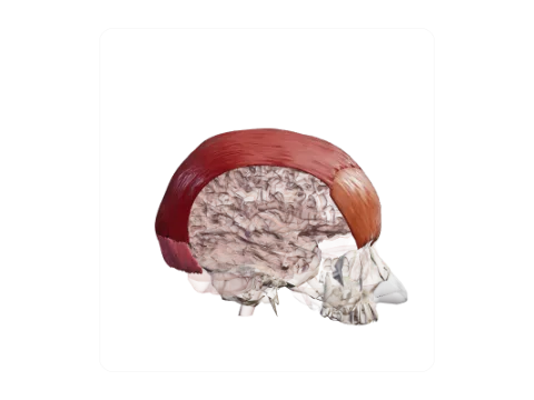 Occipitofrontalis and epicranial aponeurosis
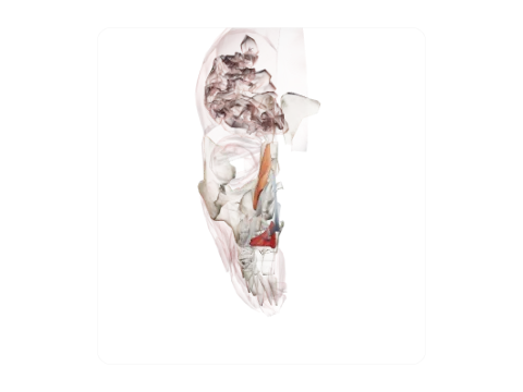 Nasalis and depressor septi nasi
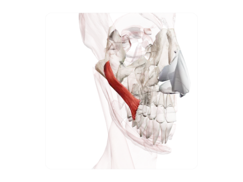 Zygomaticus major
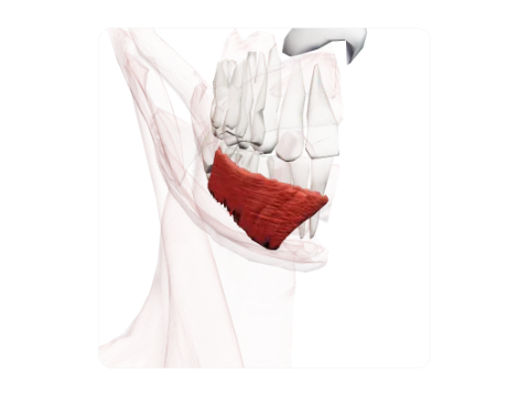 Depressor labii inferioris
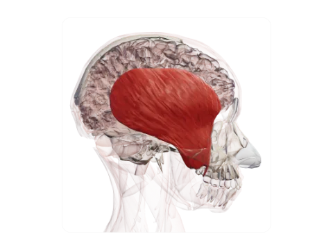 Temporalis
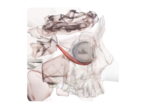 Inferior rectus
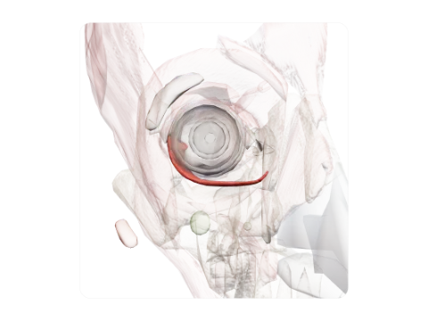 Inferior oblique
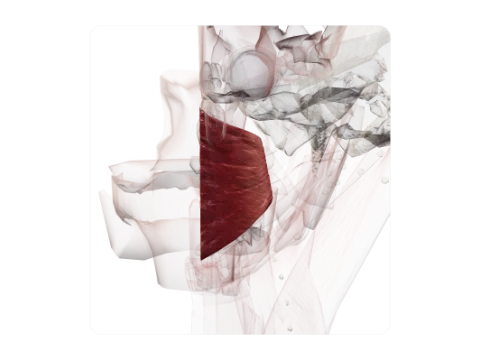 Superior pharyngeal constrictor
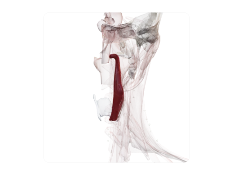 Palatopharyngeus
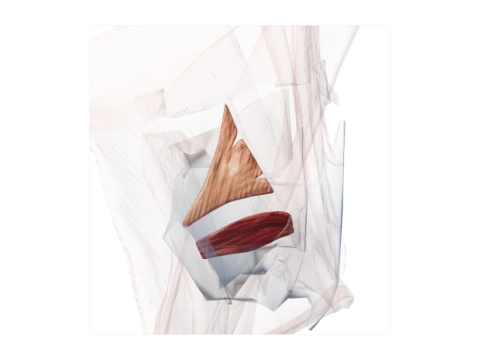 Thyroarytenoid and vocalis
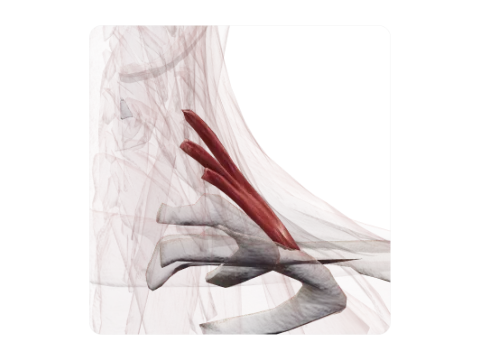 Scalenus posterior
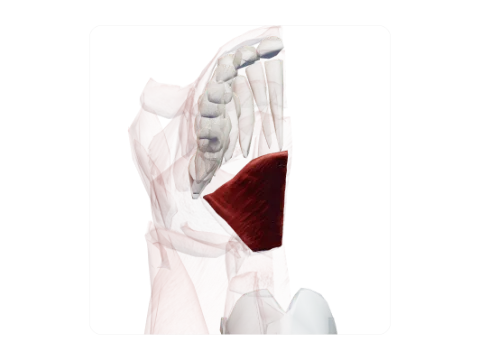 Mylohyoid
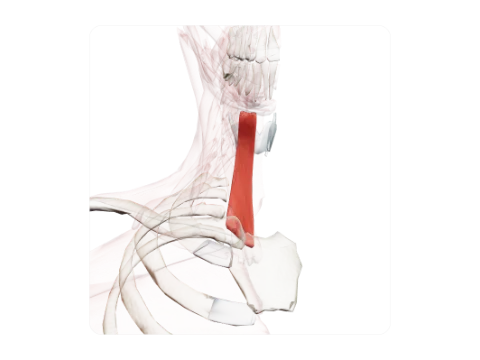 Sternohyoid
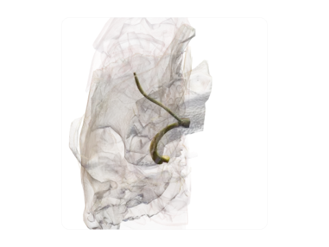 Optic nerve (II), chiasm and tract
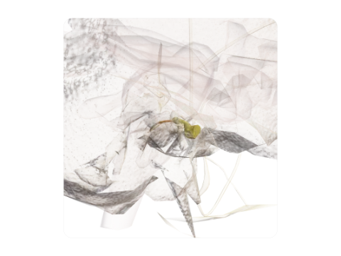 Vestibulocochlear nerve (VIII)
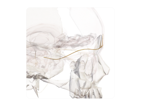 Ophthalmic nerve (V1)
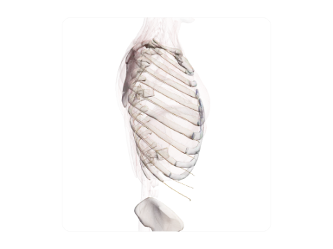 Intercostal nerves
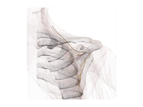 Suprascapular nerve
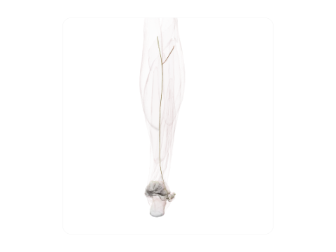 Sural nerve
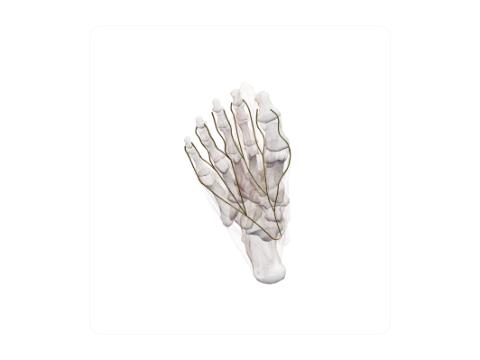 Medial and lateral plantar nerves
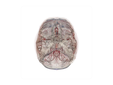 Circle of Willis
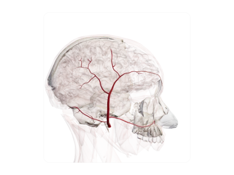 Superficial temporal artery
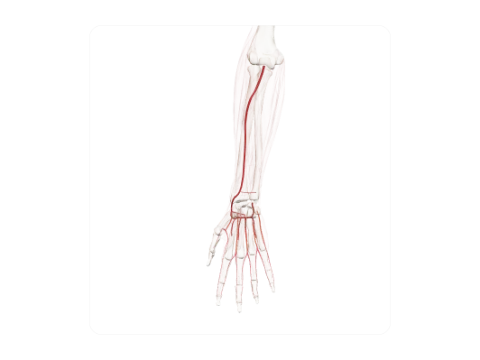 Radial artery
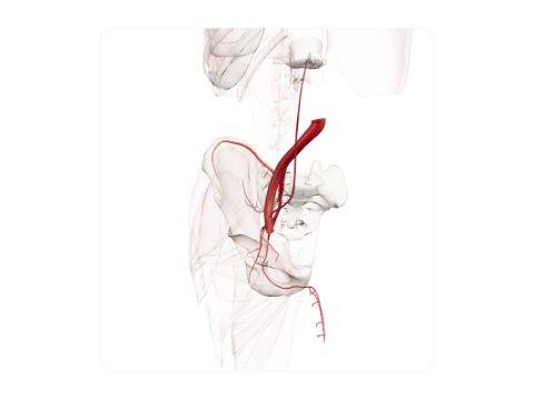 Common, external and internal iliac arteries
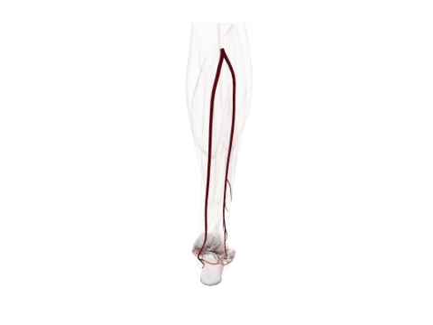 Posterior tibial and fibular arteries
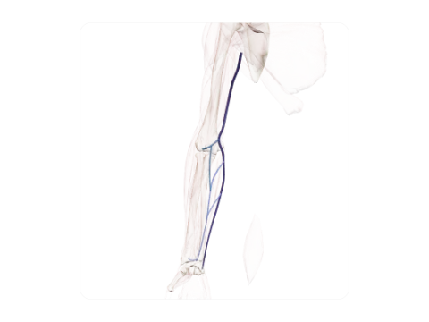 Basilic and median cubital veins
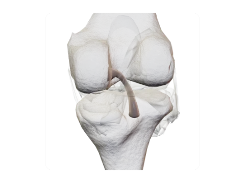 Posterior cruciate ligament
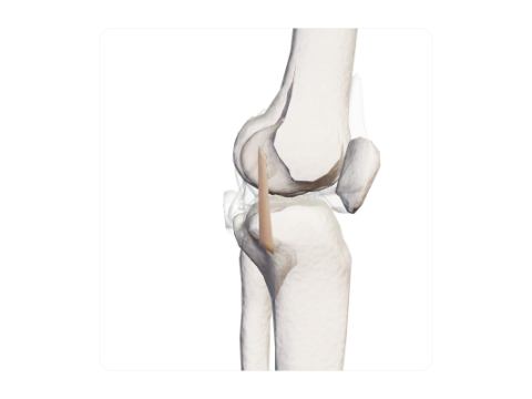 Fibular (lateral) collateral ligament
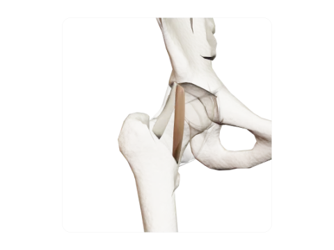 Iliofemoral ligament (Y ligament of Bigelow)
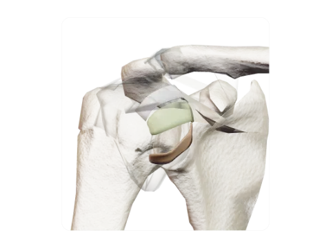 Glenohumeral ligaments
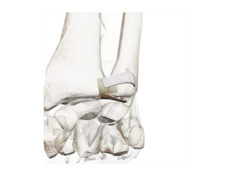 Triangular fibrocartilage complex (TFCC)
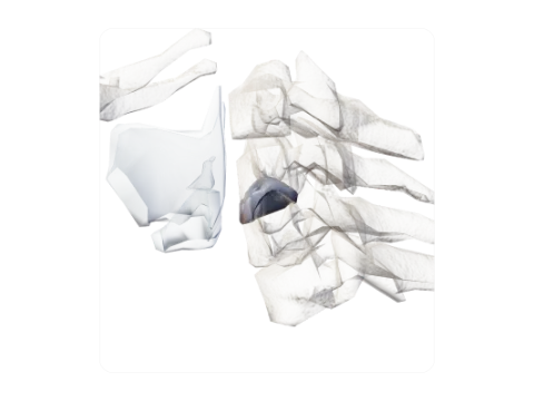 Cervical intervertebral disc C5–C6
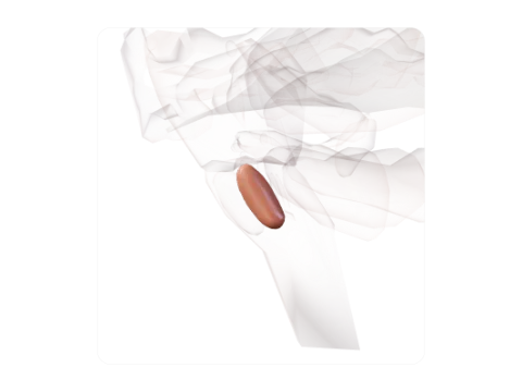 Inferior olive
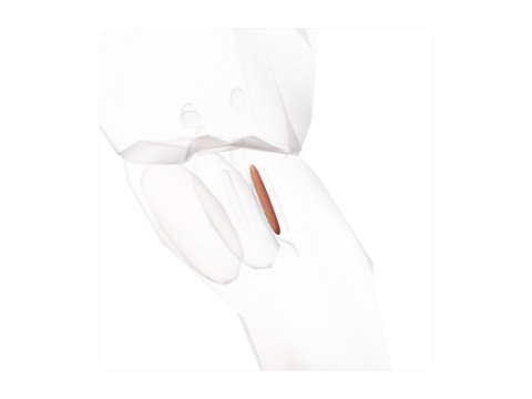 Hypoglossal nucleus
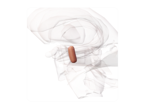 Red nucleus
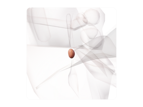 Trochlear nucleus
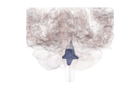 Fourth ventricle
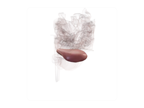 Superior semilunar lobule
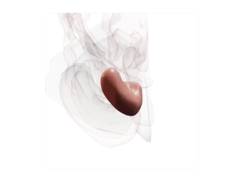 Cerebellar tonsil
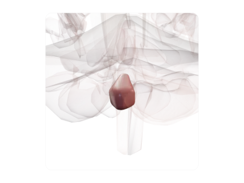 Pyramis of vermis
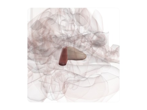 Central lobule
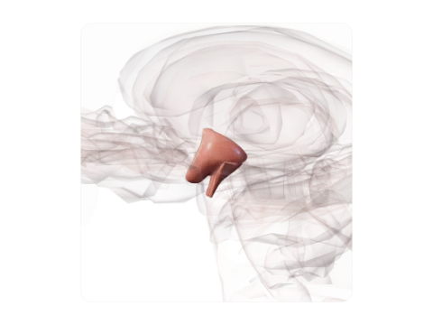 Hypothalamus
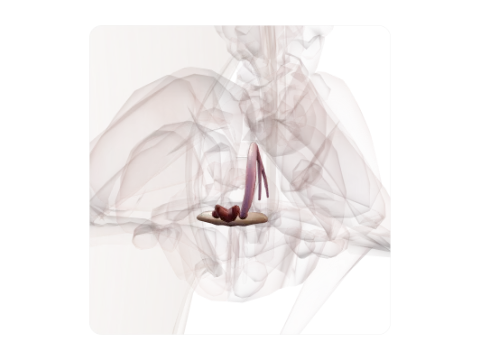 Habenula
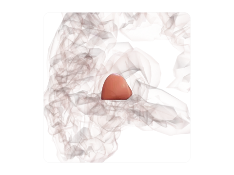 Globus pallidus
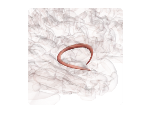 Fornix
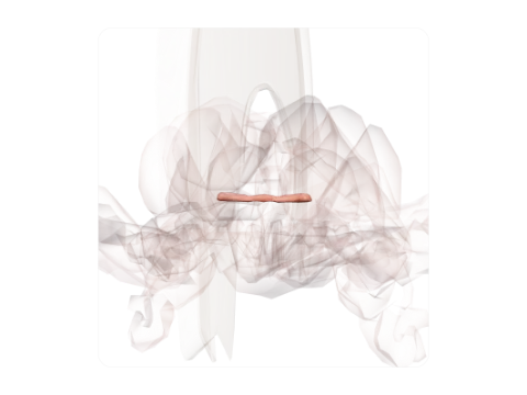 Anterior commissure
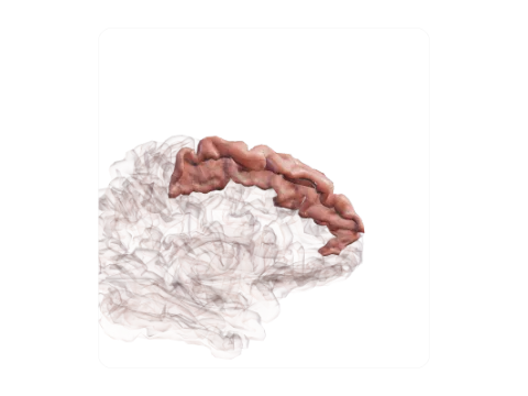 Superior frontal gyrus
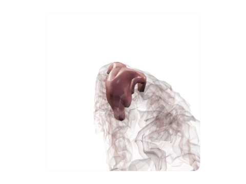 Paracentral lobule
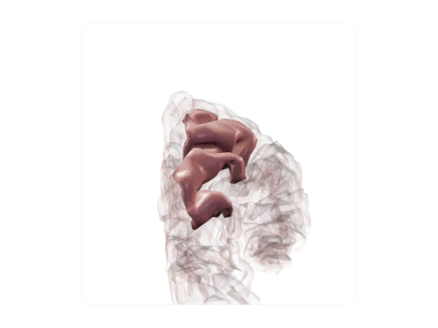 Superior parietal lobule
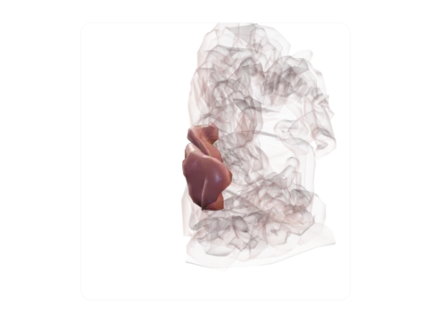 Cuneus
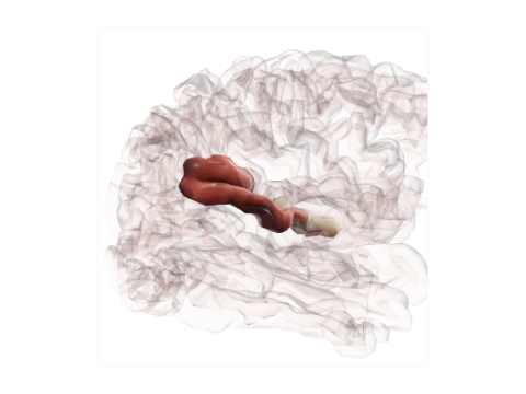 Transverse temporal gyri (Heschl)
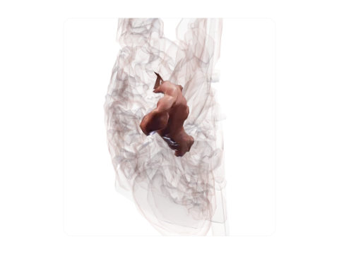 Parahippocampal gyrus
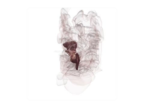 Calcarine sulcus
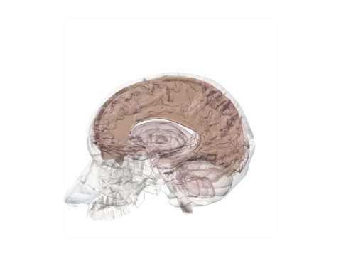 Falx cerebri
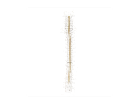 Lateral corticospinal tract
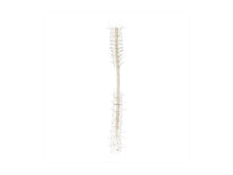 Medial vestibulospinal tract
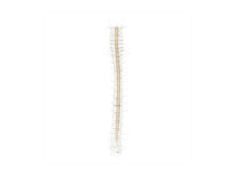 Lateral spinothalamic tract
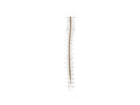 Gracile fasciculus (Goll)
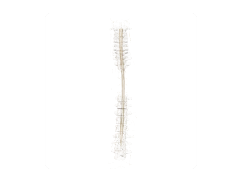 Intermediolateral nucleus (lateral horn)
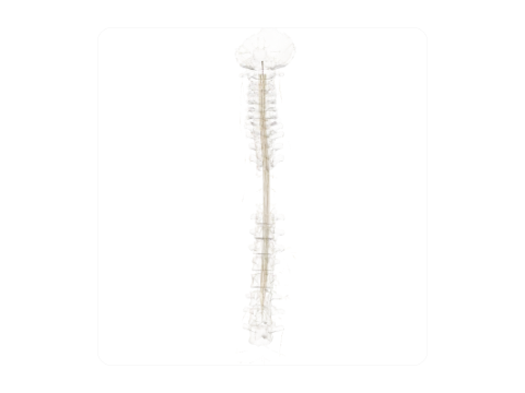 Central canal of spinal cord
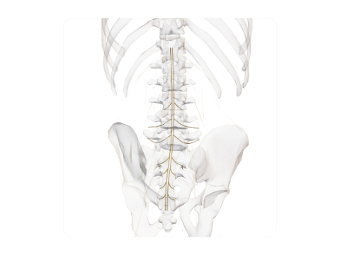 Cauda equina
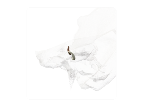 Posterior semicircular canal and common crus
 Cornea
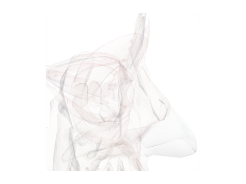 Anterior chamber of eye
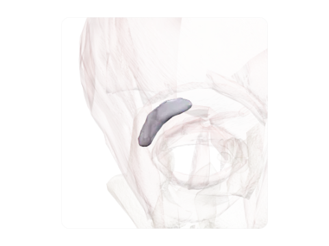 Lacrimal gland
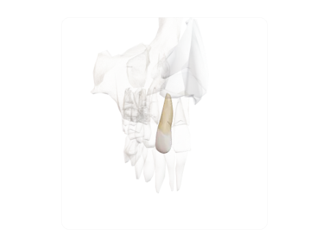 Maxillary lateral incisor
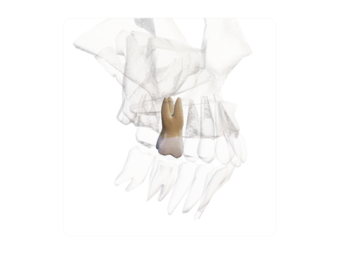 Maxillary first molar
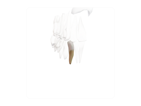 Mandibular canine
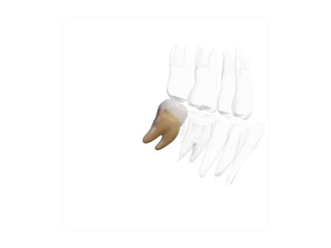 Mandibular second molar
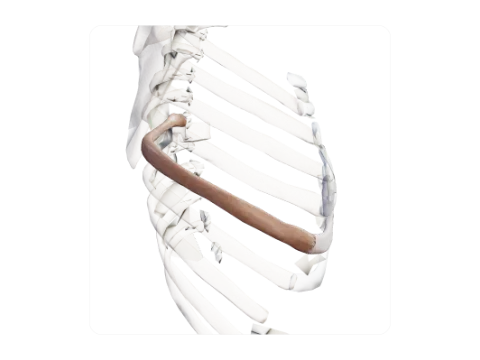 Seventh rib
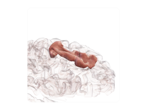 Superior frontal sulcus
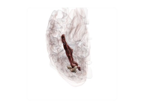 Collateral sulcus
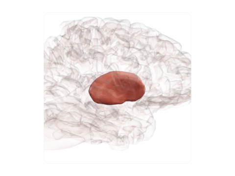 Lentiform (lenticular) nucleus
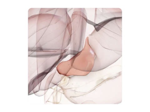 Lesser omentum
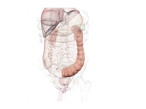 Descending colon
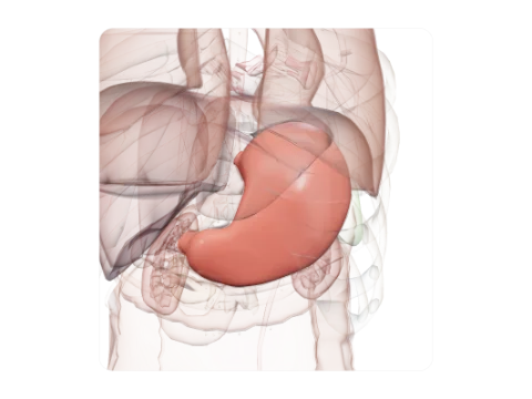 Gastric mucosa
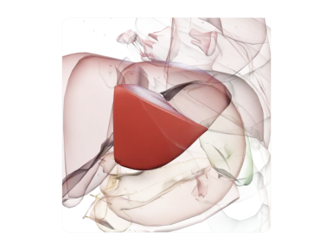 Liver segment III
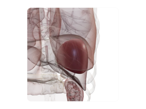 Liver segment VII
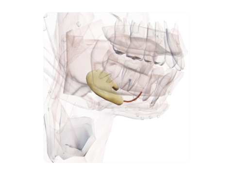 Submandibular gland and duct
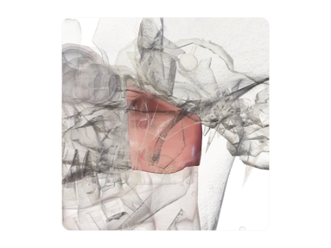 Nasopharynx
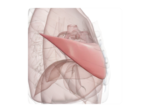 Middle lobe of right lung
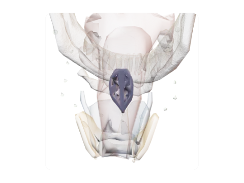 Epiglottis
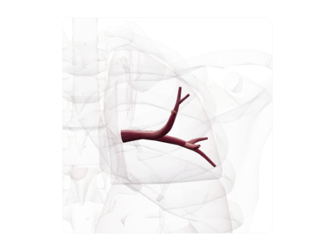 Posterior segmental bronchus, right (B2)
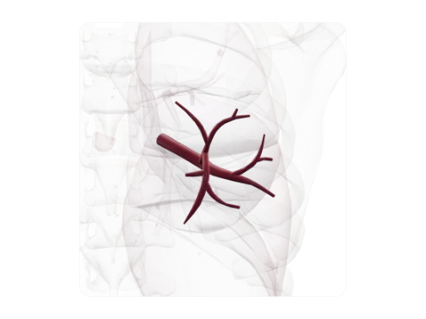 Superior segmental bronchus of right lower lobe (B6)
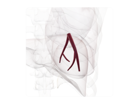 Posterior basal segmental bronchus, right (B10)
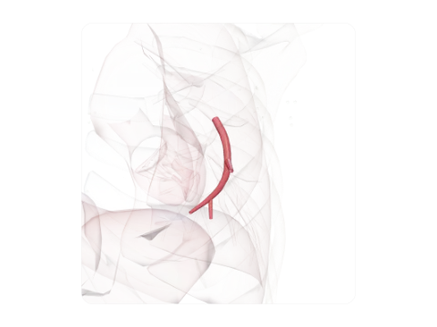 Inferior lingular bronchus (B5)
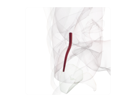 Posterior basal segmental bronchus, left (B10)
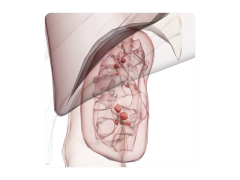 Renal papillae
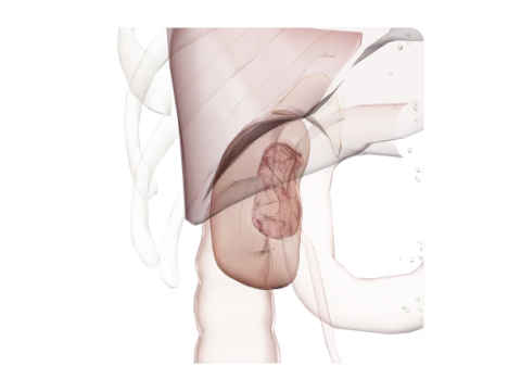 Fibrous capsule of kidney
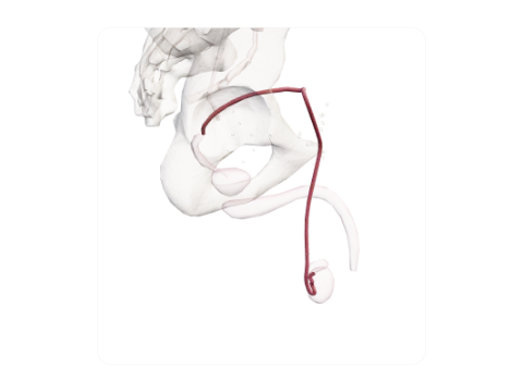 Ductus deferens (vas deferens)
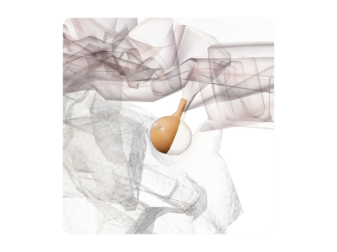 Adenohypophysis (anterior pituitary)
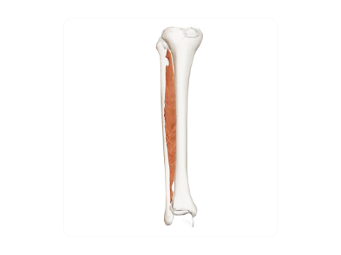 Interosseous membrane of leg
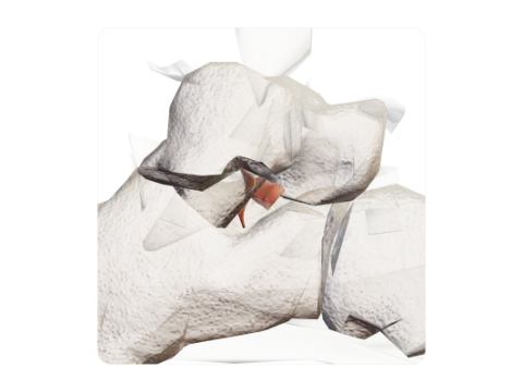 Interosseous talocalcaneal ligament
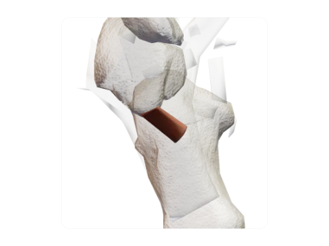 Short plantar ligament
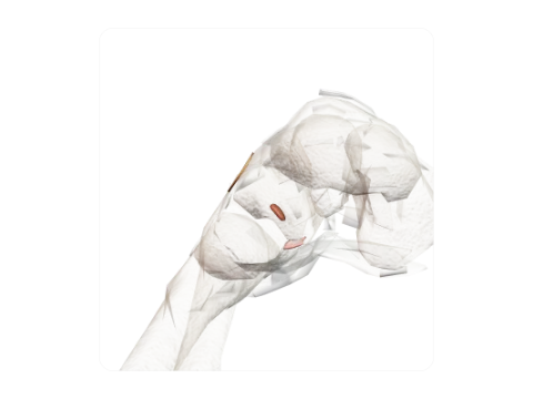 Cuneocuboid ligaments
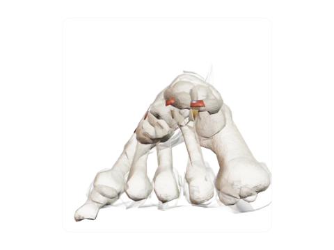 Intermetatarsal ligaments
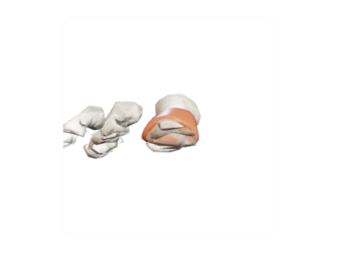 Interphalangeal joint of great toe
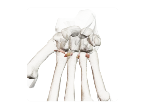 Metacarpal ligaments
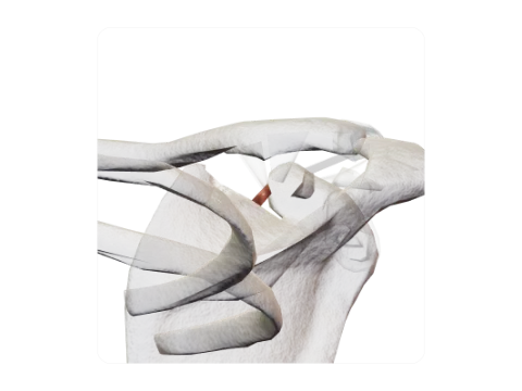 Superior transverse scapular ligament
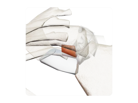 Costoclavicular ligament
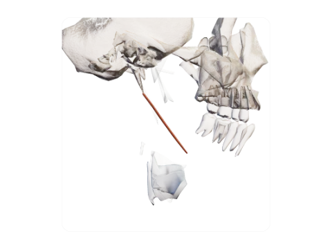 Stylohyoid ligament
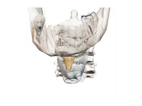 Thyrohyoid membrane and ligaments
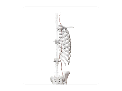 Intertransverse ligaments
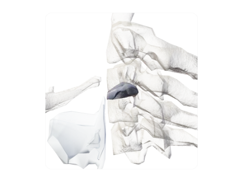 Intervertebral disc C3–C4
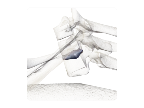 Intervertebral disc T1–T2
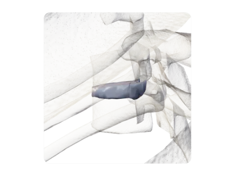 Intervertebral disc T5–T6
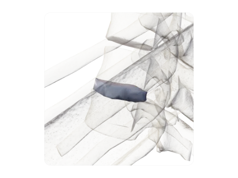 Intervertebral disc T10–T11
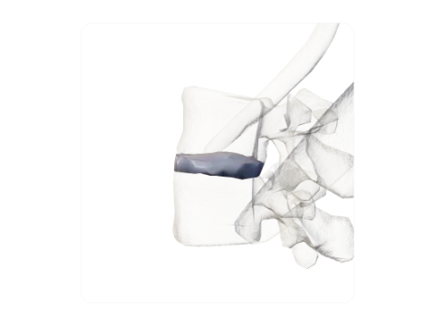 Intervertebral disc L2–L3
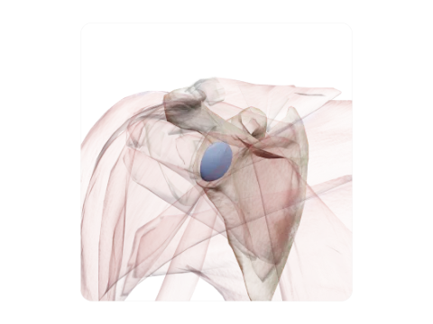 Coracobrachial bursa
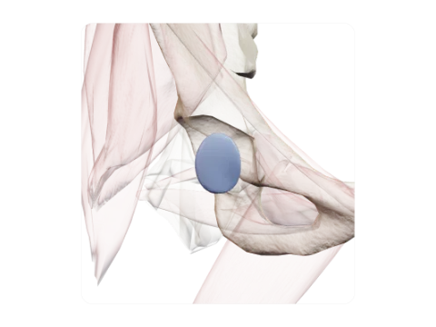 Iliopectineal bursa
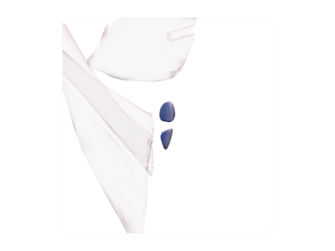 Intermuscular gluteal bursae
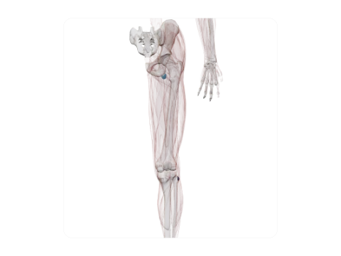 Bursae of biceps femoris
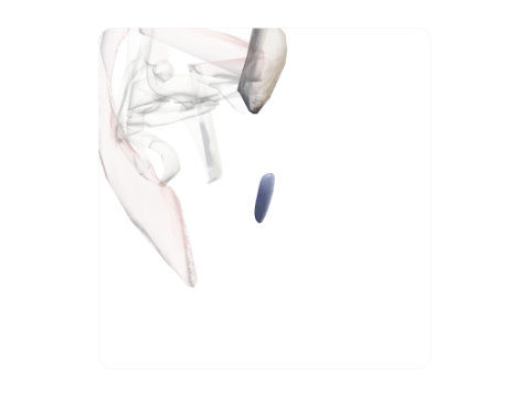 Subcutaneous bursa of tibial tuberosity
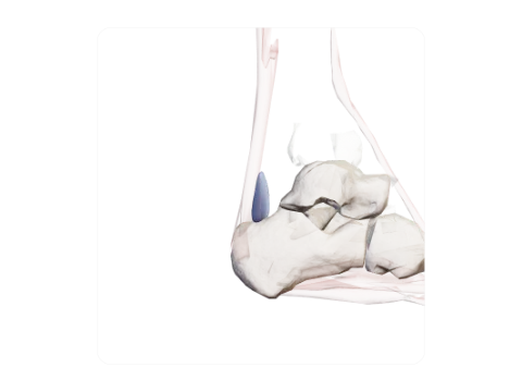 Retrocalcaneal bursa
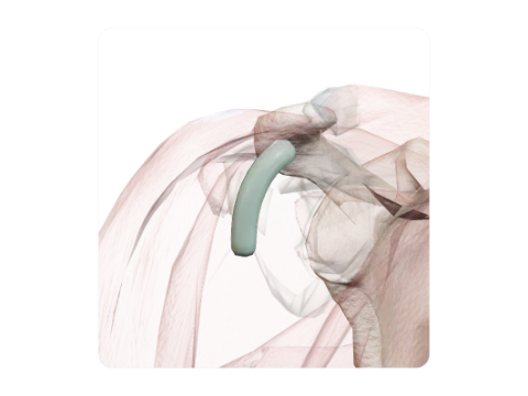 Intertubercular tendon sheath
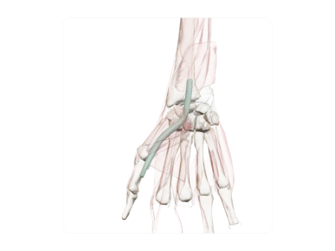 Tendon sheath of flexor pollicis longus (radial bursa)
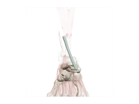 Tendon sheath of extensor hallucis longus
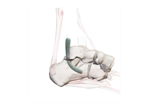 Tendon sheath of flexor digitorum longus
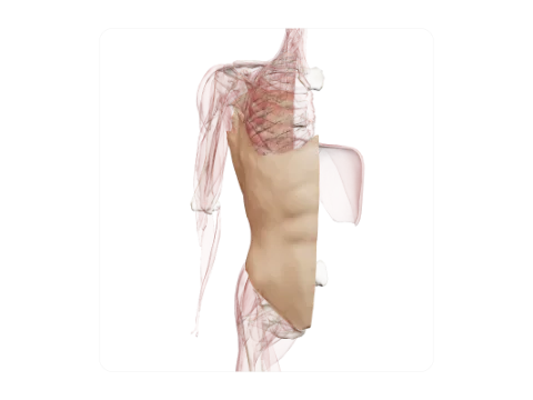 Investing abdominal fascia
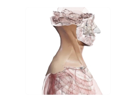 Investing layer of cervical fascia
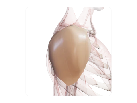 Deltoid fascia
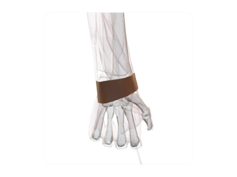 Extensor retinaculum of the wrist
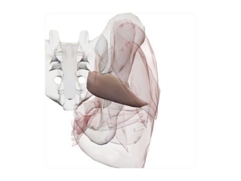 Piriformis fascia
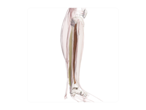 Intermuscular septa of the leg
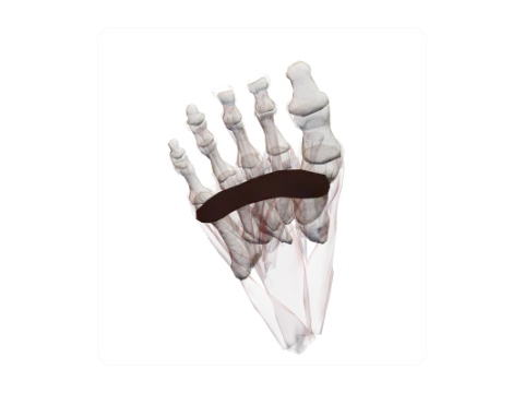 Superficial transverse metatarsal ligament
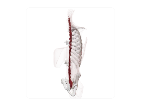 Rotatores
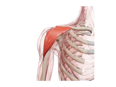 Clavicular part of deltoid
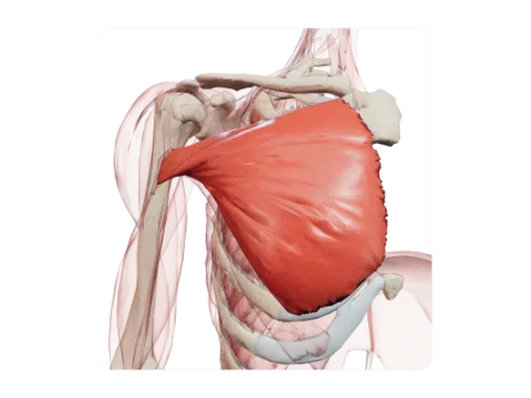 Sternocostal head of pectoralis major
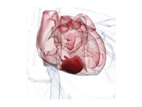 Papillary muscles of the right ventricle
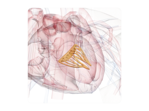 Posterior leaflet of the mitral valve
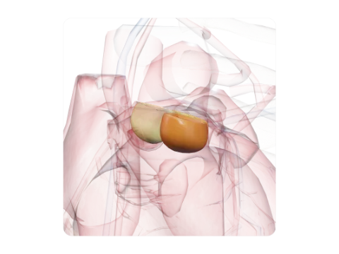 Pulmonary valve cusps
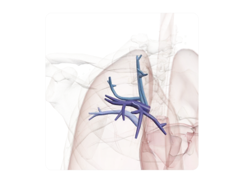 Segmental arteries of the right upper lobe
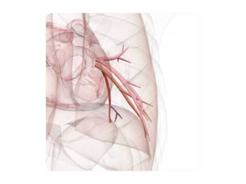 Veins of the left lower lobe
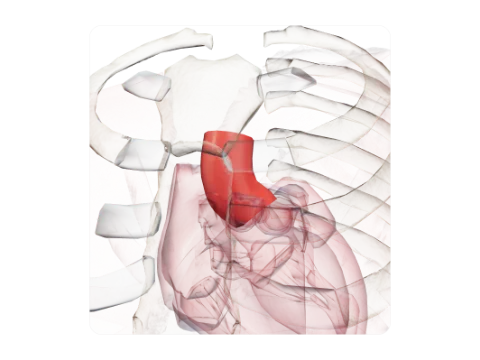 Ascending aorta
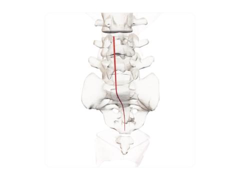 Median sacral artery
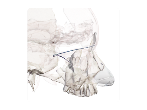 Ophthalmic veins
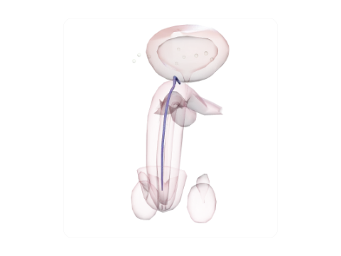 Deep dorsal vein of the penis
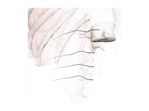 Lumbar veins
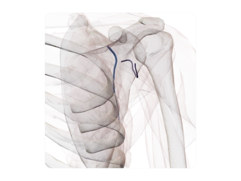 Circumflex scapular and thoracodorsal veins
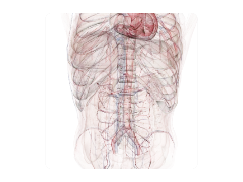 Pre-aortic nodes
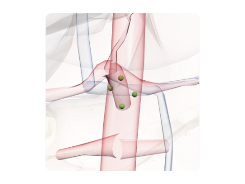 Coeliac nodes
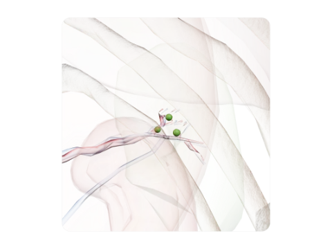 Splenic nodes
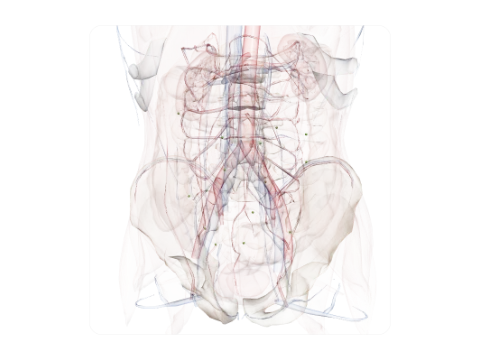 Juxta-intestinal mesenteric nodes
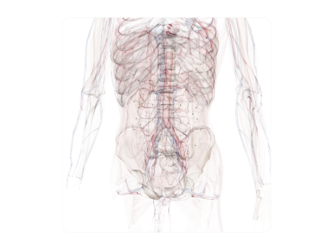 Right colic and paracolic nodes
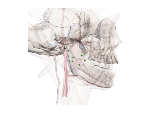 Parotid nodes
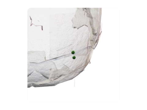 Occipital nodes
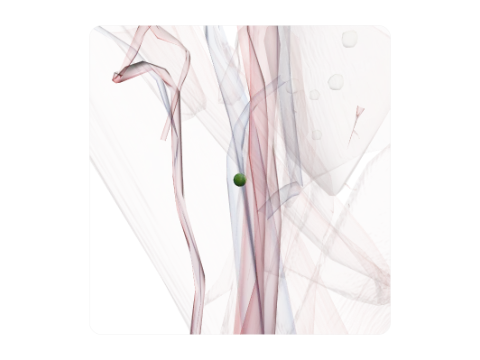 Jugulodigastric node
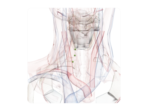 Pretracheal nodes
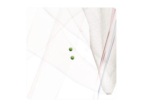 Lateral axillary nodes
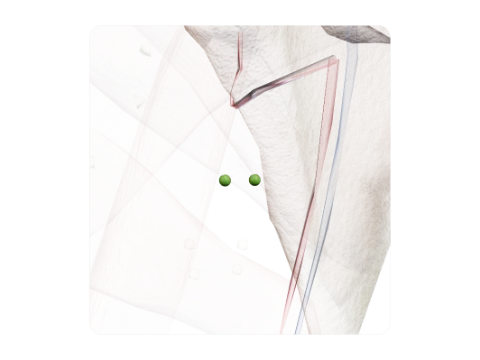 Apical axillary nodes
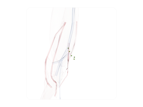 Supratrochlear nodes
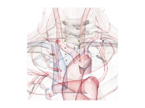 Thoracic paratracheal nodes
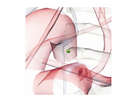 Node of the ligamentum arteriosum
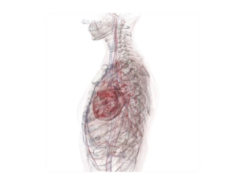 Prevertebral nodes
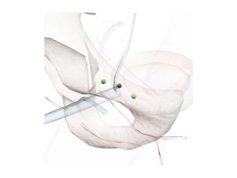 Lacunar nodes
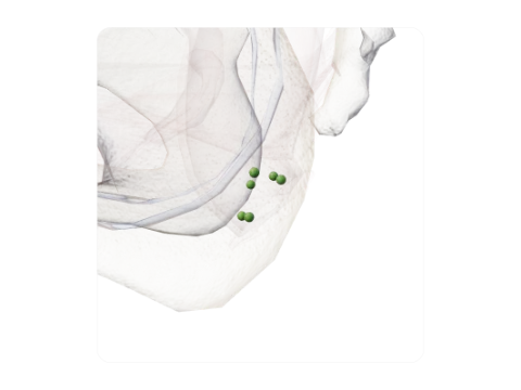 Pararectal nodes
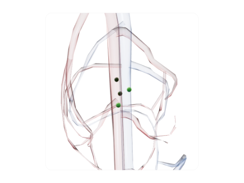 Popliteal nodes
Six levels of observation
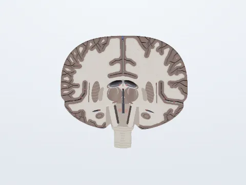
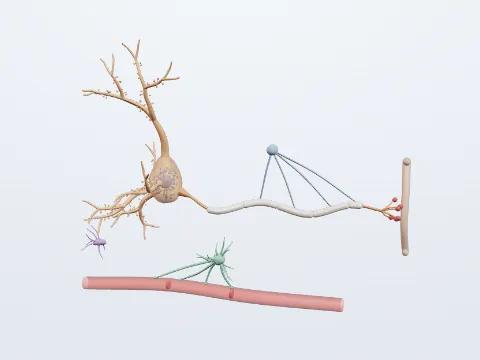
Zoom from organ system to cell
The same subject at every scale: the system across the body, the region, the organ, its section, the tissue and the cell. Each level is a model you can rotate and pick apart structure by structure.
How it works
Read it like a textbook. Study it like the dissection room. Every structure carries its standard Latin name and a source you can check.
Inside organs
See inside the organs
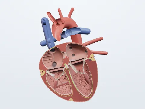
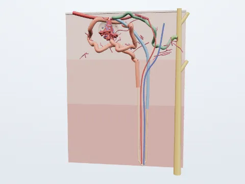
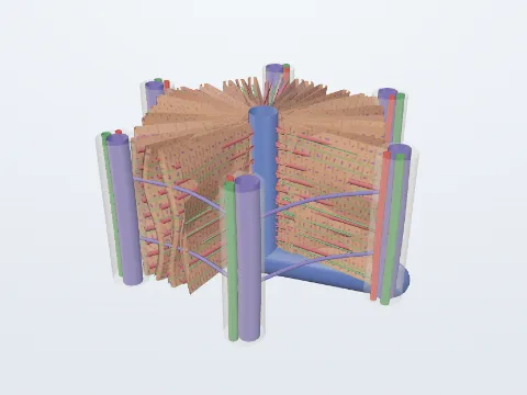
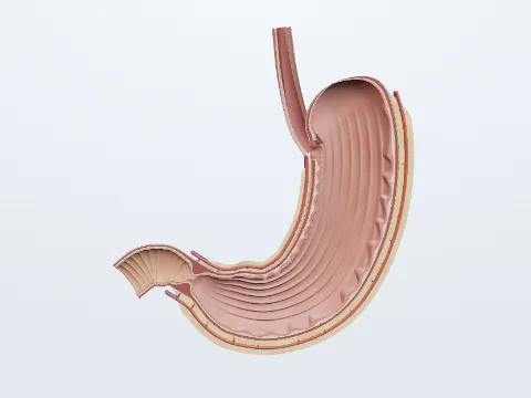
A four-chamber heart, a coronal brain, a bisected kidney down to the nephron, the liver lobule, an opened stomach and bones cut lengthwise: every part can be picked, with its Latin name and a description.
Histology
Real micrographs, not just drawings
The Cells and tissues chapter sets each model beside H&E sections, silver stains, Wright-stained blood smears and electron micrographs, all credited and openly licensed.
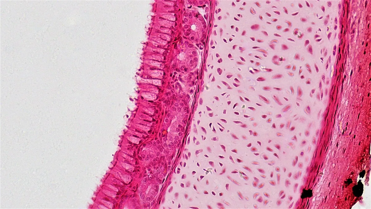 H&E · 200×
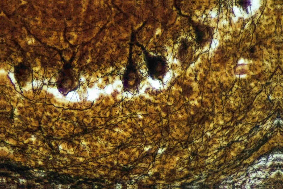 Ag
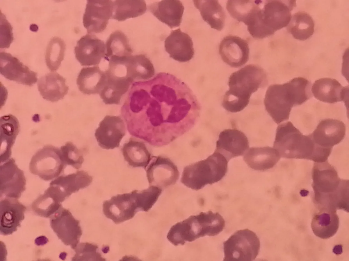 Wright
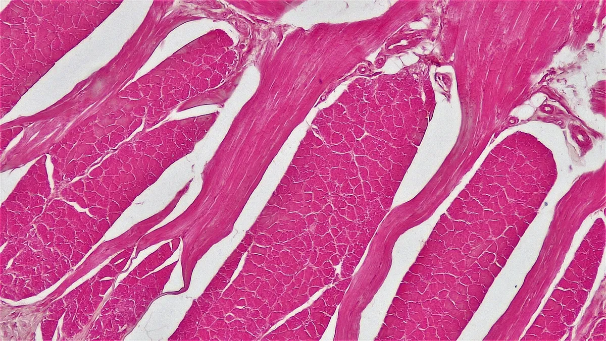 H&E · 100×
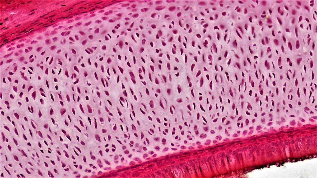 H&E · 200×
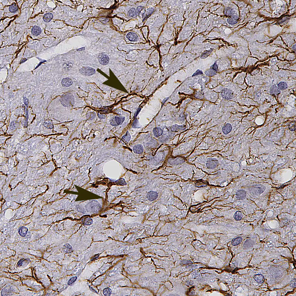 IHC · GFAP
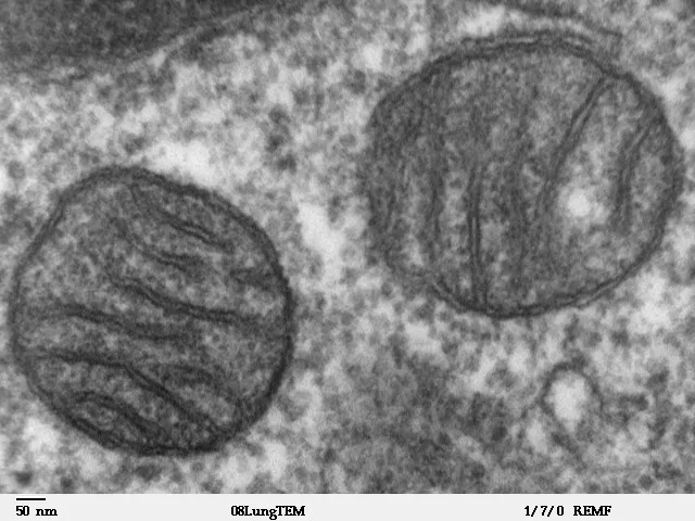 TEM
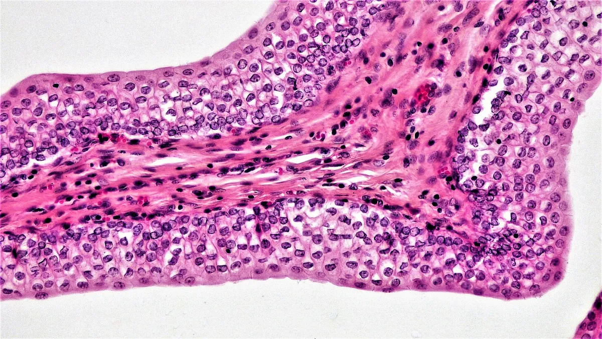 H&E · 200×
Organ systems
Every organ system, one atlas
Each chapter covers the general organisation, functions, principal organs and reference values.
languages
One structure, eight languages
The Terminologia Anatomica Latin name is the common anchor. Names, articles and the interface are available in Vietnamese, English, French, Korean, Japanese, Russian and Simplified and Traditional Chinese.
Terminologia Anatomica
Cor
Interactive anatomical atlas
Built for students of anatomy
True anatomical models
Over two thousand structures across eleven organ systems, built from BodyParts3D / Z-Anatomy data, to rotate, zoom and identify in the browser.
Dissection-room tools
Peel layers, cut along three planes, search for a structure, snap to standard views, use keyboard shortcuts and share a link to the exact view.
Every organ, part by part
Each organ page lists its parts with a description; tap one to fly straight to it on the model.
423 described parts
Referenced
Articles are written from PubMed reviews and StatPearls full texts; statements carry numbered citations that link to the original record, so readers can check them.
365 cited PubMed sources
Standard terms in eight languages
Every structure carries its Terminologia Anatomica Latin name, in Vietnamese, English, French, Korean, Chinese (Simplified and Traditional), Japanese and Russian.
Made for iPad and phone
An app-style layout on small screens with a bottom dock; models load only when needed and lower their resolution on touch devices.
Start with an organ system
Each chapter moves from the general organisation to each organ, with an anatomical model and references.
Data and terminology
Built on open data and international standards
Models, nomenclature and content can all be traced to their origin, so that readers can verify them.