Cells and tissues
Model
Hover or tap to see names, click to read a description; click a name in the legend to fly to it. Procedurally built to histological proportions; colours follow illustration conventions.
Overview
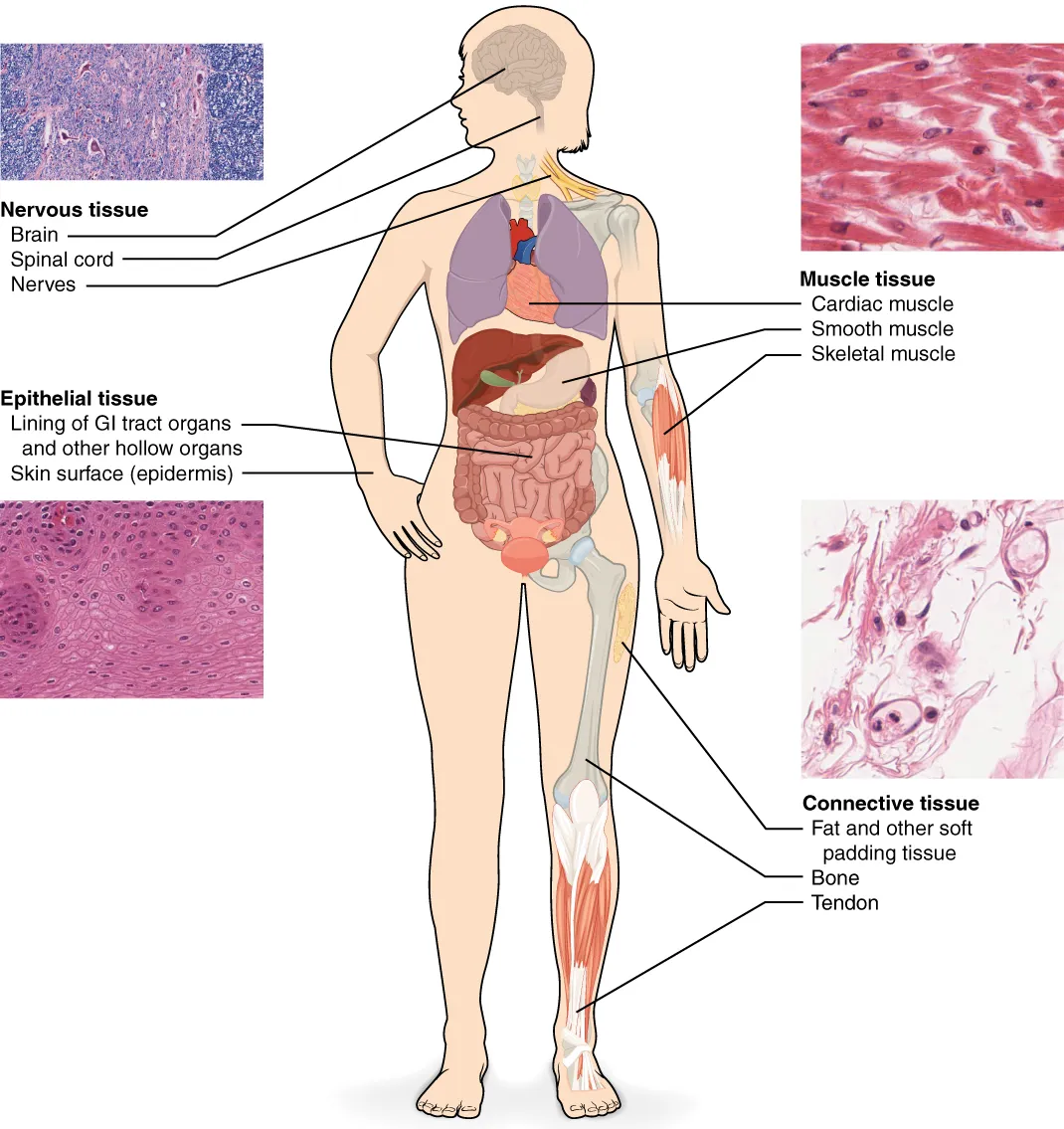
The cell is the smallest structural and functional unit of the body; an adult human has some 30–40 trillion cells of more than 200 types. Cells of common origin and function, together with the extracellular matrix they produce, form tissues. Histology groups every tissue into four basic types, epithelium, connective tissue (including blood, cartilage and bone), muscle and nervous tissue, from which all organs are built.
Functions
- 01Epithelium: covers surfaces, lines cavities and forms glands; barrier, absorption and secretion
- 02Connective tissue: support, binding, storage, transport (blood) and immune defence
- 03Muscle: contraction for movement, pumping blood and propelling contents of hollow organs
- 04Nervous tissue: receives, conducts and processes information by electrochemical signals
From cells to organs
The adult body contains some 30–40 trillion cells of a few hundred kinds, yet they assemble into only four basic tissues: epithelium, connective tissue, muscle and nervous tissue. Blood and immune cells alone number about 10¹³, most of them red cells, and the marrow must replace hundreds of billions of cells every day.[1]
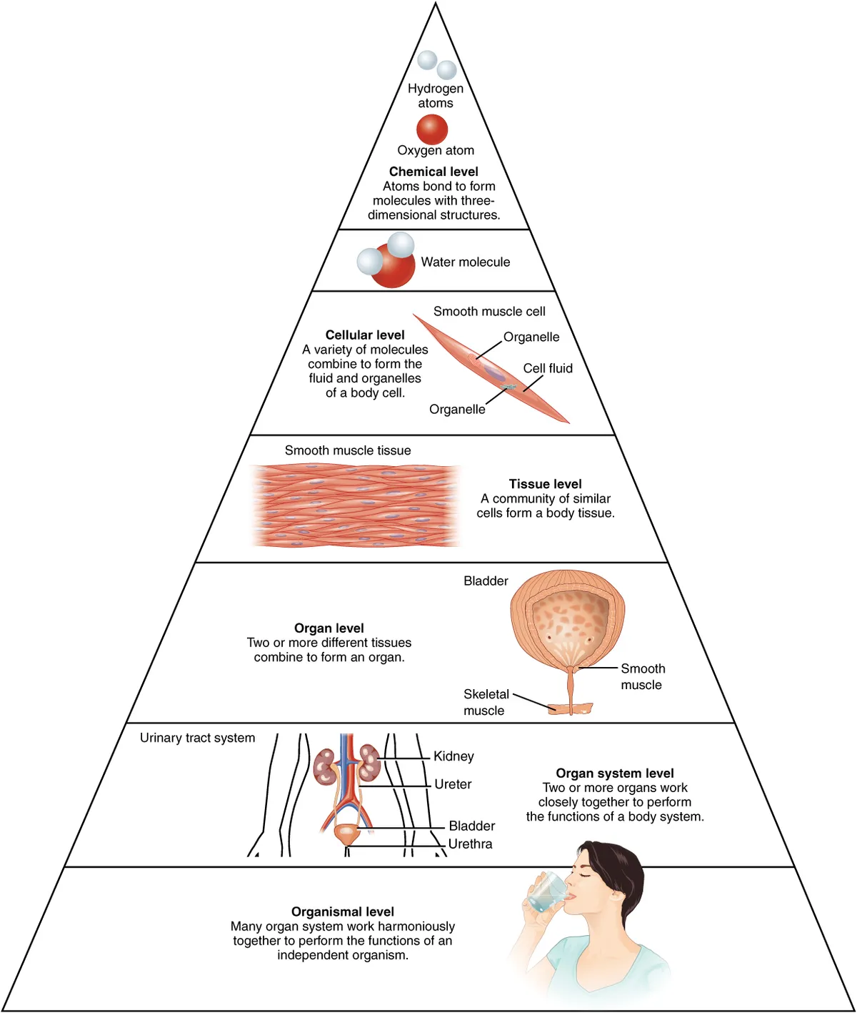
A tissue is a group of cells together with the extracellular matrix they secrete. Epithelium has almost no matrix, its cells adhering tightly into sheets that line surfaces; connective tissue is the reverse, with few cells in abundant ground substance and fibres; muscle and nervous tissue are defined by function, contraction and electrical signalling. Every organ combines several tissues: the gut wall has epithelium, connective tissue, smooth muscle and nerve plexuses.[2][3]
Epithelium
- Main cells
- Polarised epithelial cells joined by junctional complexes
- Matrix
- Scant; rests on a basement membrane
- Function
- Covering, barrier, absorption, secretion
- Examples
- Epidermis, gut lining, glands
Connective tissue
- Main cells
- Fibroblasts, adipocytes, chondrocytes, bone cells, blood cells
- Matrix
- Abundant: ground substance, collagen and elastic fibres
- Function
- Support, binding, storage, transport, defence
- Examples
- Tendon, fat, cartilage, bone, blood
Muscle
- Main cells
- Skeletal fibres, cardiomyocytes, smooth-muscle cells
- Matrix
- Basal lamina and surrounding connective sheaths
- Function
- Contraction producing force and movement
- Examples
- Skeletal muscles, heart, walls of hollow organs
Nervous tissue
- Main cells
- Neurons and glia
- Matrix
- Very little; narrow extracellular space
- Function
- Receiving, integrating and transmitting signals
- Examples
- Brain, spinal cord, nerves, ganglia
| Tissue | Main cells | Matrix | Function | Examples |
|---|---|---|---|---|
| Epithelium | Polarised epithelial cells joined by junctional complexes | Scant; rests on a basement membrane | Covering, barrier, absorption, secretion | Epidermis, gut lining, glands |
| Connective tissue | Fibroblasts, adipocytes, chondrocytes, bone cells, blood cells | Abundant: ground substance, collagen and elastic fibres | Support, binding, storage, transport, defence | Tendon, fat, cartilage, bone, blood |
| Muscle | Skeletal fibres, cardiomyocytes, smooth-muscle cells | Basal lamina and surrounding connective sheaths | Contraction producing force and movement | Skeletal muscles, heart, walls of hollow organs |
| Nervous tissue | Neurons and glia | Very little; narrow extracellular space | Receiving, integrating and transmitting signals | Brain, spinal cord, nerves, ganglia |
How tissues are studied
Tissue is fixed (usually in formalin), embedded in paraffin, cut into 4–5 µm sections and stained. Haematoxylin and eosin (H&E) colours nuclei and other nucleic-acid-rich structures (such as rough ER) blue-purple because they are basophilic, while cytoplasm and collagen, which are acidophilic, stain pink. Phrases such as “basophilic cytoplasm” or “eosinophilic granules” on these pages are the vocabulary of H&E.[4]
Immunohistochemistry uses labelled antibodies to locate a given protein on the section, for example cytokeratins in epithelium, desmin in muscle or GFAP in astrocytes, and is a daily tool for diagnosing tumours. Electron microscopy (resolution below 1 nm) reveals organelles, membranes and junctions; super-resolution fluorescence and cryo-electron tomography now resolve mitochondrial networks, nuclear pores and synaptic active zones in intact cells.[5][6][7]
Tissue renewal and repair
Tissues renew at very different rates. The intestinal lining is replaced in 3–5 days, the epidermis in about a month, red cells live 120 days, whereas neurons and cardiomyocytes hardly divide after maturity. Every fast-renewing tissue has its own stem-cell population in a niche: haematopoietic stem cells in marrow, basal cells of the epidermis, stem cells at the base of intestinal crypts.[8][9][10]
Skeletal muscle is a stable tissue that repairs well: satellite cells, lying between the sarcolemma and basal lamina, are normally quiescent but after injury proliferate, differentiate and fuse into new fibres while renewing their own pool. When damage exceeds regenerative capacity, tissue is replaced by scar: fibroblasts become myofibroblasts that lay down collagen and contract the wound, and persistent fibrosis of heart, liver or lung destroys organ function.[11][12][13]
Principal organs
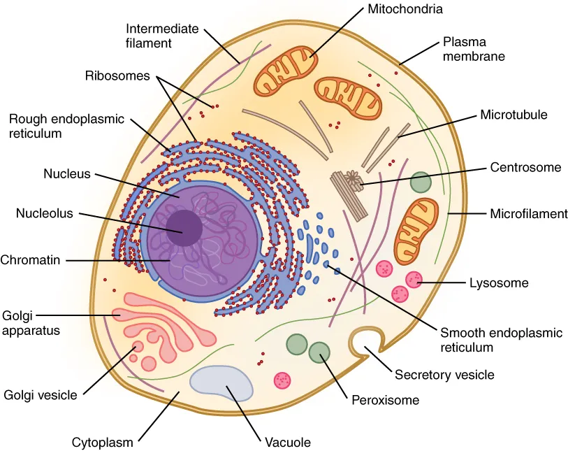
A eukaryotic cell consists of a plasma membrane enclosing the cytoplasm and nucleus. The cytoplasm contains membrane-bound organelles (endoplasmic reticulum, Golgi apparatus, mitochondria, lysosomes, peroxisomes), non-membranous organelles (ribosomes, centrosome) and the cytoskeleton. Each organelle is a specialised compartment, and together they synthesise, transport, generate energy, degrade and divide.
Epithelial tissueTextus epithelialisSheets of closely packed, polarised cells resting on a basement membrane and lacking blood vessels. Covering epithelia are named by the number of layers (simple, stratified, pseudostratified) and the shape of the surface cells (squamous, cuboidal, columnar); glandular epithelium forms exocrine and endocrine glands.
Connective tissue, cartilage and boneTextus connectivusTissue with relatively few cells scattered in abundant extracellular matrix (fibres and ground substance). The properties of the matrix set the function: soft and hydrated in loose connective tissue, tough in tendons and ligaments, compression-resistant in cartilage and rigid with calcium salts in bone.
Blood and blood cellsSanguisA fluid connective tissue, about 5 litres in an adult, made of plasma (about 55%) and formed elements: red cells that carry oxygen, five kinds of white cells that defend the body and platelets that stop bleeding. All arise from haematopoietic stem cells in the bone marrow.
Muscle tissueTextus muscularisTissue of elongated cells rich in actin and myosin filaments and able to contract. There are three kinds: skeletal muscle (multinucleate striated fibres under voluntary control), cardiac muscle (branched striated cells joined by intercalated discs, contracting automatically) and smooth muscle (spindle-shaped unstriated cells under involuntary control).
Nervous tissueTextus nervosusTissue made of neurons, which conduct electrical impulses, and neuroglia, which support, insulate, nourish and protect them. The human brain holds about 86 billion neurons and a roughly similar number of glial cells.
Reference values
- The bone marrow makes about 2–3 million new red cells every second.
- Red cells make up about 84% of all the cells in the human body, although they are only one of more than 200 cell types.
- The longest human axons, from the spinal cord to the big toe, are over a metre long.
References
Sources for the values and descriptions on this page, indexed in PubMed (US National Library of Medicine).
- [1]Hematopoiesis in numbersCosgrove J et al., Trends in immunology 2021PMID 34742656
- [2]Anatomy, Connective TissueKamrani P et al., StatPearls 2023PMID 30860769
- [3]Histology, Epithelial CellKurn H et al., StatPearls 2023PMID 32644489
- [4]Histology, Rough Endoplasmic ReticulumSanvictores T et al., StatPearls 2023PMID 33085273
- [5]Principles and Methods of ImmunohistochemistryRamos-Vara JA, Methods in molecular biology 2017PMID 28748460
- [6]Mitochondrial Structure, Dynamics, and Physiology: Light Microscopy to Disentangle the NetworkLandoni JC et al., Annual review of cell and developmental biology 2024PMID 38976811
- [7]Visualizing presynaptic functionKavalali ET et al., Nature neuroscience 2014PMID 24369372
- [8]Concise review: how do red blood cells born, live, and die?Corrons JLV et al., Annals of hematology 2021PMID 34342697
- [9]HematopoiesisRieger MA et al., Cold Spring Harbor perspectives in biology 2012PMID 23209149
- [10]The emergence of the stem cell nicheHicks MR et al., Trends in cell biology 2023PMID 35934562
- [11]Satellite cells and the muscle stem cell nicheYin H et al., Physiological reviews 2013PMID 23303905
- [12]The Role of Myofibroblasts in Physiological and Pathological Tissue RepairSchuster R et al., Cold Spring Harbor perspectives in biology 2023PMID 36123034
- [13]Cardiac fibrosisFrangogiannis NG, Cardiovascular research 2021PMID 33135058