Diaphragm
Model
Layers
Z-Anatomy anatomical model based on BodyParts3D, licensed CC BY-SA 4.0.
Description
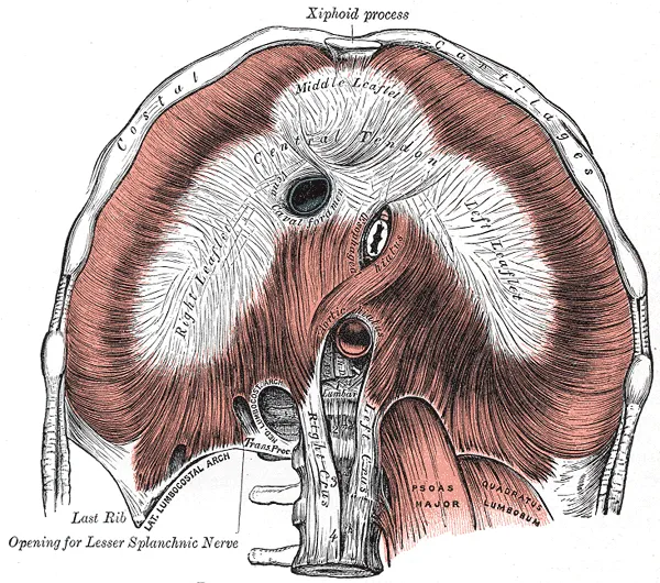
A dome-shaped musculotendinous partition between the thorax and the abdomen, whose muscle fibres arise from the sternum, the lower six costal cartilages and the lumbar spine (crura) and converge on the central tendon. It is the chief muscle of inspiration: on contraction the dome descends and enlarges the thoracic cavity. It has three main openings: the caval opening (T8), the oesophageal hiatus (T10) and the aortic hiatus (T12).
Parts
Each part has its own card; press “Show on the model” to fly to it and select it in the 3D model above.
- 01
Central tendon
Centrum tendineum
A trefoil-shaped aponeurosis at the centre of the dome, with no bony attachment, into which all the muscle fibres converge. It is fused above to the fibrous pericardium. The caval opening lies within it at T8, so inspiration widens rather than narrows it, aiding venous return.
- 02
Sternal and costal parts
Pars sternalis et pars costalis
The sternal part is two small slips from the back of the xiphoid process; the costal part arises from the inner surfaces of the lower six costal cartilages and ribs. Gaps between them (sternocostal triangles of Larrey) transmit the superior epigastric vessels and are the site of Morgagni hernias.
- 03
Lumbar part and crura
Pars lumbalis et crura
The right crus (L1–L3) is longer than the left (L1–L2); they are joined by the median arcuate ligament arching in front of the aorta. The medial and lateral arcuate ligaments arch over psoas major and quadratus lumborum. Fibres of the right crus loop around the oesophagus as a physiological anti-reflux sphincter.
- 04
Diaphragmatic openings
Hiatus diaphragmatis
The three main openings follow the mnemonic “8, 10, 12”: the caval opening (T8, with branches of the right phrenic nerve), the oesophageal hiatus (T10, with the two vagal trunks) and the aortic hiatus (T12, behind the median arcuate ligament, with the thoracic duct and azygos vein). A widened oesophageal hiatus leads to sliding hiatal hernia.
- 05
Right and left domes
Cupulae diaphragmatis
The right dome sits higher than the left (because of the liver), reaching the fourth or fifth intercostal space in full expiration. In quiet inspiration the domes descend about 1.5 cm, up to 10 cm in deep breathing, providing about 75% of tidal volume. A raised dome on a radiograph suggests paralysis of the phrenic nerve on that side.
- 06
Nerve and blood supply
Innervatio et vascularisatio
Its sole motor supply is the phrenic nerve (C3–C5: “C3, 4, 5 keep the diaphragm alive”), so cord injuries above C3 require ventilation. The phrenic nerve also carries sensation from the pleura and peritoneum over the central dome, which is why irritation there is felt as shoulder-tip pain. Blood comes from the superior and inferior phrenic, musculophrenic and lower intercostal arteries.
Position and relations
The diaphragm has two domes, the right higher (at the fifth intercostal space in expiration) because of the liver beneath. Its upper surface is related to the pleurae and pericardium, its lower surface to the liver, stomach, spleen, kidneys and suprarenal glands. Posteriorly, arcuate ligaments arch over psoas and quadratus lumborum, and the two crura attach to the lumbar vertebral bodies.
Form and structure
The muscle has sternal, costal and lumbar parts by origin (the right crus is larger than the left), all inserting into the trefoil-shaped central tendon. The caval opening lies in the central tendon and is pulled open in inspiration; the oesophageal hiatus is encircled by fibres of the right crus, which help prevent reflux; the aortic hiatus lies behind the median arcuate ligament. Congenital diaphragmatic hernia occurs most often through the left lumbocostal triangle (Bochdalek).
Vessels and nerves
Its only motor nerve is the phrenic nerve (C3–C5), so a high cervical cord injury above C3 stops breathing; the periphery also receives sensory fibres from the lower intercostal nerves. Irritation of the central diaphragm refers pain to the shoulder through segments C3–C5. Arteries come from the superior and inferior phrenic, pericardiacophrenic and musculophrenic arteries and the lower intercostal arteries.
Clinical correlations
The diaphragm is the main muscle of ventilation, and its dysfunction is an under-recognised cause of breathlessness, arising from surgery, trauma, tumour or infection. Chest radiographs usually show only an elevated dome; fluoroscopy or ultrasound distinguishes paralysis (paradoxical motion on sniffing), weakness and eventration (congenital thinning of a segment).[1]
Functions and openings of the diaphragm
The diaphragm is the most important muscle of inspiration: as it contracts it pulls the central tendon down, increasing the vertical diameter of the thorax and the negative intrathoracic pressure, so air flows in; in inspiration it flattens into the abdomen while the external intercostals raise the anterior chest wall like a bucket handle. In expiration the chest wall sags back and the diaphragm relaxes and rises, expelling air. It also helps the anterior abdominal muscles raise intra-abdominal pressure in micturition, defecation and parturition, and when lifting with a held breath (the Valsalva manoeuvre, used to tell right- from left-sided heart murmurs). It is a thoracoabdominal pump: as it descends, intrathoracic pressure falls and intra-abdominal pressure rises, squeezing blood in the inferior vena cava up to the right atrium and helping lymph up the thoracic duct, whose valves prevent backflow. Fibres of the right crus loop around the oesophageal opening like a sling and act as a sphincter against reflux.[2]
The sternal part consists of two fleshy slips from the back of the xiphoid process; the costal part arises from the inner surfaces of the lower six costal cartilages and interdigitates with transversus abdominis. The medial lumbocostal arch is a tendinous band over psoas major from the body to the transverse process of L1; the lateral arch covers the upper quadratus lumborum from the L1 transverse process to the lower border of rib 12. The right crus arises from the bodies of the upper three lumbar vertebrae and their discs, the left from the upper two; their medial margins form the median arcuate ligament across the front of the aorta. Besides the three major openings there are minor ones: apertures in the right and left crura for the greater and lesser splanchnic nerves (on the left also the hemiazygos vein), the sympathetic trunk passing behind the medial lumbocostal arch, and the foramen of Morgagni between the sternal and costal parts, carrying the superior epigastric artery and abdominal-wall lymphatics, which is also the site of anterior diaphragmatic hernia.[2]
Caval opening
- Level
- T8
- Location
- Central tendon
- Transmits
- Inferior vena cava, branches of right phrenic nerve
Oesophageal hiatus
- Level
- T10
- Location
- Muscular sling of the right crus, left of midline
- Transmits
- Oesophagus, vagal trunks, oesophageal branches of left gastric vessels, lymphatics
Aortic hiatus
- Level
- T12
- Location
- Between the crura, behind the median arcuate ligament
- Transmits
- Aorta, thoracic duct, azygos vein
| Opening | Level | Location | Transmits |
|---|---|---|---|
| Caval opening | T8 | Central tendon | Inferior vena cava, branches of right phrenic nerve |
| Oesophageal hiatus | T10 | Muscular sling of the right crus, left of midline | Oesophagus, vagal trunks, oesophageal branches of left gastric vessels, lymphatics |
| Aortic hiatus | T12 | Between the crura, behind the median arcuate ligament | Aorta, thoracic duct, azygos vein |
Full-text reading (StatPearls)
The StatPearls chapters on this topic, reproduced verbatim. Their CC BY-NC-ND 4.0 licence allows unmodified copies only, so they are not translated or shortened.
Anatomy, Thorax: DiaphragmBains KNS, Kashyap S, Lappin SL · StatPearls 2023 · NBK519558 · 11k characters
Loading…
Bains KNS, Kashyap S, Lappin SL. Anatomy, Thorax: Diaphragm. StatPearls [Internet]. Treasure Island (FL): StatPearls Publishing; 2023. NBK519558 · PMID 30137842 · CC BY-NC-ND 4.0. Reproduced verbatim, unmodified.
Organs in this system
References
Sources for the values and descriptions on this page, indexed in PubMed (US National Library of Medicine).