Duodenum
Model
Layers
Z-Anatomy anatomical model based on BodyParts3D, licensed CC BY-SA 4.0.
Description
The first part of the small intestine, about 25 cm long, curving in a C around the head of the pancreas and mostly retroperitoneal. It has four parts, superior, descending, horizontal and ascending; the bile and pancreatic ducts open on the major duodenal papilla in the descending part. The duodenum neutralises gastric acid and is where pancreatic enzymes and bile begin digestion.
Parts
Each part has its own card; press “Show on the model” to fly to it and select it in the 3D model above.
- 01
Superior part (D1)
Pars superior duodeni
About 5 cm long from the pylorus at L1; its first 2 cm (the duodenal bulb or cap) is mobile, intraperitoneal and smooth on contrast studies, and is the site of about 95% of duodenal ulcers. A posterior ulcer can erode the gastroduodenal artery and bleed massively.
- 02
Descending part (D2)
Pars descendens duodeni
About 7–10 cm long, it descends to the right of L1–L3 retroperitoneally, curving round the head of the pancreas. Its posteromedial wall bears the major duodenal papilla (ampulla of Vater, about 7–10 cm from the pylorus) and, about 2 cm above, the minor papilla for the accessory pancreatic duct.
- 03
Horizontal part (D3)
Pars horizontalis duodeni
Crosses L3 from right to left, sandwiched between the superior mesenteric artery in front and the abdominal aorta behind. A narrow aortomesenteric angle, as after marked weight loss, can compress it (superior mesenteric artery syndrome).
- 04
Ascending part (D4) and duodenojejunal flexure
Pars ascendens duodeni; Flexura duodenojejunalis
Ascends to the left of the aorta to L2 and turns forward at the duodenojejunal flexure, held by the suspensory muscle (ligament of Treitz), the landmark dividing upper from lower gastrointestinal bleeding. Duodenal fossae around the flexure can harbour internal hernias.
- 05
Mucosa and Brunner glands
Plicae circulares; Glandulae duodenales
From D2 onwards the mucosa has circular folds (of Kerckring) and villi; the submucosa contains duodenal (Brunner) glands secreting alkaline mucus that neutralises acid chyme, the histological hallmark of the duodenum. Enteroendocrine cells release secretin and cholecystokinin.
Position and relations
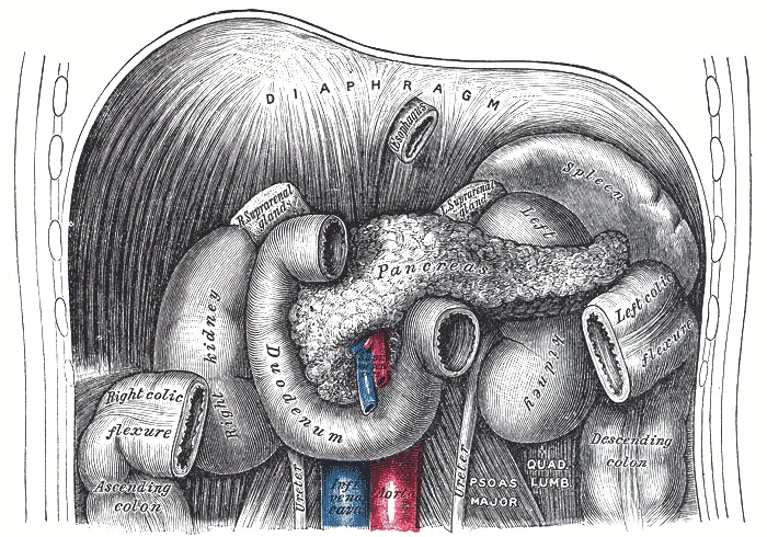
The superior part (duodenal bulb) lies at L1 and is mobile; the descending part runs beside the head of the pancreas in front of the right kidney; the horizontal part crosses in front of the inferior vena cava and aorta, behind the superior mesenteric vessels; the ascending part reaches the duodenojejunal flexure at L2, held by the suspensory muscle (ligament of Treitz), the landmark separating upper from lower gastrointestinal bleeding.
Form and structure
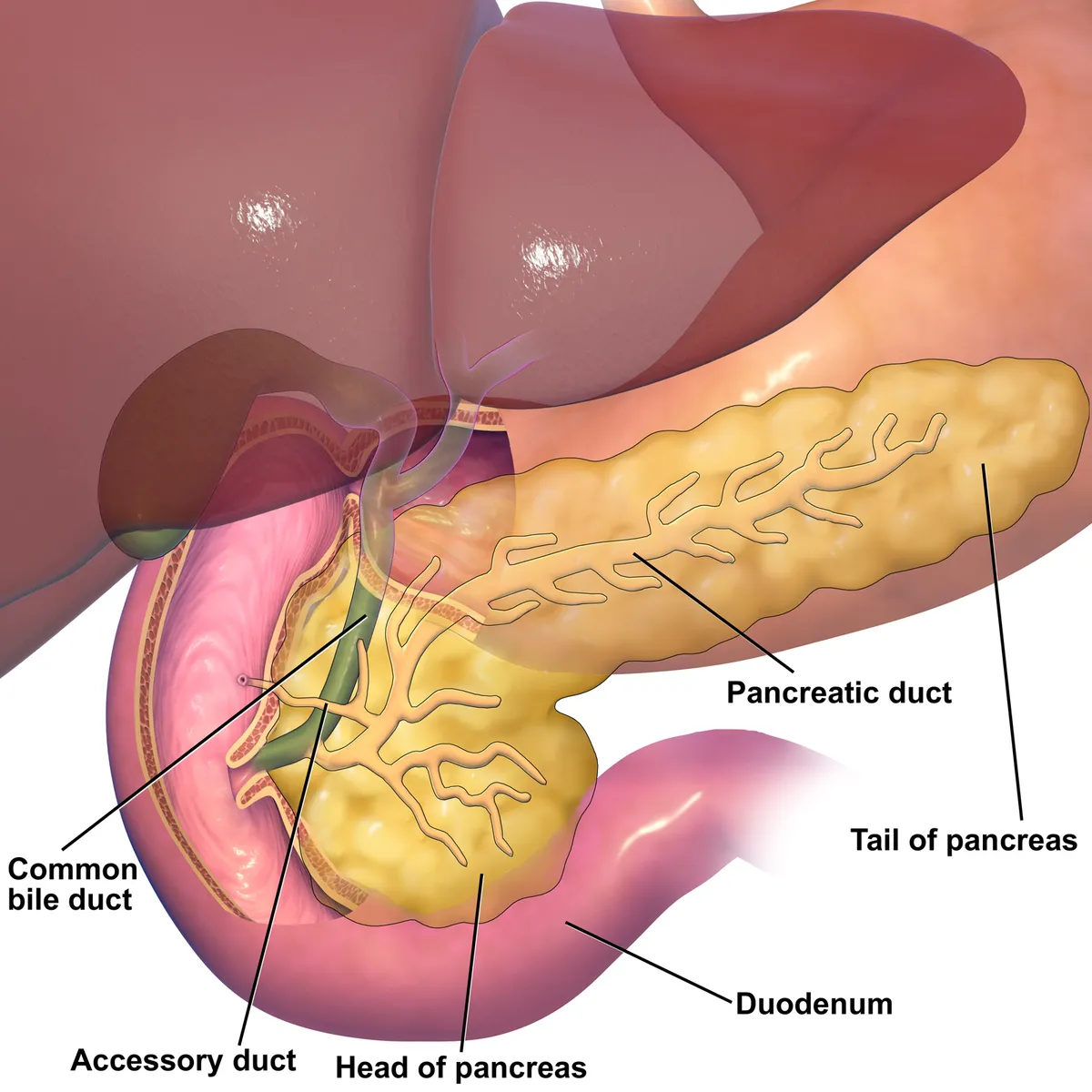
The mucosa has circular folds and villi like the rest of the small intestine, but its submucosa contains Brunner’s glands, which secrete alkaline mucus that protects against gastric acid. The smooth duodenal bulb is the usual site of duodenal ulcers. The major papilla lies on the posteromedial wall of the descending part, about 7–10 cm from the pylorus; the minor papilla above it receives the accessory pancreatic duct.
Vessels and nerves
The duodenum straddles the foregut–midgut boundary: its pancreaticoduodenal arcades are formed by the superior pancreaticoduodenal arteries (from the gastroduodenal, a coeliac branch) and the inferior ones (from the superior mesenteric). The gastroduodenal artery runs directly behind the bulb, so a posterior ulcer can bleed massively. Superior mesenteric artery syndrome occurs when the horizontal part is compressed between that artery and the aorta.
Physiology
The duodenum controls and coordinates gastroduodenal function. Its mucosa defends itself against gastric acid and pepsin with several layers: mucus and bicarbonate, epithelial junctions, mucosal blood flow, nitric oxide, antimicrobial peptides (β-defensins) and immune cells such as macrophages and dendritic cells.[1][2]
The duodenal mucosa also “chemosenses” the lumen: long-chain fatty acids, bile acids, bacterial short-chain fatty acids and lipopolysaccharide bind receptors on enteroendocrine and inflammatory cells. These signals adjust mucosal protection, motility, visceral sensation and nutrient uptake, and reach systemically to metabolism, energy balance and food intake.[3]
Clinical correlations
Growing evidence points to the duodenum as a key site in functional dyspepsia: impaired mucosal integrity and low-grade inflammation, with altered neuronal signalling and systemic immune activation. Acid, bile, the microbiota and food antigens are suspected triggers, but none has been causally linked to symptoms.[1]
The major duodenal papilla (ampulla of Vater) is where the common bile duct and pancreatic duct open into the descending duodenum. Ampullary carcinoma is rare (0.5–0.9 per 100,000), with intestinal and pancreaticobiliary subtypes (the latter with worse prognosis); the treatment of choice is pancreatoduodenectomy with lymphadenectomy. Localised papillary adenomas can be removed endoscopically (papillectomy).[4][5]
Duodenal atresia and intestinal malrotation cause neonatal bowel obstruction and are often suspected antenatally; malrotation is particularly dangerous because it can lead to volvulus requiring emergency surgery.[6]
Full-text reading (StatPearls)
The StatPearls chapters on this topic, reproduced verbatim. Their CC BY-NC-ND 4.0 licence allows unmodified copies only, so they are not translated or shortened.
Anatomy, Abdomen and Pelvis: DuodenumOmole AE, Gogna S, Khorasani-Zadeh A · StatPearls 2026 · NBK482390 · 11k characters
Loading…
Omole AE, Gogna S, Khorasani-Zadeh A. Anatomy, Abdomen and Pelvis: Duodenum. StatPearls [Internet]. Treasure Island (FL): StatPearls Publishing; 2026. NBK482390 · PMID 29494012 · CC BY-NC-ND 4.0. Reproduced verbatim, unmodified.
Organs in this system
References
Sources for the values and descriptions on this page, indexed in PubMed (US National Library of Medicine).
- [1]Novel concepts in the pathophysiology and treatment of functional dyspepsiaWauters L et al., Gut 2020PMID 31784469
- [2]Gastroduodenal mucosal defenseKemmerly T et al., Current opinion in gastroenterology 2014PMID 25229259
- [3]Duodenal chemosensingIwasaki M et al., Current opinion in gastroenterology 2018PMID 30124472
- [4]The Diagnosis and Treatment of Ampullary CarcinomaWalter D et al., Deutsches Arzteblatt international 2023PMID 37656482
- [5]Endoscopic PapillectomyCampos ST et al., Gastrointestinal endoscopy clinics of North America 2022PMID 35691696
- [6]Malrotation and intestinal atresiasAdams SD et al., Early human development 2014PMID 25448782
- [7]Gastrointestinal mean transit times in young and middle-aged healthy subjectsPMID 11318834Supports: Gastrointestinal transit time
- [8]Revised Estimates for the Number of Human and Bacteria Cells in the BodySender R, Fuchs S, Milo R, 2016PMID 27541692Supports: Colonic bacterial count