Skeletal system
Model
Z-Anatomy anatomical model based on BodyParts3D, licensed CC BY-SA 4.0.
Overview
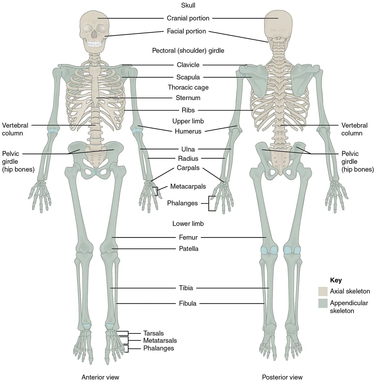
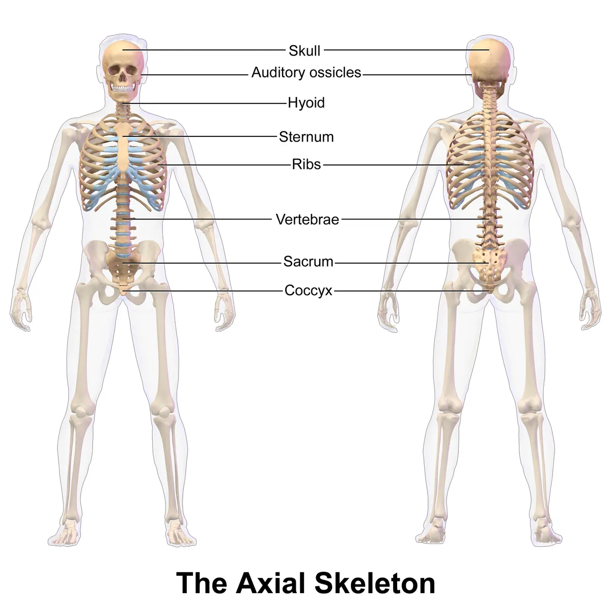
The adult skeleton has 206 bones, divided into the axial skeleton (skull, vertebral column, thoracic cage) and the appendicular skeleton (pectoral and pelvic girdles, limb bones). Together with joints and ligaments it supports the body, protects the viscera and provides attachment for muscles.
Functions
- 01Mechanical support
- 02Protection of the brain, heart, lungs and viscera
- 03Haematopoiesis in red bone marrow
- 04Mineral reservoir (calcium, phosphate)
Bone modelling and remodelling
Bone tissue has three main cell types: osteoblasts that form bone, osteoclasts that resorb it, and osteocytes embedded in the matrix, which act as mechanosensors and orchestrate remodelling. An imbalance between resorption and formation leads to osteoporosis.[1]
In childhood, modelling predominates: bones lengthen at the growth plates and widen radially. In adulthood, remodelling predominates, replacing old bone with new to preserve strength and mineral balance. Osteoblasts and osteoclasts communicate directly and through secreted factors such as the RANKL/OPG system; bone formation is also tightly coupled to angiogenesis (type H vessels).[2][3][4]
Bone cells and the remodelling cycle
Osteoblasts are cuboidal cells lining bone surfaces and make up only 4–6% of bone cells. They derive from mesenchymal stem cells under BMP and Wnt signalling and the transcription factors Runx2, Dlx5 and osterix, Runx2 being the master gene. They build matrix in two steps: secretion of organic matrix (mainly type I collagen, plus osteocalcin, osteonectin, bone sialoprotein, osteopontin and proteoglycans), then mineralisation, in which 30–200 nm matrix vesicles accumulate calcium and phosphate (released by alkaline phosphatase), nucleate hydroxyapatite and rupture so the crystals spread into the matrix. Mature osteoblasts then undergo apoptosis or become osteocytes or bone lining cells.[1]
Osteocytes make up 90–95% of bone cells and are the longest-lived, up to 25 years. Each sits in a lacuna and sends up to about 50 processes through canaliculi, joined by gap junctions to each other and to surface cells; this lacunocanalicular system has a surface about 400 times that of all Haversian and Volkmann systems. Through it osteocytes act as mechanosensors: loading makes them release anabolic PGE₂, NO and IGF-1, while unloading induces sclerostin (SOST gene) and DKK-1, which inhibit osteoblasts. Osteocytes are thought to be the main source of RANKL, and osteocyte apoptosis is a chemotactic signal for osteoclasts.[1]
Osteoclasts are terminally differentiated multinucleated cells of haematopoietic origin, formed under M-CSF and RANKL. Remodelling takes place in basic multicellular units, with osteoclasts in front forming a cutting cone and osteoblasts behind forming a closing cone, together with vessels and nerves. The cycle runs through initiation (resorption), reversal and formation. Factors stored in the matrix (IGFs, TGF-β, BMPs, FGF, PDGF) are released by resorption and couple the phases. Early on, osteoclasts express Sema4D, which inhibits bone formation, while osteoblasts express Sema3A, which suppresses resorption; bidirectional ephrinB2/EphB4 signalling may help end resorption and start formation.[1]
The vertebral column: composition, functions and curves
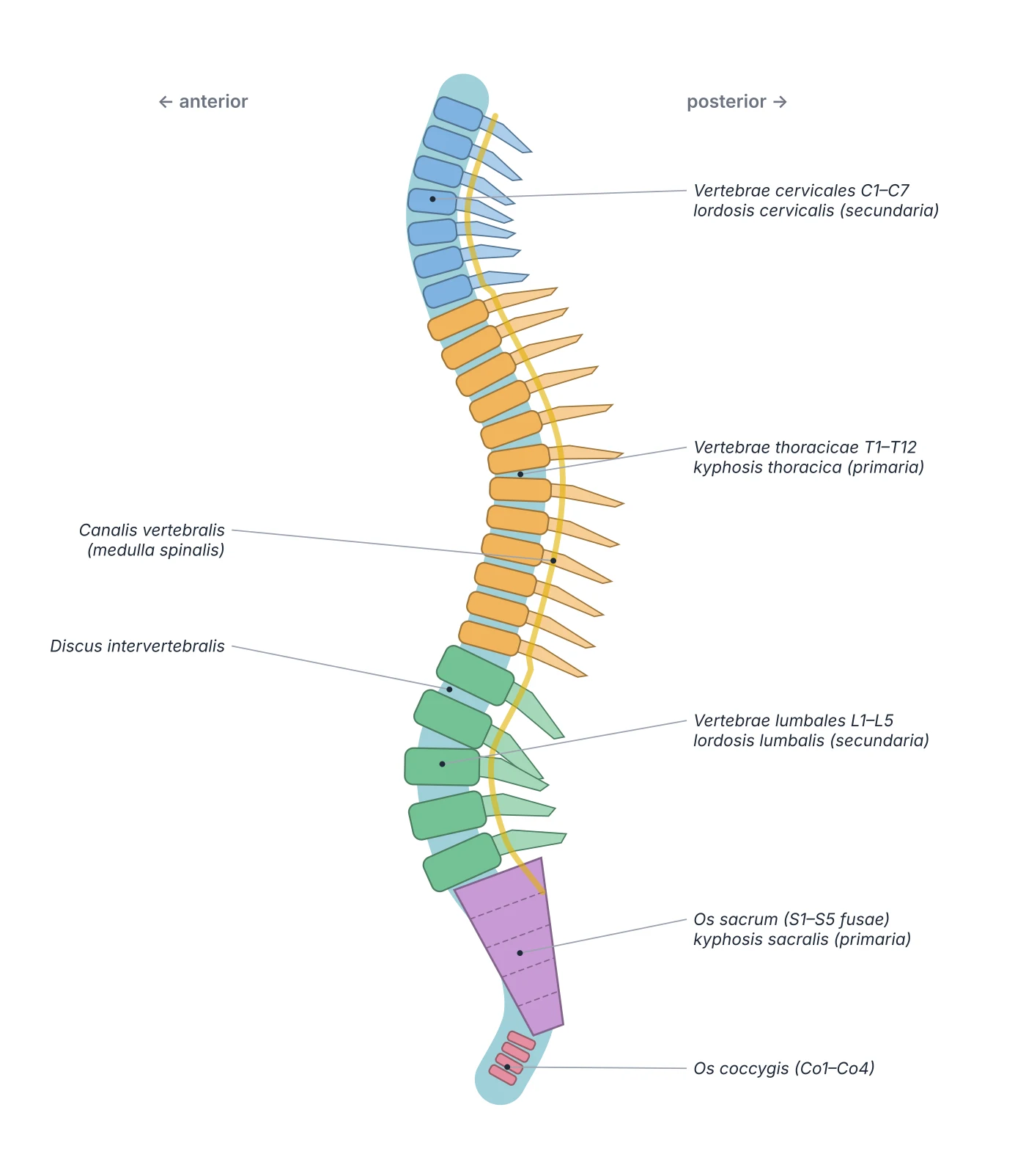
The vertebral column defines the subphylum Vertebrata. In humans it has 33 vertebrae: 7 cervical, 12 thoracic, 5 lumbar, 5 sacral (fused into the sacrum) and 4 coccygeal; with the skull, ribs and sternum they make up the axial skeleton. The column has three tasks: protecting the spinal cord and nerve roots in the vertebral canal (wider in the cervical and lumbar regions, narrower in the thoracic); acting as the weight-bearing axis that supports the head and transfers the weight of the trunk to the legs; and allowing flexion, side-bending and rotation through its chain of joints, while giving attachment to the ribs and many muscles. The intervertebral discs make up about 25% of the length of the column and support the anterior and posterior longitudinal ligaments.[5]
The adult column has four curves: cervical lordosis, thoracic kyphosis, lumbar lordosis and the sacral curve. The thoracic and sacral curves are primary, present in fetal life (the “fetal position”); the cervical curve appears when the infant starts to hold up its head and the lumbar curve when the child starts to stand, so they are called secondary. Disease can exaggerate the curves: increased thoracic kyphosis (often from osteoporosis and poor posture in the elderly), increased lumbar lordosis (from weak anterior abdominal muscles, common in late pregnancy and obesity), and scoliosis, a lateral deviation with vertebral rotation, classically in adolescent girls and genetic, post-traumatic or idiopathic. The column is supplied by branches of the vertebral and ascending cervical arteries (neck), posterior intercostal arteries (thorax), lumbar and lateral sacral arteries; each divides into an anterior branch to the vertebral body and a posterior branch to the arch.[5]
Development of the spine and its variants
The notochordal process begins to form around day 17 and folds into the notochord, the axis that orients the embryo. The notochord secretes Sonic Hedgehog to keep alive and pattern the sclerotome cells of the paraxial mesoderm, which grow around the neural tube and notochord to form the vertebral bodies, arches and ribs. Each vertebral body forms from the fusion of two adjacent half-sclerotomes (the caudal half of one and the cranial half of the next), a process called resegmentation. Where vertebral bodies will form, the notochord regresses under Pax1 and Pax9; in between it expands transversely into the nucleus pulposus, while the surrounding annular mesenchyme differentiates into the annulus fibrosus and cartilaginous endplates. The vertebrae ossify endochondrally: cartilage appears around week 6, is replaced by bone from weeks 7–8, and ossification is complete only around age 26.[5][6]
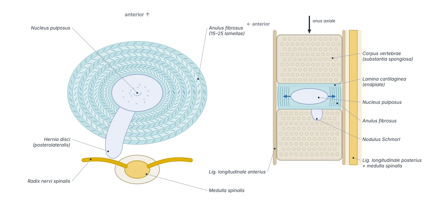
Faults of segmentation and closure produce many anomalies. In the VACTERL association (vertebral, anal, cardiac, tracheo-oesophageal, renal and limb anomalies), 60–80% of patients have vertebral defects such as fused, missing, extra or misshapen vertebrae. Spina bifida is a neural tube defect, in increasing severity spina bifida occulta, meningocele and myelomeningocele (often with Arnold–Chiari malformation and a tethered cord). Block vertebrae result from failed segmentation. Lumbosacral transitional vertebrae occur in about 35% of people (sacralisation of L5 or lumbarisation of S1, Castellvi types I–IV): the disc at the transitional level moves little and is protected, but the rigid lever arm transfers torque to the segment just above, which degenerates and herniates earlier; vertebrae must be counted on whole-spine imaging to avoid wrong-level surgery. Schmorl nodes (vertical herniation of nucleus pulposus through the endplate into the vertebral spongiosa) occur in up to 75% of people and are mostly asymptomatic.[5][6]
The intervertebral disc: a composite load-bearing structure
The intervertebral disc gives the spine flexibility while transmitting large compressive, tensile and shear loads; mechanically it behaves as a viscoelastic solid with time-dependent responses. The nucleus pulposus is the main load-distributing component: its matrix is rich in proteoglycans (mainly aggrecan) bearing highly anionic glycosaminoglycan chains, creating a high fixed charge density that draws in water by osmotic pressure. The nucleus is about 70–80% water and acts hydrostatically: under axial load it pressurises uniformly, converting vertical compression into radially directed stress.[6]
The annulus fibrosus contains the pressurised nucleus with 15–25 concentric lamellae of type I collagen; the fibres of successive lamellae cross at about 30–60° to the spinal axis, like plywood. This angle-ply structure converts radial stress into circumferential tension (“hoop stress”), and different fibre populations tighten in flexion, extension and torsion. The cartilaginous endplates on the upper and lower faces of the vertebral bodies both stop the nucleus from herniating into cancellous bone and act as semipermeable membranes through which glucose and oxygen diffuse from vertebral capillaries into the avascular disc centre; when the endplates calcify, nutrition falls and degeneration begins. After a day of standing, hydrostatic pressure exceeds osmotic swelling pressure and water is squeezed out (creep), so the disc loses 10–20% of its height by evening; during recumbent sleep it reimbibes fluid. A tear of the annulus (itself fibrocartilage) through degeneration, straining or lifting lets the nucleus herniate into the canal, compressing nerve roots and causing pain, numbness, weakness and reduced reflexes; most cases settle with conservative treatment.[6][7]
Cartilage: three types and why it heals slowly
Cartilage resists compression, adds resilience to bone and supports places that need flexibility. Its main cell, the chondrocyte, sits in lacunae; the matrix consists of fibres and proteoglycan–glycosaminoglycan complexes, rich in chondroitin rather than calcium. It arises from mesoderm: mesenchyme differentiates into chondroblasts that secrete aggrecan and type II collagen; after initial chondrification cartilage hardly divides, so its size changes little. The key point is that cartilage has no blood vessels, lymphatics or nerves: chondrocytes live by diffusion from surrounding tissues (helped by repeated compression), the perichondrium regenerates poorly, so cartilage injuries heal very slowly and are not painful in themselves; pain in cartilage disease comes from neighbouring structures such as the inflamed joint capsule and bone.[7]
Hyaline cartilage is the most abundant, pale blue-white, made of type II collagen and proteoglycans, and found in the trachea, nose, growth plates, sternum, costal cartilages and articular surfaces, where it gives a low-friction, compression-resistant surface; it becomes dry, thin and yellow with age. Elastic cartilage is dull yellow, found in the larynx, auricle, epiglottis and auditory tube, and is flexible and pressure-resistant. Fibrocartilage is rich in type I collagen with little proteoglycan, lacks a perichondrium, resists strong tension and compression, and is found in tendons, ligaments, intervertebral discs, menisci and some articular surfaces. Fibrocartilage also forms the enthesis where tendon inserts into bone, grading from tendon with longitudinal fibroblasts through fibrocartilage, the tidemark and calcified fibrocartilage to bone. Osteoarthritis mainly wears away the articular cartilage of load-bearing joints, reducing range of motion and producing “bone on bone” contact; achondroplasia is a genetic disorder of cartilage formation and the commonest cause of dwarfism.[7]
Hyaline
- Main collagen
- Type II
- Perichondrium
- Yes (except articular surfaces)
- Typical sites
- Trachea, nose, costal cartilage, growth plate, joint surfaces
Elastic
- Main collagen
- Type II and elastic fibres
- Perichondrium
- Yes
- Typical sites
- Auricle, epiglottis, auditory tube
Fibrocartilage
- Main collagen
- Type I
- Perichondrium
- No
- Typical sites
- Intervertebral discs, menisci, pubic symphysis, entheses
| Type | Main collagen | Perichondrium | Typical sites |
|---|---|---|---|
| Hyaline | Type II | Yes (except articular surfaces) | Trachea, nose, costal cartilage, growth plate, joint surfaces |
| Elastic | Type II and elastic fibres | Yes | Auricle, epiglottis, auditory tube |
| Fibrocartilage | Type I | No | Intervertebral discs, menisci, pubic symphysis, entheses |
Synovial joints: the elbow and ankle as examples
A synovial joint has articular surfaces covered with hyaline cartilage, a capsule lined by synovial membrane and reinforcing ligaments; its stability comes from bone shape, ligaments and muscles. The elbow shows this clearly. Flexion and extension occur at the ulnohumeral joint, where the trochlear notch of the olecranon wraps almost 180° around the humeral trochlea like a wrench, giving a highly congruent joint; pronation and supination occur at the radioulnar joints. The triangular medial (ulnar) collateral ligament has an anterior oblique band (the strongest, resisting valgus stress), a posterior oblique band and a transverse (Cooper) band. The lateral complex comprises the annular, radial collateral and lateral ulnar collateral ligaments (the last resisting posterolateral rotatory instability). The annular ligament encircles the radial neck, attached at both ends to the radial notch of the ulna; a sudden pull on the extended arm of a child aged 6 months to 5 years lets the radial head slip out of it (“nursemaid’s elbow”), reduced by supination–flexion or hyperpronation. The ulnar nerve runs superficially in the cubital tunnel beneath Osborne’s ligament and is easily compressed.[8]
The ankle joint lets the foot adapt to the ground in walking, with dorsiflexion, plantarflexion, inversion and eversion, each produced by a compartment of leg muscles. The superficial posterior compartment (gastrocnemius, soleus) is the main plantarflexor; the deep posterior compartment (tibialis posterior, flexor digitorum longus, flexor hallucis longus) assists plantarflexion, and tibialis posterior inverts the foot; tibialis anterior in the anterior compartment is the main dorsiflexor; peroneus longus and brevis in the lateral compartment evert the foot. Three tests examine the ligaments: the anterior drawer test (anterior talofibular ligament, the most often sprained), the talar tilt or inversion stress test (calcaneofibular ligament) and the eversion stress test (medial deltoid ligament). The elbow is the second most commonly dislocated large joint after the shoulder; complex dislocations with fracture or ligament injury may need surgery.[9][8]
Principal organs
Composed of 22 bones (8 neurocranial and 14 facial, excluding the auditory ossicles), it encloses and protects the brain and the special sense organs.
Thoracic cageCavea thoracisFormed by 12 pairs of ribs, the sternum and the 12 thoracic vertebrae; it protects the heart and lungs and takes part in the mechanics of breathing.
Vertebral columnColumna vertebralisThe axial skeleton of the trunk, made of 33–34 vertebrae (7 cervical, 12 thoracic, 5 lumbar, the sacrum and coccyx) joined by intervertebral discs and ligaments. It supports the head and trunk, allows movement and protects the spinal cord.
Hip boneOs coxaeA large flat bone formed by three bones (ilium, ischium, pubis) that fuse at the acetabulum by adulthood. The two hip bones and the sacrum form the bony pelvis, which transmits body weight to the lower limbs and protects the pelvic organs.
HumerusHumerusThe long bone of the arm, articulating with the scapula at the shoulder and with the radius and ulna at the elbow. Many important muscles and nerves attach to or run close to it.
FemurFemurThe longest and strongest bone, about one quarter of body height; it articulates with the hip bone at the hip joint and with the tibia at the knee.
TibiaTibiaThe large weight-bearing bone of the leg, medial to the fibula. Its proximal end forms part of the knee and its distal end part of the ankle; the medial surface of the shaft lies just under the skin.
Reference values
- A newborn has about 270 bones, some of which fuse during ossification.
- About 99% of the body’s calcium is stored in bone.
References
Sources for the values and descriptions on this page, indexed in PubMed (US National Library of Medicine).
- [1]Biology of Bone Tissue: Structure, Function, and Factors That Influence Bone CellsFlorencio-Silva R et al., BioMed research international 2015PMID 26247020
- [2]The biology of normal bone remodellingKatsimbri P, European journal of cancer care 2017PMID 28786518
- [3]Osteoblast-Osteoclast Communication and Bone HomeostasisKim JM et al., Cells 2020PMID 32927921
- [4]Type H blood vessels in bone modeling and remodelingPeng Y et al., Theranostics 2020PMID 31903130
- [5]Anatomy, Back, Vertebral ColumnDeSai C et al., StatPearls 2023PMID 30247844
- [6]Anatomy, Back, Intervertebral DiscsWaxenbaum JA et al., StatPearls 2026PMID 29262063
- [7]Anatomy, CartilageChang LR et al., StatPearls 2022PMID 30422559
- [8]Anatomy, Shoulder and Upper Limb, Elbow JointCard RK et al., StatPearls 2025PMID 30422543
- [9]Anatomy, Bony Pelvis and Lower Limb: Ankle JointManganaro D et al., StatPearls 2023PMID 31424742
- [10]Anatomy, BonesStatPearls (NCBI Bookshelf)PMID 30725884Supports: Number of bones
- [11]Calcium metabolism in health and diseasePeacock M, 2010PMID 20089499Supports: Skeletal calcium store