Pituitary gland
Model
Layers
Z-Anatomy anatomical model based on BodyParts3D, licensed CC BY-SA 4.0.
Description
A small gland of about 0.5 g in the hypophysial fossa of the sphenoid, joined to the hypothalamus by the pituitary stalk. The anterior lobe (adenohypophysis) secretes GH, prolactin, ACTH, TSH, FSH and LH; the posterior lobe (neurohypophysis) releases ADH and oxytocin made by hypothalamic nuclei.
Parts
Each part has its own card; press “Show on the model” to fly to it and select it in the 3D model above.
- 01
Pars distalis (anterior lobe)
Pars distalis adenohypophysis
About 75% of the gland, derived from Rathke’s pouch (oral ectoderm). Five cell types secrete GH, prolactin, ACTH, TSH and FSH/LH under hypothalamic control via the portal system. Prolactinoma is the commonest secreting pituitary adenoma.
- 02
Pars intermedia and pars tuberalis
Pars intermedia; Pars tuberalis
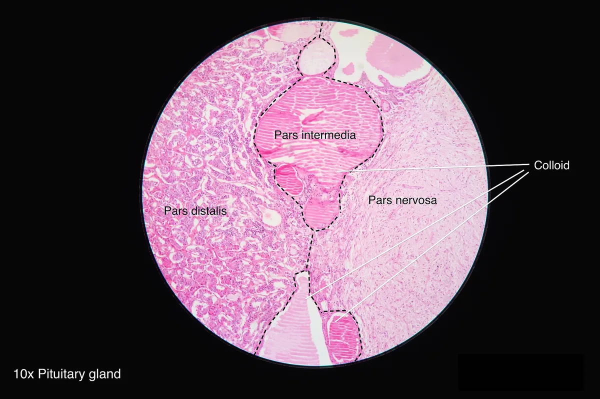
The pars intermedia is a thin band between the lobes, rudimentary in humans and often containing small colloid cysts (Rathke cleft cysts). The pars tuberalis wraps round the pituitary stalk like a collar and carries the portal vessels.
- 03
Neurohypophysis (posterior lobe)
Neurohypophysis; Pars nervosa
A downgrowth of the diencephalic floor made of axon terminals from the supraoptic and paraventricular nuclei and glial pituicytes. It stores and releases ADH (vasopressin) and oxytocin; on T1-weighted MRI it shows a characteristic “bright spot”.
- 04
Infundibulum and median eminence
Infundibulum; Eminentia mediana
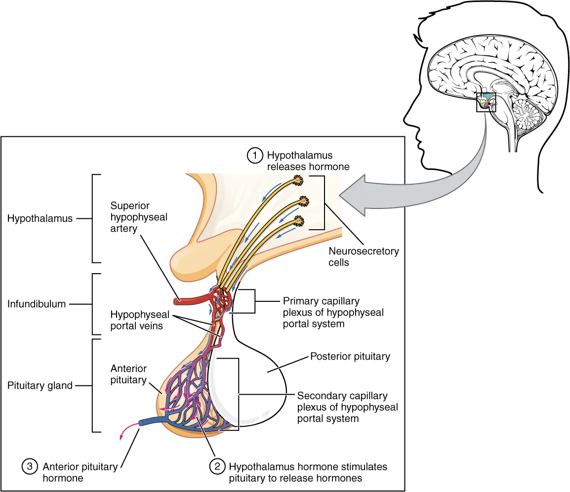
The stalk joining the pituitary to the hypothalamus and carrying the hypothalamo-hypophyseal tract. At the median eminence hypothalamic releasing hormones enter the primary capillary plexus of the portal system; stalk damage removes dopaminergic inhibition and raises prolactin (stalk effect).
- 05
Hypophyseal portal system
Systema portale hypophysiale
The superior hypophyseal arteries form a capillary plexus in the median eminence; long portal veins on the stalk carry blood to a second capillary plexus in the anterior lobe, delivering releasing hormones at high concentration. The anterior lobe has no direct arterial supply and can infarct after postpartum hypotension (Sheehan syndrome).
- 06
Sella turcica and relations
Sella turcica; Diaphragma sellae
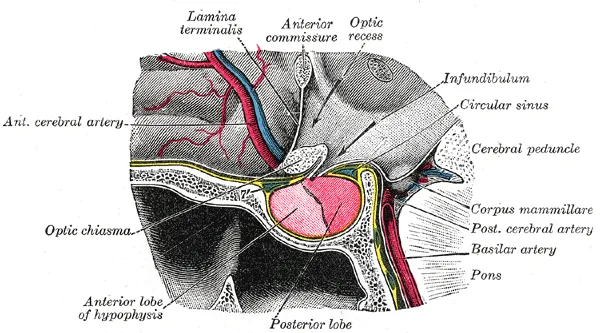
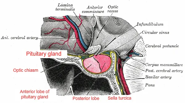
The gland weighs about 0.5 g and lies in the sella turcica of the sphenoid, roofed by the diaphragma sellae; above is the optic chiasm, on each side the cavernous sinus (nerves III, IV, V1, V2, VI and the internal carotid), below the sphenoid sinus. A large tumour compressing the chiasm causes bitemporal hemianopia; surgery is usually transsphenoidal.
Position and relations
The pituitary sits in the hypophysial fossa of the sella turcica, roofed by the diaphragma sellae (a dural fold) pierced by the stalk. Above lies the optic chiasm, so a pituitary tumour growing upwards typically causes bitemporal hemianopia; on each side lies the cavernous sinus containing the internal carotid artery and cranial nerves III, IV, V1, V2 and VI; below lies the sphenoid sinus, the route for transsphenoidal surgery.
Form and structure
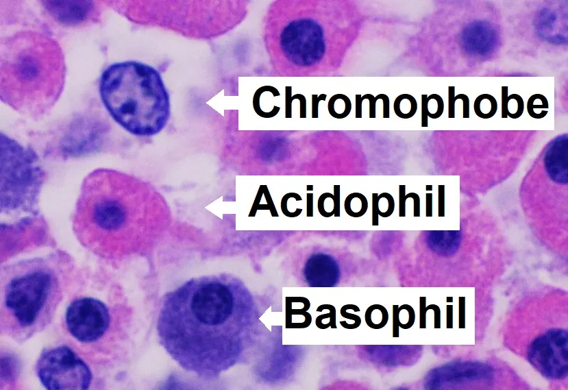
The anterior lobe arises from Rathke’s pouch (oral ectoderm) and comprises the pars distalis, pars intermedia and pars tuberalis; acidophils secrete GH and prolactin, basophils ACTH, TSH, FSH and LH. The posterior lobe is an extension of the hypothalamus, containing the axon terminals of the supraoptic and paraventricular nuclei, where ADH and oxytocin are released into the blood.
Vessels and nerves
The superior and inferior hypophysial arteries arise from the internal carotid. The superior arteries form a first capillary bed in the median eminence, where hypothalamic releasing hormones enter the blood; long portal veins run down the stalk to a second capillary bed in the anterior lobe. This hypophysial portal system lets the hypothalamus control the anterior lobe. The veins drain into the cavernous sinuses.
Development
The pituitary has a dual embryological origin: the anterior lobe from oral ectoderm, the posterior lobe from the diencephalon. Its terminology, intricate vasculature and position within the sella have been reinterpreted many times; understanding its blood supply helps explain pituitary injury after traumatic brain injury.[1]
The sellar region: size, boundaries and development
The pituitary grows rapidly from birth to adulthood, reaching about 500 mg, with an anteroposterior diameter of 8 mm, a transverse diameter of 12 mm and a height of 3–11 mm; it is larger in women and almost doubles in pregnancy as the pars distalis enlarges. It sits in the sella turcica, a hollow in the sphenoid bone: the tuberculum sellae forms the anterior wall and the dorsum sellae the posterior wall, whose margins form the posterior clinoid processes, while the anterolateral margins form the anterior clinoid processes that anchor dural folds. Above is the diaphragma sellae, anteroinferiorly the sphenoid sinus and laterally the cavernous sinuses; the optic chiasm lies anterosuperiorly, 1.5–8 mm from the tuberculum. The infundibulum, arising from the tuber cinereum and median eminence of the hypothalamus, connects the gland to the brain. The shape and pneumatisation of the sphenoid sinus (the sellar type is commonest) vary greatly and must be assessed before transsphenoidal surgery.[2]
The pituitary has a dual origin. Around week 4 a hypophysial placode in the oral ectoderm forms Rathke’s pouch, which grows up towards the neural ectoderm to become the anterior lobe; at the same time the floor of the diencephalon grows down to form the posterior lobe, which is therefore an extension of the central nervous system. Around weeks 6–8 the stalk of Rathke’s pouch constricts and closes, separating it from the oral epithelium. The process requires tight coordination of signals such as BMP4, FGF8, the Notch pathway and SOX2; mutations of early transcription factors such as Rpx (HESX1), Prop-1 and Pit-1 cause various forms of congenital hypopituitarism. The superior hypophysial artery (from the internal carotid or posterior communicating artery) forms the primary plexus in the median eminence, from which portal veins carry blood to the secondary plexus of the anterior lobe; the inferior hypophysial artery (from the meningohypophysial trunk) supplies the posterior lobe, and the intermediate lobe is fed by anastomoses between the two.[2]
Pituitary tumours and mass effects
Pituitary tumours are the commonest disease of the sellar region and are divided into microadenomas (under 10 mm) and macroadenomas (over 10 mm); any cell line can form an adenoma. Macroadenomas compress neighbouring structures: upwards into the optic chiasm causing bitemporal hemianopia, laterally into the cavernous sinus causing ophthalmoplegia and diplopia, or simply causing headache. Prolactinoma is the commonest functioning adenoma, often silent for a long time until it causes hormonal dysfunction, visual loss, hydrocephalus and hypogonadism. ACTH-secreting adenomas cause Cushing disease (proximal myopathy, psychiatric disturbance, central obesity, purple striae, a buffalo hump and hypertension); GH-secreting adenomas cause acromegaly or gigantism, with carpal tunnel syndrome and proximal myopathy. A mass compressing the stalk raises prolactin because hypothalamic dopamine can no longer reach the gland (stalk effect). Pituitary apoplexy is acute haemorrhagic infarction of a previously silent adenoma, with sudden severe headache and visual disturbance. The standard operative route is through the posterior wall of the sphenoid sinus (transsphenoidal adenectomy).[2]
Prolactinoma
- Hormone
- Prolactin
- Main features
- Hypogonadism, galactorrhoea, mass effect when large
Corticotroph adenoma
- Hormone
- ACTH
- Main features
- Cushing disease
Somatotroph adenoma
- Hormone
- GH
- Main features
- Acromegaly (adults), gigantism (children)
Non-functioning macroadenoma
- Hormone
- —
- Main features
- Bitemporal hemianopia, headache, stalk-effect hyperprolactinaemia
| Adenoma | Hormone | Main features |
|---|---|---|
| Prolactinoma | Prolactin | Hypogonadism, galactorrhoea, mass effect when large |
| Corticotroph adenoma | ACTH | Cushing disease |
| Somatotroph adenoma | GH | Acromegaly (adults), gigantism (children) |
| Non-functioning macroadenoma | — | Bitemporal hemianopia, headache, stalk-effect hyperprolactinaemia |
Full-text reading (StatPearls)
The StatPearls chapters on this topic, reproduced verbatim. Their CC BY-NC-ND 4.0 licence allows unmodified copies only, so they are not translated or shortened.
Anatomy, Head and Neck, Pituitary GlandGanapathy MK, Tadi P · StatPearls 2023 · NBK551529 · 21k characters
Loading…
Ganapathy MK, Tadi P. Anatomy, Head and Neck, Pituitary Gland. StatPearls [Internet]. Treasure Island (FL): StatPearls Publishing; 2023. NBK551529 · PMID 31855373 · CC BY-NC-ND 4.0. Reproduced verbatim, unmodified.
Organs in this system
References
Sources for the values and descriptions on this page, indexed in PubMed (US National Library of Medicine).
- [1]Anatomy of the pituitary glandSav A et al., Best practice & research. Clinical endocrinology & metabolism 2025PMID 40500594
- [2]Anatomy, Head and Neck, Pituitary GlandGanapathy MK et al., StatPearls 2023PMID 31855373
- [3]Physiology, Pituitary HormonesStatPearls (NCBI Bookshelf)PMID 32491488Supports: Pituitary and endocrine axes
- [4]Physiology, Anterior PituitaryStatPearls (NCBI Bookshelf)PMID 29763073