Deltoid
Model
Layers
Z-Anatomy anatomical model based on BodyParts3D, licensed CC BY-SA 4.0.
Description
A triangular muscle covering the shoulder joint and forming its rounded contour. Its three parts (clavicular, acromial, spinal) flex, abduct and extend the arm.
Parts
Each part has its own card; press “Show on the model” to fly to it and select it in the 3D model above.
- 01
Clavicular (anterior) part
Pars clavicularis musculi deltoidei
Arises from the lateral third of the anterior border of the clavicle. It flexes and medially rotates the arm, working with pectoralis major.
- 02
Acromial (middle) part
Pars acromialis musculi deltoidei
Arises from the lateral border of the acromion and is multipennate and powerful. It abducts the arm from about 15° to 90° after supraspinatus has started the movement; beyond 90° the scapula must rotate upwards through trapezius and serratus anterior.
- 03
Spinal (posterior) part
Pars spinalis musculi deltoidei
Arises from the lower lip of the spine of the scapula. It extends and laterally rotates the arm, opposing the clavicular part.
- 04
Insertion: deltoid tuberosity
Tuberositas deltoidea
The three parts converge on a tendon attached to the deltoid tuberosity midway down the lateral humerus. The muscle wraps the shoulder and gives its rounded contour; in dislocation the head leaves its place beneath the muscle and the shoulder looks “squared off”. Its middle part is a common intramuscular injection site, 3–5 cm below the acromion to avoid the axillary nerve.
- 05
Nerve and blood supply
Innervatio et vascularisatio
The axillary nerve (C5–C6) passes through the quadrangular space with the posterior circumflex humeral artery, winds around the surgical neck of the humerus and enters the deep surface of the muscle. Injury (surgical neck fracture, shoulder dislocation) weakens abduction and numbs the outer shoulder.
Position and relations
It arises from the lateral third of the clavicle, the acromion and the spine of the scapula (mirroring trapezius) and converges onto the deltoid tuberosity on the lateral humeral shaft. The subacromial–subdeltoid bursa separates it from the humeral head and rotator cuff.
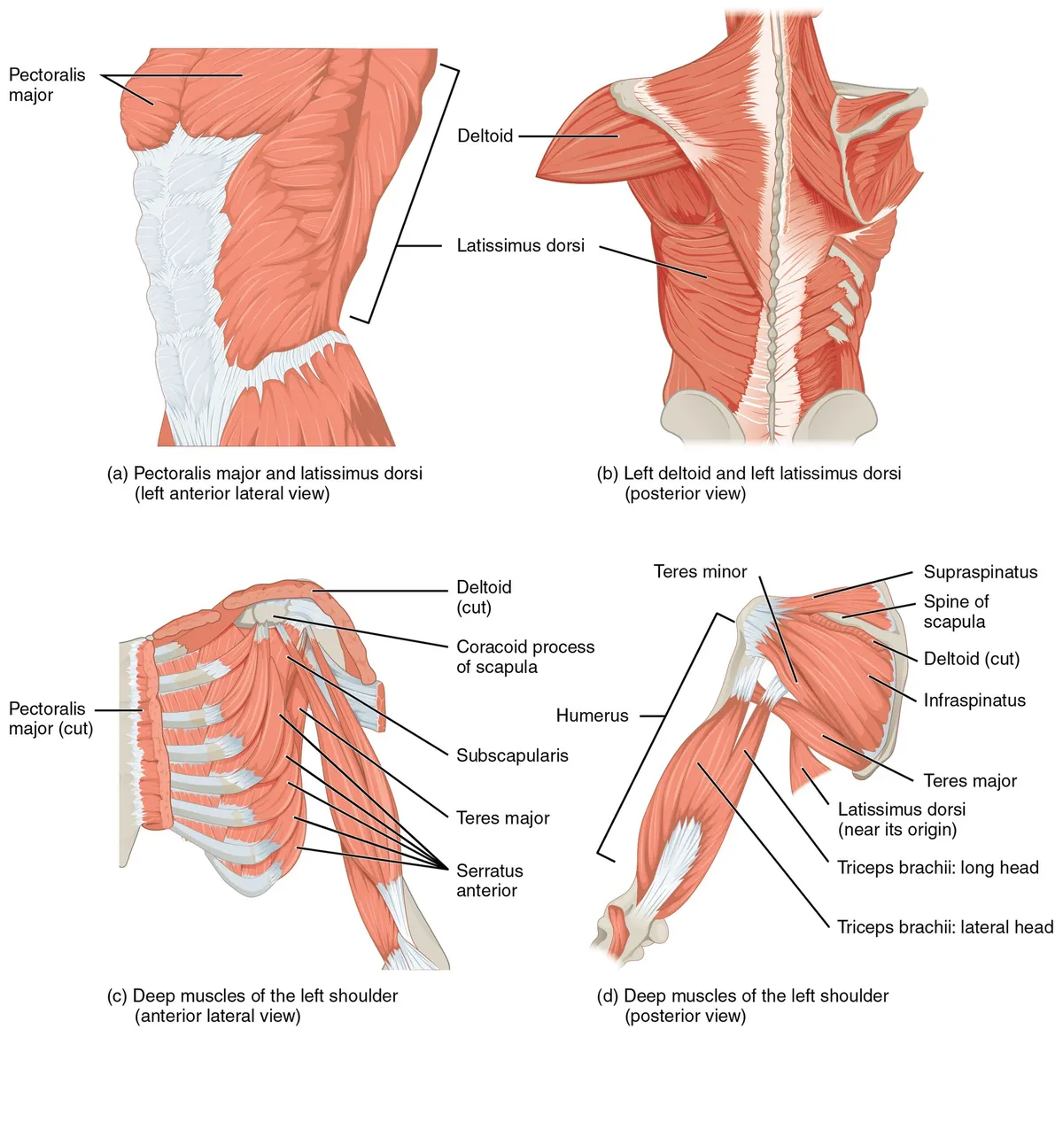
Form and structure
The acromial part is multipennate and powerful, the main abductor from about 15° to 90° after supraspinatus initiates the movement; the anterior part flexes and medially rotates, the posterior part extends and laterally rotates the arm. The deltoid is a common intramuscular injection site, using its upper part to avoid the axillary nerve.
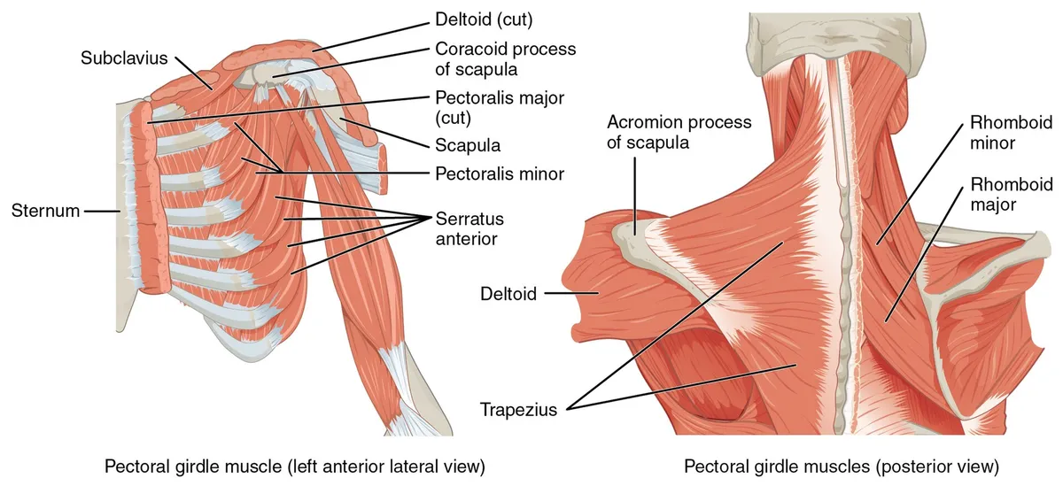
Vessels and nerves
It is supplied by the axillary nerve (C5–C6), which travels with the posterior circumflex humeral artery through the quadrangular space and winds around the surgical neck. Surgical-neck fractures or shoulder dislocation can injure it: the deltoid wastes, abduction is lost and the “regimental badge” skin over the lateral shoulder goes numb.
The deltoid in shoulder motion and stability
The deltoid is a large triangular intrinsic shoulder muscle, named for its resemblance to an inverted Greek delta, that forms the rounded contour of the shoulder, being thick and spread anteroposteriorly. It is a common site for intramuscular injections, including vaccines. It is key to shoulder motion and stability, and after rotator cuff injury it may have to compensate for the weakness. Deltoid flaps can repair shoulder defects after breast cancer treatment.[1]
Glenohumeral stability depends on static stabilisers (bony anatomy, negative intra-articular pressure, the labrum, the glenohumeral ligaments and capsule) and dynamic stabilisers (the rotator cuff and the other muscles around the shoulder, including the deltoid). Together they support the joint’s many degrees of motion, and failure of some mechanisms leads to shoulder pathology.[2]
Three parts, abduction mechanics and neurovascular supply
The deltoid is a large, triangular intrinsic shoulder muscle, named after the inverted Greek letter delta, that forms the rounded contour of the shoulder. Its U-shaped origin from the scapular spine, the upper surface of the acromion and the lateral third of the clavicle mirrors the insertion of trapezius; it inserts on the deltoid tuberosity of the humerus. The anterior and posterior parts are unipennate and the middle (acromial) part multipennate. The deltoid cannot initiate abduction because it pulls parallel to the humeral shaft: supraspinatus and the rotator cuff abduct from 0° to 15°, after which the middle part raises the arm to about 100° while the anterior and posterior parts steady it. In walking, the anterior part flexes the arm with pectoralis major and the posterior part extends it with latissimus dorsi. The deltoid also prevents inferior subluxation of the glenohumeral joint when the arm hangs adducted under a heavy load (as in a deadlift), and a 2018 study found it can compensate for abduction weakness from rotator cuff tears by as much as 108.1% of normal. Developmentally, like all striated trunk and limb muscles, it comes from paraxial mesoderm: in week 5 myoblasts from the somites migrate into the limb buds, and the deltoid develops from the dorsal muscle mass.[1]
Blood comes mainly from the thoracoacromial branch of the axillary artery (from its second part, behind pectoralis minor), which runs with the cephalic vein in the deltopectoral groove, with smaller contributions from the posterior circumflex humeral and profunda brachii arteries. Terminal branches of the posterior circumflex artery cross the space between the deltoid and the proximal humerus and are prone to bleeding or haematoma during anterior approaches to the shoulder. Lymph drains to the deltopectoral nodes beside the cephalic vein. The muscle is supplied by the anterior and posterior branches of the axillary nerve (C5–C6, a terminal branch of the posterior cord of the brachial plexus), which passes with the posterior circumflex artery through the quadrangular space and winds around the surgical neck of the humerus. It can be tested by raising the patient’s arm past 15° or by resisted abduction while watching the middle part contract. Axillary nerve palsy is commonly caused by excessive crutch use, surgery or shoulder dislocation from severe trauma. Rare variants include a posterior part with its own fascial sheath or completely separate and resembling teres minor, aberrant straps running perpendicular to the posterior fibres, and an abnormal insertion on the medial epicondyle, in which the fibres pass superficial to the brachial artery and the ulnar and median nerves and raise the risk of iatrogenic neurovascular injury. The thoracoacromial artery may also tunnel into the deltoid (type I) or run with the cephalic vein and cross back to pectoralis major (type II).[1]
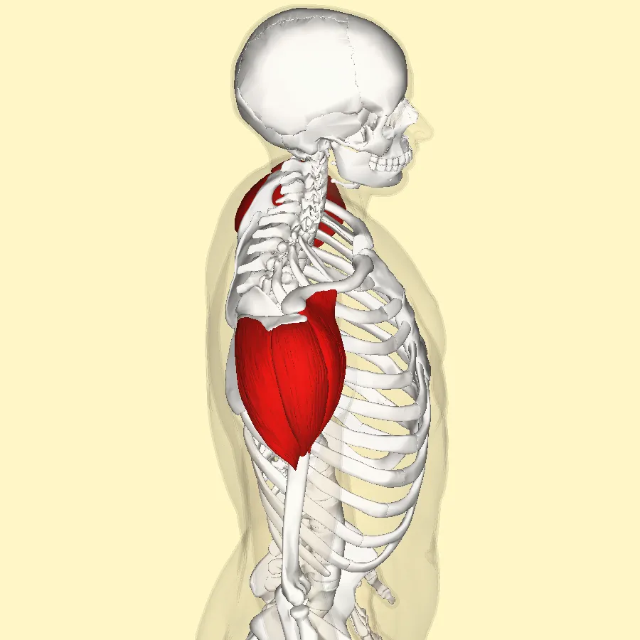
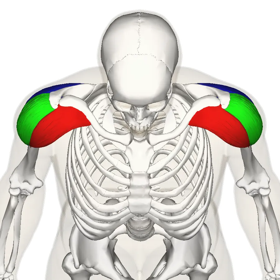
Anterior (clavicular)
- Origin
- Lateral third of clavicle
- Architecture
- Unipennate
- Main action
- Flexion and medial rotation; steadies abduction
Middle (acromial)
- Origin
- Upper surface of acromion
- Architecture
- Multipennate
- Main action
- Abduction from 15° to about 100°
Posterior (spinal)
- Origin
- Scapular spine
- Architecture
- Unipennate
- Main action
- Extension and lateral rotation; steadies abduction
| Part | Origin | Architecture | Main action |
|---|---|---|---|
| Anterior (clavicular) | Lateral third of clavicle | Unipennate | Flexion and medial rotation; steadies abduction |
| Middle (acromial) | Upper surface of acromion | Multipennate | Abduction from 15° to about 100° |
| Posterior (spinal) | Scapular spine | Unipennate | Extension and lateral rotation; steadies abduction |
Shoulder approaches, complications and differential diagnosis
Many shoulder operations pass through or retract the deltoid: open Bankart capsular reconstruction for recurrent anterior instability; shoulder arthroplasty (hemiarthroplasty, total and reverse total) for post-traumatic deformity, advanced degenerative arthritis and avascular necrosis; repair, tenotomy or tenodesis of the long head of biceps; and rotator cuff repair, now mostly arthroscopic or by a mini-open lateral deltoid-splitting approach. In the deltopectoral approach, the coracoid process is marked on the skin to orient the incision along the deltopectoral groove, the “fat stripe” containing the cephalic vein is found between the two muscles, and the deltoid is retracted laterally and pectoralis major medially. When this approach is impractical, an anterolateral approach (landmarks coracoid and acromion, splitting the deltoid for cuff repair and exposing the posterior shoulder as well) or a direct lateral approach can be used: a 5 cm longitudinal incision from the acromion and a 5 cm deltoid split, with a stay suture at the apex to protect the axillary nerve. The lateral minimally invasive technique uses one proximal and one distal deltoid incision, with the axillary nerve running between them.[1]
Complications of deltoid manipulation include detachment from the clavicle, which needs full-thickness transosseous sutures and 4–6 weeks to heal completely; axillary nerve injury from anterior retraction or incision, since the nerve runs beneath the muscle from posterior to anterior, weakening abduction beyond 15° and numbing the skin over the deltoid; and rupture of the cephalic vein with upper-limb oedema. The deltoid is also used as a flap to cover shoulder defects after breast cancer treatment, and posterior-part variants or aberrant straps can cause confusion when raising the flap. Inability to abduct the arm does not necessarily mean deltoid disease, because it also occurs in systemic and proximal neuromuscular disorders: Lambert–Eaton myasthenic syndrome, polymyositis and dermatomyositis, polymyalgia rheumatica (usually stiffness rather than weakness), side effects of aluminium-hydroxide-containing vaccines, and cachexia from chronic disease or malnutrition. Careful examination separates these causes. The deltoid is also a common intramuscular injection site, including for vaccines, using its upper part to avoid the axillary nerve.[1]
Full-text reading (StatPearls)
The StatPearls chapters on this topic, reproduced verbatim. Their CC BY-NC-ND 4.0 licence allows unmodified copies only, so they are not translated or shortened.
Anatomy, Shoulder and Upper Limb, Deltoid MuscleElzanie A, Varacallo MA · StatPearls 2024 · NBK537056 · 11k characters
Loading…
Elzanie A, Varacallo MA. Anatomy, Shoulder and Upper Limb, Deltoid Muscle. StatPearls [Internet]. Treasure Island (FL): StatPearls Publishing; 2024. NBK537056 · PMID 30725741 · CC BY-NC-ND 4.0. Reproduced verbatim, unmodified.
Organs in this system
References
Sources for the values and descriptions on this page, indexed in PubMed (US National Library of Medicine).
- [1]Anatomy, Shoulder and Upper Limb, Deltoid MuscleElzanie A et al., StatPearls 2024PMID 30725741
- [2]Shoulder biomechanicsLugo R et al., European journal of radiology 2008PMID 18511227
- [3]Anatomy, Bony Pelvis and Lower Limb, Gluteus Maximus MuscleStatPearls (NCBI Bookshelf)PMID 30855781Supports: Gluteus maximus