Parathyroid glands
Model
Layers
Z-Anatomy anatomical model based on BodyParts3D, licensed CC BY-SA 4.0.
Description
Usually four small glands (each about 30–50 mg) on the back of the thyroid that secrete parathyroid hormone (PTH), which regulates blood calcium and phosphate.
Parts
Each part has its own card; press “Show on the model” to fly to it and select it in the 3D model above.
- 01
Superior parathyroid glands
Glandulae parathyroideae superiores
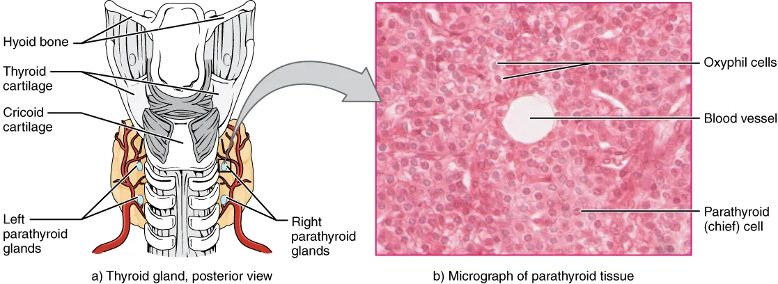
Usually on the back of the thyroid lobe at the level of the cricoid, posterior to the recurrent laryngeal nerve, in a fairly constant position. They arise from the fourth pharyngeal pouch and migrate little, so are seldom ectopic.
- 02
Inferior parathyroid glands
Glandulae parathyroideae inferiores
Arising from the third pharyngeal pouch with the thymus, they migrate further and vary more: usually near the lower pole of the thyroid but possibly in the thyrothymic ligament, within the thymus or in the mediastinum. This is why a parathyroid adenoma sometimes has to be sought in the chest.
- 03
Chief cells
Endocrinocyti principales
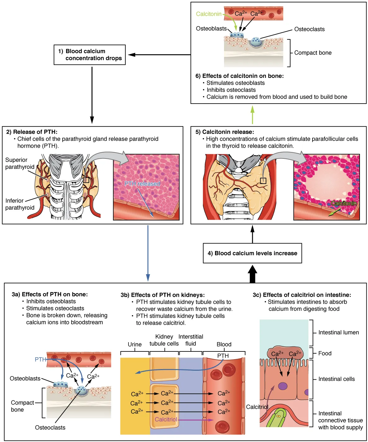
The predominant cells, which sense ionised calcium through the calcium-sensing receptor and secrete parathyroid hormone (PTH) when it falls. PTH increases bone resorption and renal calcium reabsorption and activates vitamin D; a single-gland adenoma is the main cause of primary hyperparathyroidism.
- 04
Oxyphil cells
Cellulae oxyphilae
Larger cells with eosinophilic, mitochondria-rich cytoplasm that appear at puberty and increase with age; their function is unclear. The stroma also gains fat cells with age; each gland weighs only about 30–50 mg.
Position and relations
The superior glands (from the fourth pharyngeal pouch) are fairly constant on the back of the thyroid lobe at the level of the cricoid. The inferior glands (from the third pouch, like the thymus) migrate further in the embryo and vary widely: at the lower thyroid pole, in the thyrothymic ligament or in the mediastinum, which matters when searching for an ectopic gland.
Form and structure
The parenchyma consists of chief cells that secrete PTH and oxyphil cells that increase with age, interspersed with fat. Chief cells carry the calcium-sensing receptor (CaSR): when ionised calcium falls, PTH rises, mobilising calcium from bone, increasing renal calcium reabsorption and phosphate excretion and activating vitamin D in the kidney.
Vessels and nerves
All four glands are usually supplied by the inferior thyroid artery (thyrocervical trunk), the superior ones sometimes also by the superior thyroid artery. Because their vessels are tiny, thyroidectomy can devascularise them and cause postoperative hypocalcaemia.
Position, ectopia and calcium control
There are normally four parathyroids, although supernumerary glands are found in up to 13% of autopsies. The superior glands derive from the fourth pharyngeal pouch; they lie on the posterolateral aspect of the upper thyroid pole, about 1 cm above the crossing of the recurrent laryngeal nerve and inferior thyroid artery, and deep to the nerve. The inferior glands derive from the third pouch; they lie near the lower pole within 1–2 cm of the inferior thyroid artery’s entry, superficial to the nerve, and are far more variable in position.[1]
A meta-analysis found up to 16% of glands in ectopic sites: mediastinum, retro-oesophageal space, lateral neck, within the thyroid or thymus, even along the aortic arch. Imaging therefore precedes parathyroidectomy, and persistent hypercalcaemia with raised PTH after surgery points to residual, ectopic or supernumerary tissue; the clinical picture mirrors hypercalcaemia: kidney stones, osteoporosis, confusion, constipation and weakness.[1][2]
Extracellular calcium binds the calcium-sensing receptor (CaSR) on parathyroid cells and lowers PTH release; hypocalcaemia does the opposite. PTH rapidly raises renal calcium reabsorption and, over hours to days, increases osteoclastic bone resorption and renal conversion of 25(OH)D to 1,25(OH)₂D, which boosts intestinal calcium absorption. FGF23 from bone joins the loop by lowering 1,25(OH)₂D and possibly PTH.[3]
Hypoparathyroidism is rare and shows hypocalcaemia with low or undetectable PTH, plus hyperphosphataemia and low 1,25(OH)₂D. Removal of or damage to the glands during anterior neck surgery is by far the commonest cause, which is why thyroid surgery requires identifying and preserving both the parathyroids and the laryngeal nerves.[4][5]
Full-text reading (StatPearls)
The StatPearls chapters on this topic, reproduced verbatim. Their CC BY-NC-ND 4.0 licence allows unmodified copies only, so they are not translated or shortened.
Histology, Parathyroid GlandBrown MB, Limaiem F · StatPearls 2023 · NBK546596 · 17k characters
Loading…
Brown MB, Limaiem F. Histology, Parathyroid Gland. StatPearls [Internet]. Treasure Island (FL): StatPearls Publishing; 2023. NBK546596 · PMID 31536203 · CC BY-NC-ND 4.0. Reproduced verbatim, unmodified.
Anatomy, Head and Neck, ParathyroidIlahi A, Muco E, Ilahi TB · StatPearls 2023 · NBK537203 · 12k characters
Loading…
Ilahi A, Muco E, Ilahi TB. Anatomy, Head and Neck, Parathyroid. StatPearls [Internet]. Treasure Island (FL): StatPearls Publishing; 2023. NBK537203 · PMID 30725888 · CC BY-NC-ND 4.0. Reproduced verbatim, unmodified.
Anatomy, Head and Neck, Parathyroid, Ectopic GlandsGeorgakopoulos B, Al Khalili Y · StatPearls 2023 · NBK540972 · 7k characters
Loading…
Georgakopoulos B, Al Khalili Y. Anatomy, Head and Neck, Parathyroid, Ectopic Glands. StatPearls [Internet]. Treasure Island (FL): StatPearls Publishing; 2023. NBK540972 · PMID 31082016 · CC BY-NC-ND 4.0. Reproduced verbatim, unmodified.
Organs in this system
References
Sources for the values and descriptions on this page, indexed in PubMed (US National Library of Medicine).
- [1]Anatomy, Head and Neck, ParathyroidIlahi A et al., StatPearls 2023PMID 30725888
- [2]Anatomy, Head and Neck, Parathyroid, Ectopic GlandsGeorgakopoulos B et al., StatPearls 2023PMID 31082016
- [3]Physiology of the Calcium-Parathyroid Hormone-Vitamin D AxisGoltzman D et al., Frontiers of hormone research 2018PMID 29597231
- [4]HypoparathyroidismBilezikian JP, The Journal of clinical endocrinology and metabolism 2020PMID 32322899
- [5]Anatomy of thyroid and parathyroid glands and neurovascular relationsMohebati A et al., Clinical anatomy 2012PMID 21800365
- [6]Physiology, Pituitary HormonesStatPearls (NCBI Bookshelf)PMID 32491488Supports: Pituitary and endocrine axes
- [7]Physiology, Anterior PituitaryStatPearls (NCBI Bookshelf)PMID 29763073