Epithelial tissue
Model
Hover or tap to see names, click to read a description; click a name in the legend to fly to it. Procedurally built to histological proportions; colours follow illustration conventions.
Description
Sheets of closely packed, polarised cells resting on a basement membrane and lacking blood vessels. Covering epithelia are named by the number of layers (simple, stratified, pseudostratified) and the shape of the surface cells (squamous, cuboidal, columnar); glandular epithelium forms exocrine and endocrine glands.
Classifying epithelia
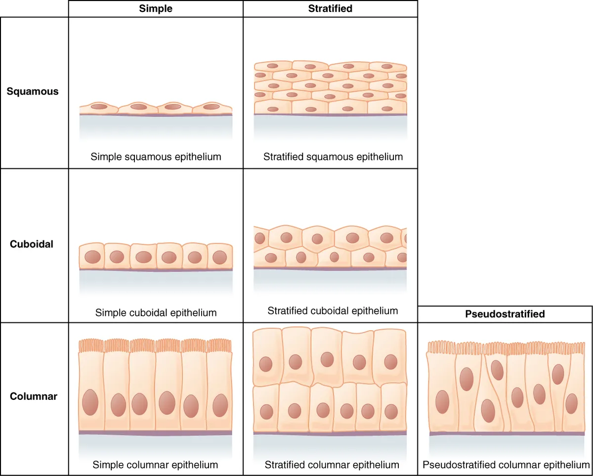
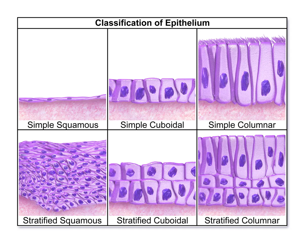
Epithelia are sheets of closely apposed cells with almost no intercellular matrix that cover the body surface, line cavities and ducts, and form the parenchyma of glands. Epithelium is avascular: its cells receive oxygen and nutrients by diffusion from capillaries in the underlying connective tissue across the basement membrane. Every epithelium is polarised: an apical surface faces the lumen or the environment, lateral surfaces adhere to neighbours and a basal surface anchors to the basement membrane, each domain carrying its own membrane proteins and organelles.[1][2]
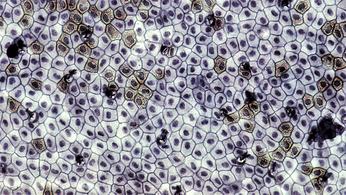
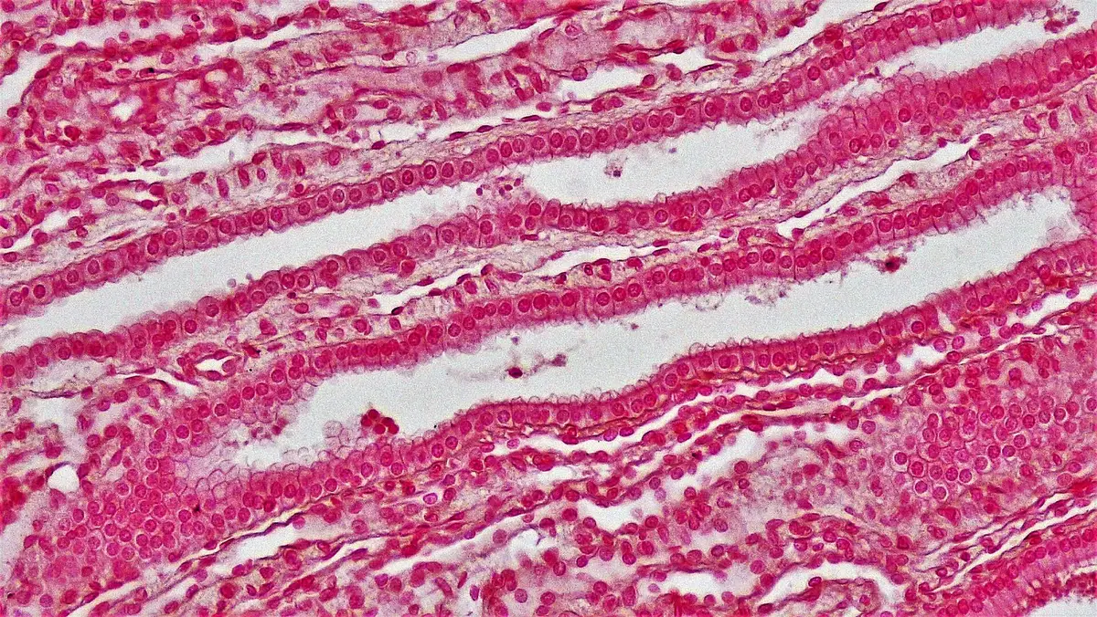
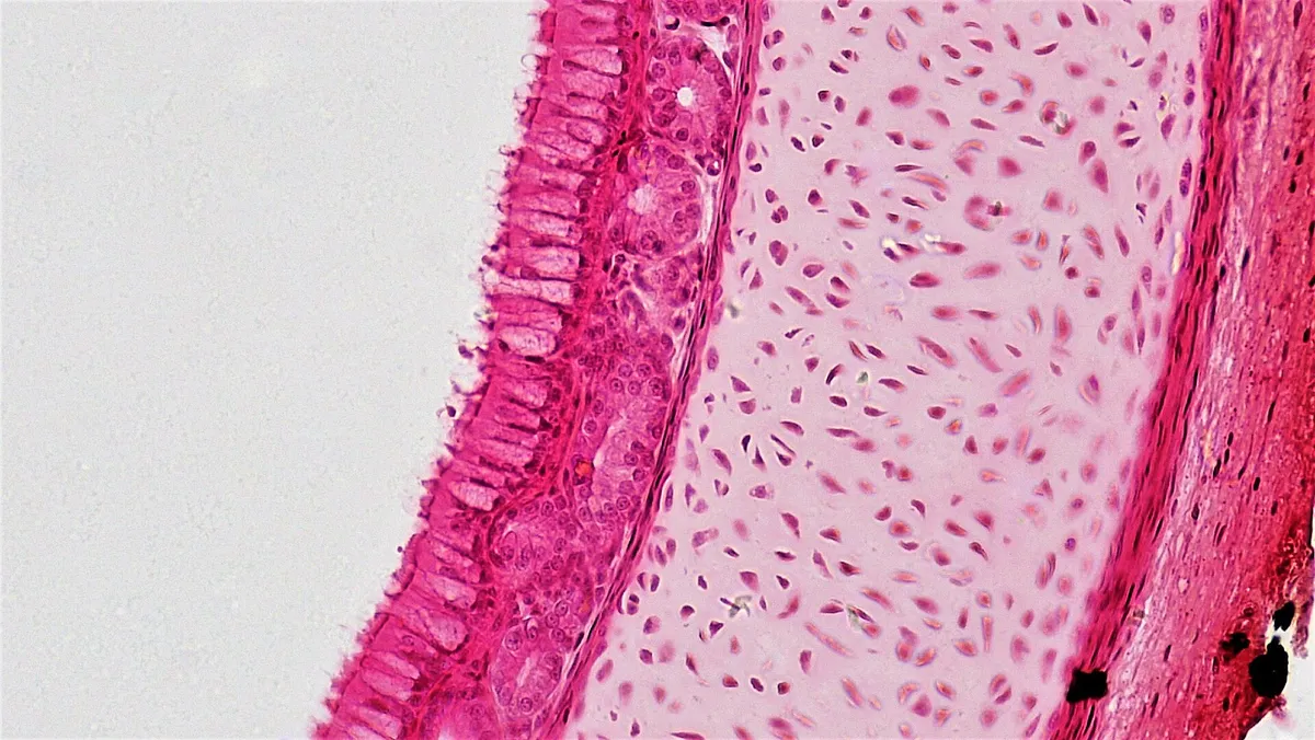
Covering epithelia are named by two criteria: the number of cell layers (simple, stratified, or pseudostratified when every cell touches the basement membrane but the nuclei lie at several levels) and the shape of the surface cells (squamous, cuboidal, columnar). Simple squamous epithelium is thin enough for gases and fluid to cross easily, so it lines alveoli, capillaries and serous cavities; simple cuboidal and columnar epithelia are taller and packed with organelles for absorption and secretion. Pseudostratified ciliated columnar epithelium with goblet cells is the respiratory epithelium; transitional epithelium (urothelium) lines the urinary tract and changes thickness as the bladder fills.[3][1]
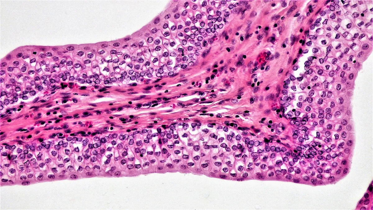
Simple squamous
- Typical sites
- Alveoli, vascular endothelium, pleural and peritoneal mesothelium
- Main function
- Gas exchange, filtration, gliding
Simple cuboidal
- Typical sites
- Kidney tubules, gland ducts, thyroid follicles
- Main function
- Reabsorption, secretion
Simple columnar
- Typical sites
- Stomach, intestine, gallbladder
- Main function
- Absorption, mucus secretion
Pseudostratified ciliated columnar
- Typical sites
- Trachea, bronchi, nasal cavity
- Main function
- Mucociliary clearance
Keratinised stratified squamous
- Typical sites
- Epidermis
- Main function
- Barrier to water loss and abrasion
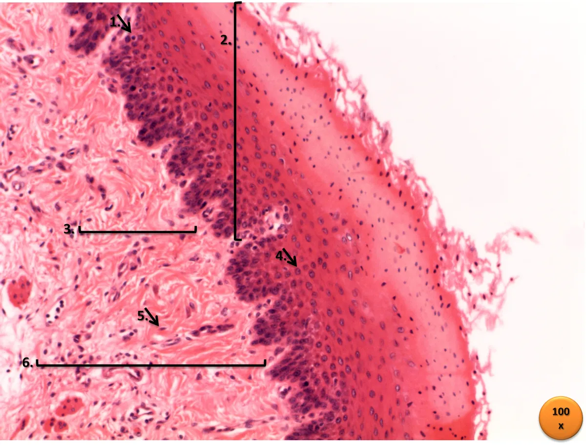
Non-keratinised stratified squamous
- Typical sites
- Mouth, oesophagus, vagina, cornea
- Main function
- Resists friction on moist surfaces
Transitional (urothelium)
- Typical sites
- Renal pelvis, ureter, bladder
- Main function
- Distensible, urine-tight
| Type | Typical sites | Main function |
|---|---|---|
| Simple squamous | Alveoli, vascular endothelium, pleural and peritoneal mesothelium | Gas exchange, filtration, gliding |
| Simple cuboidal | Kidney tubules, gland ducts, thyroid follicles | Reabsorption, secretion |
| Simple columnar | Stomach, intestine, gallbladder | Absorption, mucus secretion |
| Pseudostratified ciliated columnar | Trachea, bronchi, nasal cavity | Mucociliary clearance |
| Keratinised stratified squamous | Epidermis | Barrier to water loss and abrasion |
| Non-keratinised stratified squamous | Mouth, oesophagus, vagina, cornea | Resists friction on moist surfaces |
| Transitional (urothelium) | Renal pelvis, ureter, bladder | Distensible, urine-tight |
Apical specialisations
Microvilli are cytoplasmic projections about 1 µm long whose core of a few dozen parallel actin filaments, bundled by villin and fimbrin, inserts into the terminal web at the cell apex. In the small intestine and proximal tubule thousands of regularly packed microvilli form the brush border, enlarging the absorptive surface some twentyfold and carrying membrane-bound digestive enzymes. The stereocilia of the epididymis and of inner-ear hair cells are also actin-based projections, longer and non-motile.[1][4]
Cilia are 5–10 µm long and contain a 9 + 2 microtubule axoneme growing from a basal body. In the airways each ciliated cell carries some 200–300 cilia beating in coordinated waves, driving the mucus secreted by goblet cells and submucosal glands towards the pharynx at a few millimetres per minute. Mucus is a gel of the mucins MUC5AC and MUC5B that traps dust and bacteria; when the mucins become too viscous, as in cystic fibrosis or asthma, or the cilia are immotile, mucociliary clearance fails and infections recur.[3][5][6]
Junctional complexes
At the very top of the lateral surface, tight junctions (zonula occludens) are strands of the transmembrane proteins claudin and occludin that seal adjacent membranes and link to the cytoskeleton through ZO-1. They close the paracellular route and form selective gates: each claudin type defines a pore for ions of a given size and charge, which is why the proximal tubule is leaky to water while the collecting duct is very tight. Tight junctions also act as a fence that keeps apical and basolateral membrane proteins from mixing, maintaining polarity.[7][8][9]
Just below lies the adherens junction (zonula adherens), where E-cadherins of neighbouring cells pair in a Ca²⁺-dependent manner and connect through catenins to a perijunctional actomyosin belt; tension in this belt spreads across the sheet, letting tissues fold during development and close wounds. Desmosomes are disc-shaped spot welds built from the cadherins desmoglein and desmocollin, anchored to keratin intermediate filaments by desmoplakin, which let epithelia withstand stretch. Antibodies against desmoglein cause pemphigus, with blisters inside the epidermis. Gap junctions are connexin channels that pass ions and small molecules between cells; on the basal side hemidesmosomes fix the cell to the basement membrane through integrins.[10][11][12][13]
Freeze-fracture electron microscopy shows the tight junction as a network of protein strands encircling the cell apex; each strand is a claudin polymer arranged in two parallel rows that pair with the strand of the neighbouring cell. “Barrier-forming” claudins seal the gap, whereas “channel-forming” claudins assemble selective pores: claudin-2 passes only small cations (Na⁺, Ca²⁺) and water, with a size limit of about 0.57 nm. Beside this pore pathway lies a leak pathway for larger molecules, regulated by the actomyosin ring and myosin light-chain kinase (MLCK); where three cells meet, tricellulin and angulins seal the barrier’s weakest point. In intestinal inflammation, cytokines such as IL-1β activate NF-κB and MLCK, opening tight junctions and raising gut permeability, a vicious circle in Crohn disease.[14][15]
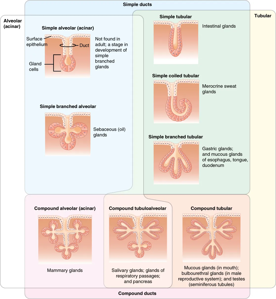
Glandular epithelium
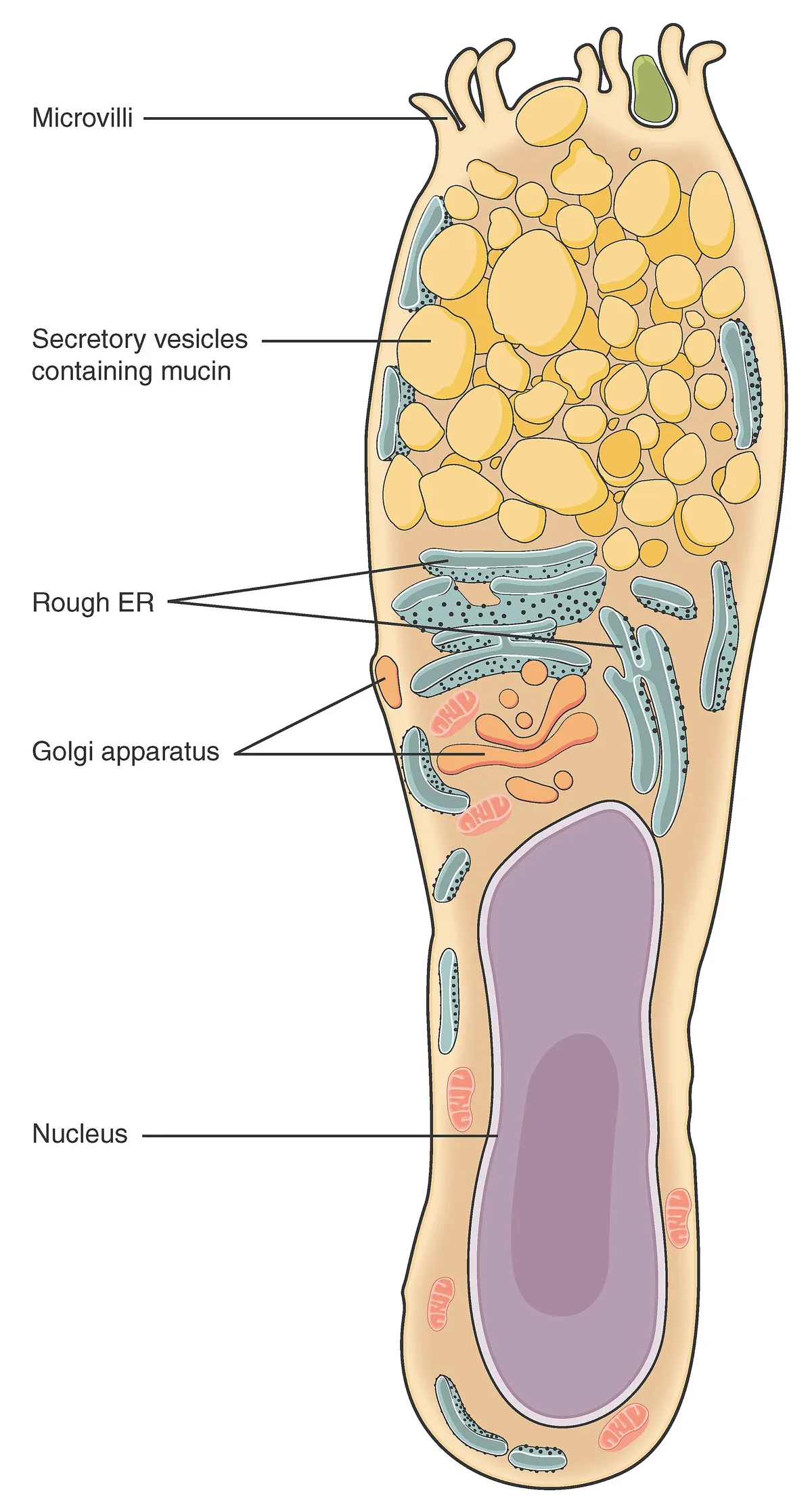
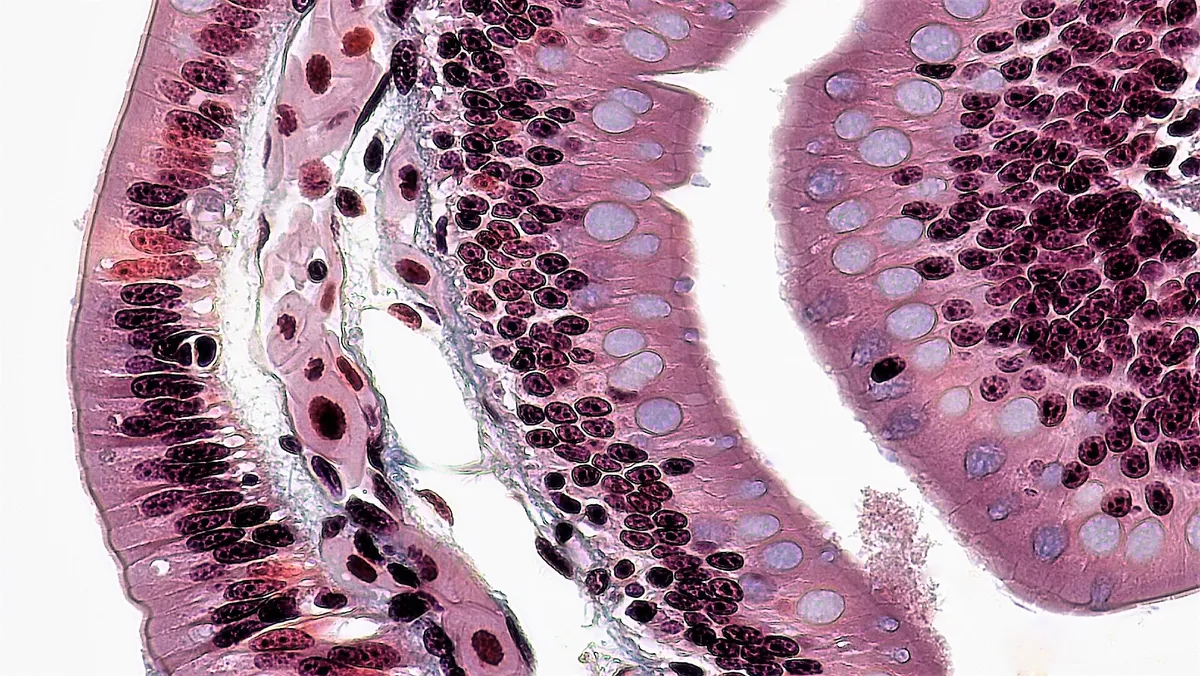
Glands form when covering epithelium invaginates into the underlying connective tissue. Exocrine glands keep a duct opening onto a surface; endocrine glands lose the duct and release hormones directly into fenestrated capillaries. Exocrine glands are classified by duct shape (simple, branched, compound) and secretory unit (tubular, acinar, tubuloacinar); goblet cells are unicellular glands scattered through intestinal and airway epithelium. By product, serous cells secrete protein-rich fluid and hold dark zymogen granules at the apex, whereas mucous cells contain pale mucin droplets that push a flattened nucleus to the base; the submandibular and sublingual salivary glands are mixed.[16][6][1]
By release mechanism, most glands are merocrine: secretory vesicles fuse with the apical membrane without loss of cytoplasm, as in the exocrine pancreas and eccrine sweat glands. Sebaceous glands are holocrine: the whole lipid-filled cell dies and becomes the secretion. The mammary gland releases milk fat droplets wrapped in a piece of apical membrane (apocrine secretion). Myoepithelial cells around the acini of salivary, mammary and sweat glands contract to squeeze the secretion into the ducts.[17][18][16]
Stratified epithelia and self-renewal
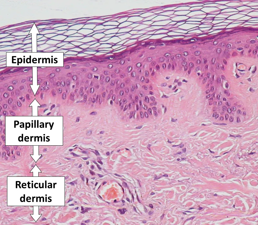
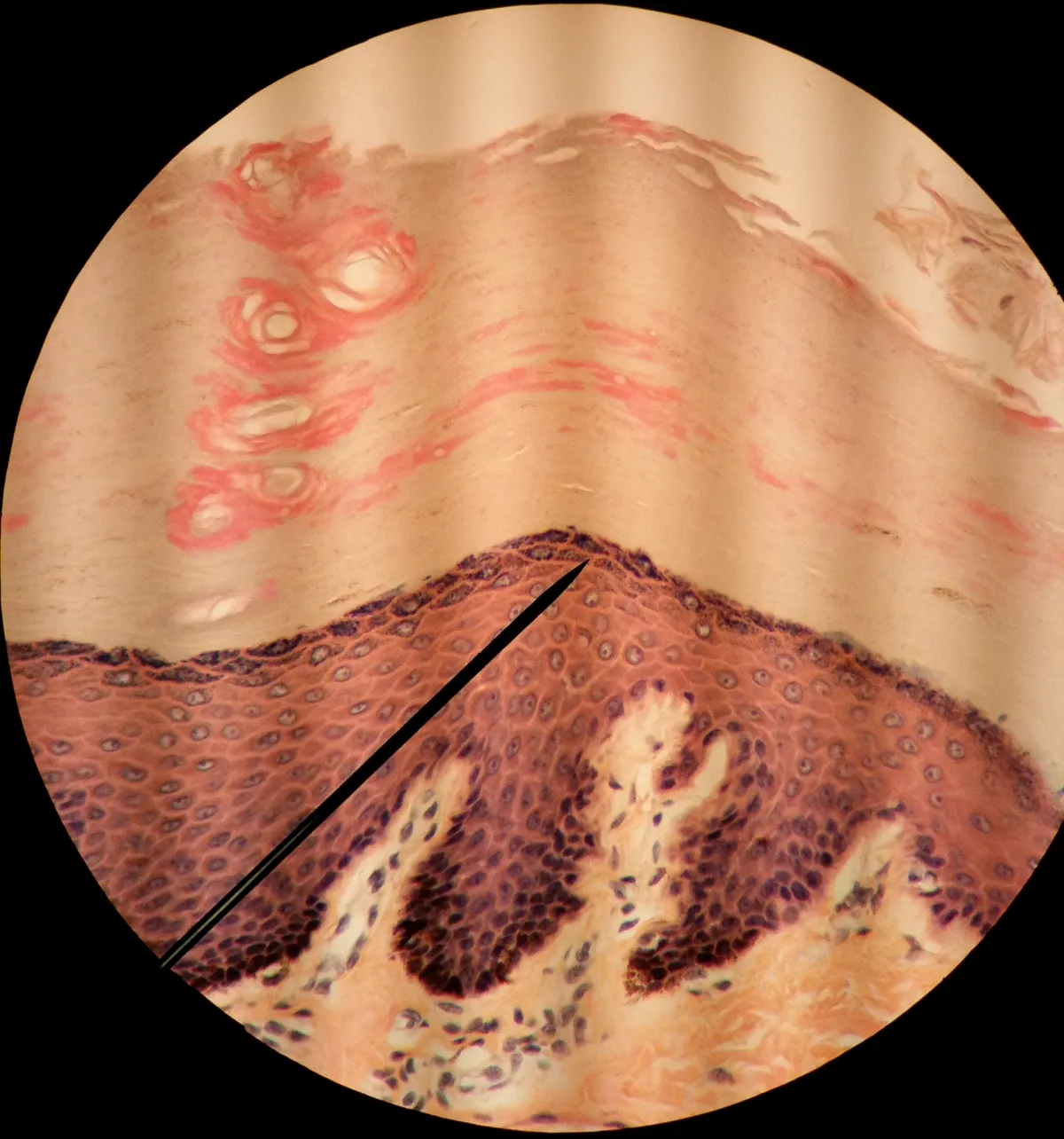
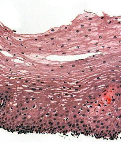
The epidermis is the archetypal keratinised stratified squamous epithelium. Stem cells in the basal layer divide; daughter cells move up through the spinous layer, where countless desmosomes join them, and the granular layer, with keratohyalin granules containing filaggrin and lamellar bodies that release lipids into the intercellular space. In the stratum corneum the cells lose nucleus and organelles, becoming flat corneocytes with a cross-linked protein envelope held together by lipid mortar; this “bricks and mortar” structure is the barrier to water loss. The whole journey takes about four weeks; loss-of-function filaggrin mutations weaken the barrier and predispose to atopic eczema.[19][2][20]
Melanocytes sit among keratinocytes, each supplying melanin through dendrites to about 36 keratinocytes; melanin gathers into a cap over the nucleus to shield it from ultraviolet light, and skin colour differs by the amount and type of melanin, not by melanocyte number. Intestinal epithelium renews fastest of all: stem cells at the base of the crypts of Lieberkühn produce cells that migrate to the villus tip and are shed after about 3–5 days. Epithelia have their own damage-control mechanisms: injured cells are extruded from the sheet and neighbours draw together to close the gap without breaching the barrier.[21][22][6]
Basement membrane and endothelium
The basement membrane is a specialised matrix layer 50–100 nm thick secreted by the epithelium itself, made of a collagen IV network, laminin, nidogen and the proteoglycan perlecan. It anchors the epithelium, sets cell polarity, scaffolds regeneration and acts as a selective filter. In the glomerulus an especially thick basement membrane lies between fenestrated endothelium and podocyte foot processes, together forming a filter that retains albumin; autoantibodies against collagen IV cause anti-glomerular basement membrane disease and mutations in collagen IV chains cause Alport syndrome. A carcinoma is called invasive once tumour cells breach the basement membrane.[23][24][25]
Endothelium, the simple squamous lining of every blood vessel with a total area of thousands of square metres, is no passive wallpaper: it regulates vascular tone through nitric oxide and endothelin, keeps blood fluid with thrombomodulin and heparan sulfate, and directs leukocyte adhesion and transmigration in inflammation. It varies widely between organs: continuous with tight junctions in the blood–brain barrier, fenestrated in the glomerulus and gut, and discontinuous with wide gaps in the sinusoids of liver, spleen and bone marrow. Endothelial dysfunction is the first step in atherosclerosis.[26][27][28]
The endothelial barrier also rests on junctional complexes, but its central component is vascular endothelial cadherin (VE-cadherin), linked to actin through α-, β- and γ-catenins. Histamine and platelet-activating factor (PAF) released by mast cells cause VE-cadherin to be phosphorylated and internalised, opening gaps between endothelial cells of the postcapillary venules; in anaphylaxis plasma leaks into the tissues so fast that up to a third of its volume can be lost within minutes, producing oedema, hypotension and shock.[29]
Full-text reading (StatPearls)
The StatPearls chapters on this topic, reproduced verbatim. Their CC BY-NC-ND 4.0 licence allows unmodified copies only, so they are not translated or shortened.
Histology, Respiratory EpitheliumKia'i N, Bajaj T · StatPearls 2023 · NBK541061 · 14k characters
Loading…
Kia'i N, Bajaj T. Histology, Respiratory Epithelium. StatPearls [Internet]. Treasure Island (FL): StatPearls Publishing; 2023. NBK541061 · PMID 31082105 · CC BY-NC-ND 4.0. Reproduced verbatim, unmodified.
Histology, Epithelial CellKurn H, Daly DT · StatPearls 2023 · NBK559063 · 10k characters
Loading…
Kurn H, Daly DT. Histology, Epithelial Cell. StatPearls [Internet]. Treasure Island (FL): StatPearls Publishing; 2023. NBK559063 · PMID 32644489 · CC BY-NC-ND 4.0. Reproduced verbatim, unmodified.
Organs in this system
References
Sources for the values and descriptions on this page, indexed in PubMed (US National Library of Medicine).
- [1]Histology, Epithelial CellKurn H et al., StatPearls 2023PMID 32644489
- [2]Building barriersTurksen K, Tissue barriers 2017PMID 29272190
- [3]Histology, Respiratory EpitheliumKia'i N et al., StatPearls 2023PMID 31082105
- [4]Microtubules and Microtubule-Associated ProteinsGoodson HV et al., Cold Spring Harbor perspectives in biology 2018PMID 29858272
- [5]Airway Mucin SecretionJaramillo AM et al., Annals of the American Thoracic Society 2018PMID 30431339
- [6]The role of goblet cells and mucus in intestinal homeostasisGustafsson JK et al., Nature reviews. Gastroenterology & hepatology 2022PMID 36097076
- [7]Tight Junction Structure and Function RevisitedOtani T et al., Trends in cell biology 2020PMID 32891490
- [8]Tight junctions: from simple barriers to multifunctional molecular gatesZihni C et al., Nature reviews. Molecular cell biology 2016PMID 27353478
- [9]Regulation of intestinal epithelial permeability by tight junctionsSuzuki T, Cellular and molecular life sciences 2013PMID 22782113
- [10]Adherens junctions as molecular regulators of emergent tissue mechanicsCampàs O et al., Nature reviews. Molecular cell biology 2024PMID 38093099
- [11]Desmosomes at a glancePerl AL et al., Journal of cell science 2024PMID 38940346
- [12]The desmosomeDelva E et al., Cold Spring Harbor perspectives in biology 2009PMID 20066089
- [13]Multiciliated cellsBrooks ER et al., Current biology 2014PMID 25291643
- [14]A short guide to the tight junctionCiti S et al., Journal of cell science 2024PMID 38712627
- [15]IL-1β and the Intestinal Epithelial Tight Junction BarrierKaminsky LW et al., Frontiers in immunology 2021PMID 34759934
- [16]Salivary gland function, development, and regenerationChibly AM et al., Physiological reviews 2022PMID 35343828
- [17]Anatomy, Skin, Sudoriferous GlandPatel BC et al., StatPearls 2023PMID 30020616
- [18]Anatomy of the human mammary gland: Current status of knowledgeHassiotou F et al., Clinical anatomy 2013PMID 22997014
- [19]Anatomy, Skin (Integument), EpidermisYousef H et al., StatPearls 2024PMID 29262154
- [20]Research Techniques Made Simple: Transepidermal Water Loss Measurement as a Research ToolAlexander H et al., The Journal of investigative dermatology 2018PMID 30348333
- [21]Melanocyte biology and skin pigmentationLin JY et al., Nature 2007PMID 17314970
- [22]Damage control of epithelial barrier function in dynamic environmentsHigashi T et al., European journal of cell biology 2024PMID 38579602
- [23]Complexities of the glomerular basement membraneNaylor RW et al., Nature reviews. Nephrology 2021PMID 32839582
- [24]Anti-Glomerular Basement Membrane DiseaseMcAdoo SP et al., Clinical journal of the American Society of Nephrology 2017PMID 28515156
- [25]Targeting podocyte-associated diseasesLeeuwis JW et al., Advanced drug delivery reviews 2010PMID 20828590
- [26]Normal endotheliumPries AR et al., Handbook of experimental pharmacology 2006PMID 16999215
- [27]Phenotypic heterogeneity of the endothelium: I. Structure, function, and mechanismsAird WC, Circulation research 2007PMID 17272818
- [28]Neuronal regulation of the blood-brain barrier and neurovascular couplingKaplan L et al., Nature reviews. Neuroscience 2020PMID 32636528
- [29]Mechanisms Governing Anaphylaxis: Inflammatory Cells, Mediators, Endothelial Gap Junctions and BeyondNguyen SMT et al., International journal of molecular sciences 2021PMID 34360549