Tibia
Model
Hover or tap to see names, click to read a description; click a name in the legend to fly to it. Procedurally built to histological proportions; colours follow illustration conventions.
Description
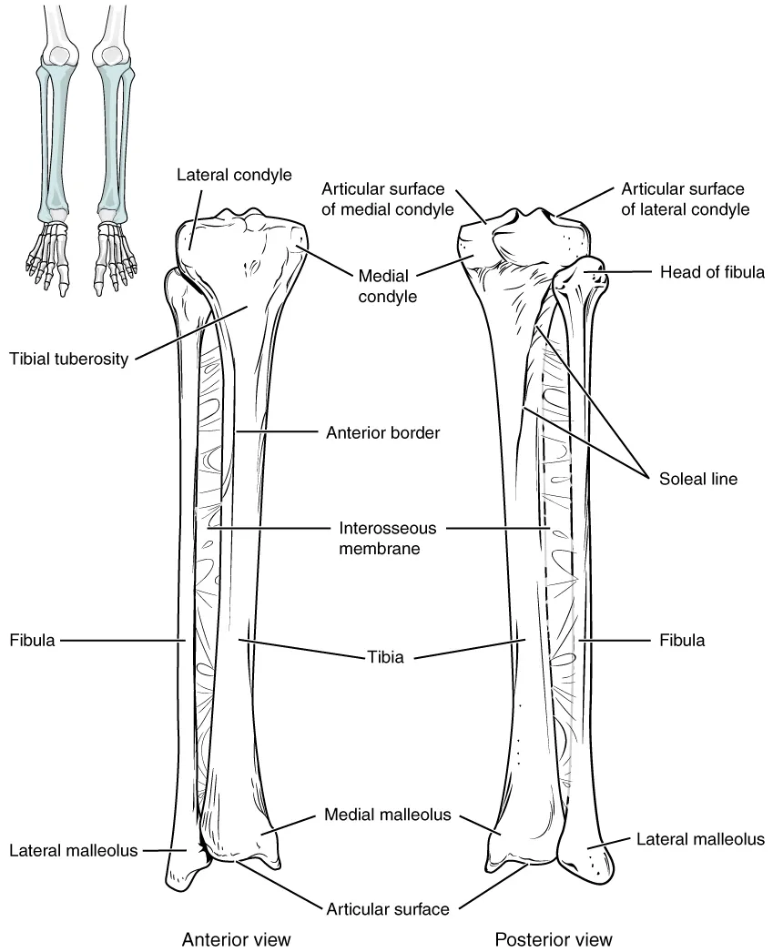
The large weight-bearing bone of the leg, medial to the fibula. Its proximal end forms part of the knee and its distal end part of the ankle; the medial surface of the shaft lies just under the skin.
Parts
Each part has its own card; press “Show on the model” to fly to it and select it in the 3D model above.
- 01
Tibial plateau (condyles)
Condyli tibiae
The broad upper end, with medial and lateral condyles whose flat upper surfaces form the tibial plateau supporting the menisci and femoral condyles. Between them lie the intercondylar eminence and the areas for the cruciate ligaments and menisci. The lateral condyle bears a facet for the fibular head.
- 02
Tibial tuberosity
Tuberositas tibiae
A bony prominence on the front of the upper end receiving the patellar ligament, through which quadriceps acts. In active adolescents its growth cartilage can become inflamed and enlarged (Osgood–Schlatter disease).
- 03
Shaft
Corpus tibiae
Triangular in section; its medial surface and anterior border (the shin) lie just under the skin, so tibial fractures are often open and heal slowly for lack of muscle cover. The posterior surface bears the soleal line. The interosseous membrane joins the lateral border to the fibula and separates the anterior and posterior compartments.
- 04
Medial malleolus
Malleolus medialis
The bony process on the medial side of the lower end that clasps the talus and anchors the deltoid ligament. Behind it the tendons of tibialis posterior and flexor digitorum longus, the posterior tibial artery and the tibial nerve pass beneath the flexor retinaculum.
- 05
Tibiofibular joints
Articulationes tibiofibulares
The superior tibiofibular joint is a plane synovial joint; the inferior one is a fibrous syndesmosis that binds the tibia and fibula into the mortise around the talus. Injury to the anterior inferior tibiofibular ligament causes a “high ankle sprain”, which heals more slowly than a common sprain.
Position and relations
The broad proximal end has two condyles forming the tibial plateau, with the intercondylar eminence between them for the cruciate ligaments and menisci; the tibial tuberosity in front receives the patellar ligament. The shaft is triangular in section with a sharp anterior border (shin). The distal end has the medial malleolus and the talar surface, which with the lateral malleolus of the fibula forms the mortise that grips the talus.
Form and structure
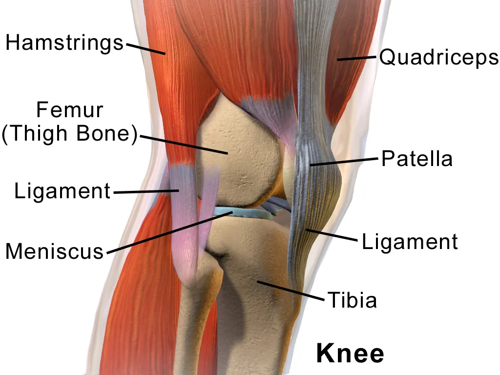
The ankle (talocrural joint) is a hinge for dorsiflexion and plantarflexion, most stable in dorsiflexion because the trochlea of the talus is wider in front. The medial deltoid ligament is very strong; the lateral ligaments (anterior talofibular, calcaneofibular, posterior talofibular) are weaker, so ankle sprains usually follow inversion.
Vessels and nerves
The tibia receives its nutrient artery (one of the largest) from the posterior tibial artery and periosteal branches from the anterior tibial. Because the medial surface is subcutaneous and the distal third poorly vascularised, tibial fractures are often open and slow to heal; leg trauma can cause compartment syndrome.
Tibial shaft and distal syndesmosis
The tibia is the weight-bearing bone of the leg, much larger and stronger than the fibula. Proximally it forms the lower part of the knee joint with the femur; distally it forms the ankle joint with the fibula and talus. It runs medial to the fibula, joined to it by the interosseous membrane. Its upper end bears medial and lateral condyles, between which the intercondylar area gives attachment to the cruciate ligaments and menisci.[1]
The tibial shaft is triangular in cross-section, with three borders (anterior, medial, interosseous) and three surfaces (lateral, medial, posterior). The medial surface is largely subcutaneous, the lateral surface faces the anterior compartment and the posterior surface the posterior compartment.[1]
About 20% of ankle fractures involve the distal tibiofibular syndesmosis. The deltoid ligament, though not part of the syndesmotic complex, contributes importantly to mortise stability and should always be assessed. Because syndesmotic anatomy varies between individuals, bilateral ankle imaging is recommended, especially for subtle instability, and tests that assess the joint dynamically in all three planes are the most reliable.[2]
Blount disease is a varus deformity of the proximal tibia with early-onset (before age four) and late-onset forms; its strong association with childhood obesity suggests a mechanical cause. The deformity combines tibial varus, procurvatum and internal torsion with limb shortening, and realignment osteotomy before age four reduces recurrence in the early-onset form.[3]
Full-text reading (StatPearls)
The StatPearls chapters on this topic, reproduced verbatim. Their CC BY-NC-ND 4.0 licence allows unmodified copies only, so they are not translated or shortened.
Anatomy, Bony Pelvis and Lower Limb: TibiaBourne M, Sinkler MA, Murphy PB · StatPearls 2023 · NBK526053 · 11k characters
Loading…
Bourne M, Sinkler MA, Murphy PB. Anatomy, Bony Pelvis and Lower Limb: Tibia. StatPearls [Internet]. Treasure Island (FL): StatPearls Publishing; 2023. NBK526053 · PMID 30252309 · CC BY-NC-ND 4.0. Reproduced verbatim, unmodified.
Organs in this system
References
Sources for the values and descriptions on this page, indexed in PubMed (US National Library of Medicine).
- [1]Anatomy, Bony Pelvis and Lower Limb: TibiaBourne M et al., StatPearls 2023PMID 30252309
- [2]The Syndesmosis, Part I: Anatomy, Injury Mechanism, Classification, and DiagnosisBejarano-Pineda L et al., The Orthopedic clinics of North America 2021PMID 34538351
- [3]Blount diseaseSabharwal S, The Journal of bone and joint surgery. American volume 2009PMID 19571101
- [4]Anatomy, BonesStatPearls (NCBI Bookshelf)PMID 30725884Supports: Number of bones
- [5]Calcium metabolism in health and diseasePeacock M, 2010PMID 20089499Supports: Skeletal calcium store