Muscular system
Model
Z-Anatomy anatomical model based on BodyParts3D, licensed CC BY-SA 4.0.
Overview
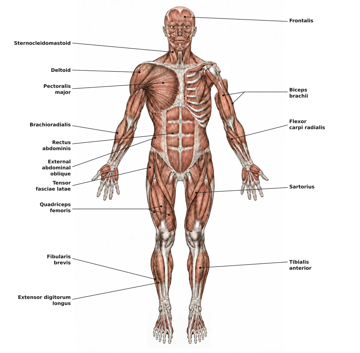
The muscular system comprises more than 600 skeletal muscles, which make up about 40% of adult body mass. Skeletal muscle contracts voluntarily and acts on the skeleton to produce movement; smooth and cardiac muscle form the walls of the viscera and the heart.
Functions
- 01Movement
- 02Maintenance of posture and joint stability
- 03Thermogenesis
- 04Assistance to venous return (muscle pump)
Microscopic organisation
Skeletal muscle makes up about 40% of body weight and contains 50–75% of all body proteins. Muscle mass depends on the balance between protein synthesis and breakdown, influenced by nutrition, hormones, activity and disease. Each fibre is a multinucleated cell; contraction begins when the action potential releases Ca²⁺ from the sarcoplasmic reticulum (excitation–contraction coupling).[1]
Fibres are classified by myosin heavy chain isoform: type I (slow-twitch, oxidative, fatigue-resistant) and type II (fast-twitch). The proportion of fibre types differs between muscles and between individuals and is modulated by neural activity and hormones.[2]
Clinical correlations
Fatigue during intense, repeated contraction is not mainly due to lactic acid accumulation, as once thought, but to ionic changes affecting the action potential, reduced Ca²⁺ release from the sarcoplasmic reticulum and reactive oxygen species. In older adults, age-related muscle wasting (sarcopenia) reduces muscle mass and strength and seriously threatens independence.[3][4]
Organisation of a skeletal muscle, from muscle to sarcomere
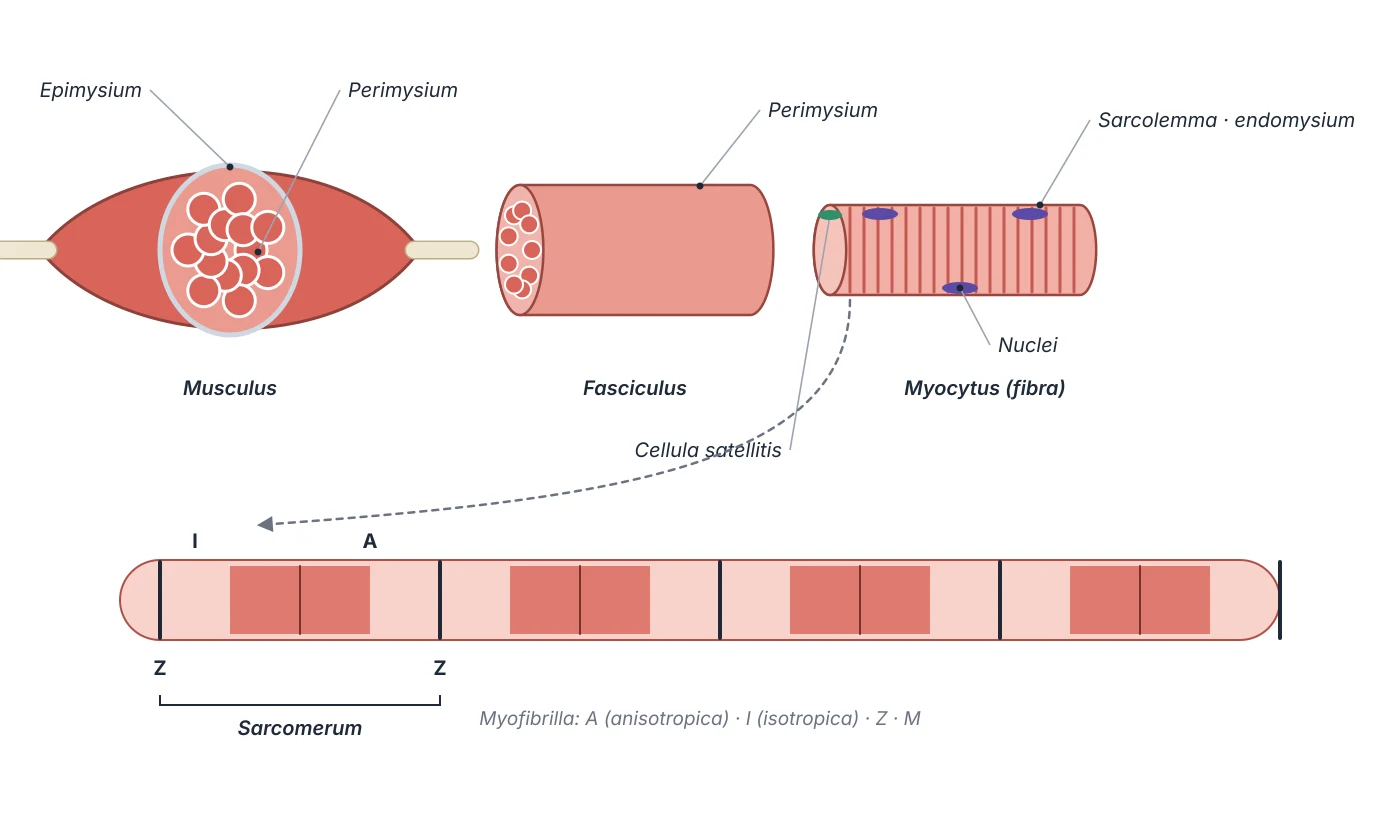
Each skeletal muscle consists of thousands of fibres wrapped in three connective tissue sheaths: the epimysium around the whole muscle, the perimysium around each fascicle and the endomysium around each fibre, carrying capillaries and nerve fibres. These sheaths are continuous with tendon and periosteum and transmit the force of contraction to bone. A skeletal fibre is a multinucleated cell 10–100 µm in diameter and many centimetres long, its nuclei lying beneath the sarcolemma. The membrane invaginates as transverse (T) tubules, sites of ion exchange that carry the action potential deep into the fibre. Satellite cells, the muscle’s stem cells, sit between the basement membrane and the sarcolemma; stimulated by growth factors they proliferate and differentiate to repair fibres or form new ones.[5][6]
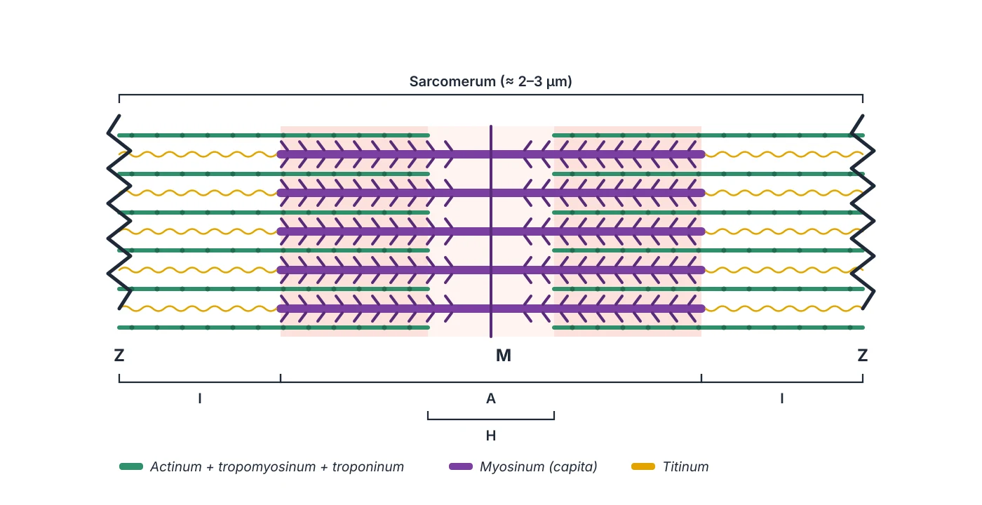
Inside the fibre are myofibrils, each a chain of sarcomeres, the basic contractile unit. A sarcomere is bounded by two Z lines, where thin actin filaments attach; in its centre the M line anchors the thick myosin filaments. The region containing myosin is the dark A band; the region with actin only is the light I band, producing the cross-striation seen under the microscope. Actin carries tropomyosin and the troponin C, I and T complex, which regulate myosin binding; titin and nebulin, giant proteins, set the filaments’ elasticity and length. This regular arrangement is repeated in every myofibril, so the whole fibre appears striated.[5]
Whole muscle
- Wrapped by
- Epimysium
- Contains
- Many fascicles, main artery
Fascicle
- Wrapped by
- Perimysium
- Contains
- Ten to a hundred or more fibres, feed arteries
Fibre
- Wrapped by
- Endomysium, basement membrane
- Contains
- Many nuclei, satellite cells, capillaries, T tubules
Myofibril
- Wrapped by
- Sarcoplasmic reticulum
- Contains
- Chain of sarcomeres
Sarcomere
- Wrapped by
- Two Z lines
- Contains
- Actin, myosin, troponin, tropomyosin, titin
| Level | Wrapped by | Contains |
|---|---|---|
| Whole muscle | Epimysium | Many fascicles, main artery |
| Fascicle | Perimysium | Ten to a hundred or more fibres, feed arteries |
| Fibre | Endomysium, basement membrane | Many nuclei, satellite cells, capillaries, T tubules |
| Myofibril | Sarcoplasmic reticulum | Chain of sarcomeres |
| Sarcomere | Two Z lines | Actin, myosin, troponin, tropomyosin, titin |
From nerve impulse to contraction
Each motor axon branches as it reaches the muscle, each branch ending on one fibre at a motor end plate (neuromuscular junction). The terminal is rich in mitochondria, endoplasmic reticulum and synaptic vesicles containing acetylcholine. When the action potential arrives, vesicles fuse with the membrane and release acetylcholine into the cleft; the postsynaptic membrane has a very high density of acetylcholine receptors, ligand-gated ion channels. Their opening rapidly depolarises the end plate and triggers an action potential that spreads along the sarcolemma. Muscle also carries sensory fibres (muscle spindles, Golgi tendon organs), and diseases of the junction such as myasthenia gravis show how critical this step is.[5]
Excitation–contraction coupling is the chain that turns an action potential into cross-bridge cycling. The action potential travels down the T tubules and changes the conformation of dihydropyridine receptors in the tubule membrane; these open ryanodine receptors on the terminal cisternae of the sarcoplasmic reticulum, releasing stored Ca²⁺ and raising cytosolic Ca²⁺ about tenfold. Each troponin C binds up to four Ca²⁺ ions cooperatively, so a small rise in Ca²⁺ saturates the binding sites. The change in troponin pulls tropomyosin away from the myosin-binding sites on actin; myosin heads attach, flex and slide the thin filaments towards the M line. When Ca²⁺ is pumped back into the reticulum the muscle relaxes.[6]
Development and microcirculation of muscle
Trunk and limb skeletal muscle arises from paraxial mesoderm. This mesoderm segments into somites, which under Wnt (from neural tube and ectoderm), Shh (from the notochord) and BMP4 (from lateral plate mesoderm) signalling divide into dermomyotome and sclerotome. The myotome differentiates when Shh induces Myf5 and then MyoD. Its dorsomedial (epaxial) part forms the deep back muscles; its ventrolateral (hypaxial) part forms the body wall and limb muscles. Embryonic myoblasts fuse into primary fibres, secondary fibres follow in the fetus, and after birth satellite cells take over growth and repair.[5]
A muscle’s main artery usually runs parallel to the fibres and gives perpendicular feed arteries to the perimysium. These branch into arterioles, then transverse arterioles and terminal arterioles; each terminal arteriole with the capillaries running along the fibres in the endomysium forms a microvascular unit, the smallest unit in which blood flow is regulated. Lymphatic capillaries start beside the blood capillaries in the endomysium and join into vessels crossing the perimysium. Their walls lack smooth muscle, so they rely on muscle contraction and arteriolar pulsation to move lymph, the principle of the “muscle pump” that also aids venous return.[5]
Muscles of respiration: diaphragm and intercostals
The diaphragm is the most important muscle of inspiration: as it contracts it pulls its central tendon down, increasing the vertical diameter of the thorax and the negative intrathoracic pressure, which draws air in. At the same time the external intercostals raise the anterior chest wall like a bucket handle, widening the chest. Quiet expiration is passive: the chest wall sinks and the diaphragm relaxes and rises. The diaphragm also works with the abdominal muscles to raise intra-abdominal pressure in micturition, defecation, childbirth and straining against a closed glottis (the Valsalva manoeuvre, also used to classify heart murmurs). The phrenic nerve (C3–C5) supplies motor and central sensory fibres; the lower six intercostal nerves supply the periphery.[7]
Clinically, skeletal muscle disorders range from cramps and muscle–tendon ruptures in athletes to myopathies, myasthenia gravis and paralysis from nerve injury. A cramp is an involuntary, painful contraction of a muscle or group of fibres lasting seconds to minutes; exercise-associated cramps are the commonest reason for medical intervention in sport, and a localised contraction does not imply a local cause. Sustained contraction of a particular muscle is seen in congenital or spasmodic torticollis (sternocleidomastoid) and in thoracic outlet syndrome (scalene hypertrophy). Maintaining muscle strength through exercise matters for bone health, balance and posture.[5][6]
Principal organs
A muscle of the anterior compartment of the arm with two heads (long and short) arising from the scapula; its main actions are elbow flexion and forearm supination.
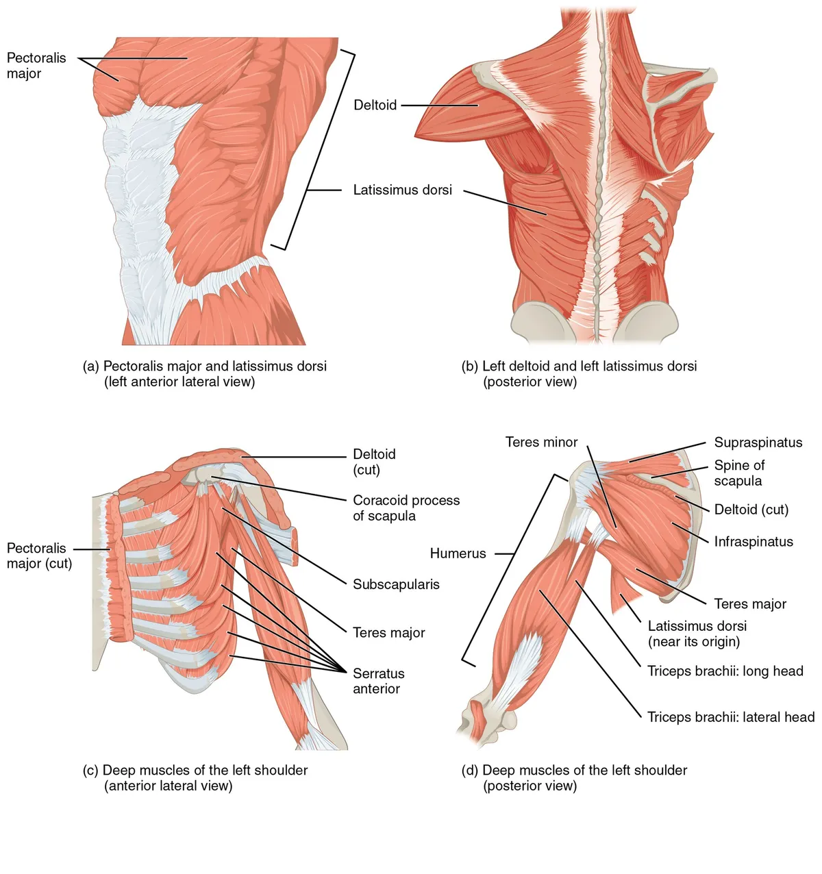
DeltoidMusculus deltoideusA triangular muscle covering the shoulder joint and forming its rounded contour. Its three parts (clavicular, acromial, spinal) flex, abduct and extend the arm.
Pectoralis majorMusculus pectoralis majorA broad fan-shaped muscle of the anterior chest wall forming the anterior axillary fold. It adducts, medially rotates and flexes the arm and is an accessory muscle of respiration.
Gluteus maximusMusculus gluteus maximusThe largest and heaviest muscle in the body, shaping the buttock. It extends and laterally rotates the hip and is essential for rising from sitting, climbing stairs, running and stabilising the trunk.
Quadriceps femorisMusculus quadriceps femorisThe anterior thigh group made up of rectus femoris, vastus lateralis, vastus medialis and vastus intermedius; inserting on the tibial tuberosity via the patellar ligament, it is the principal extensor of the knee.
Triceps suraeMusculus triceps suraeThe calf muscle, made of gastrocnemius (two heads) and soleus, inserting on the calcaneus through the calcaneal (Achilles) tendon, the largest and strongest tendon in the body. It is the main plantarflexor, propelling the body in walking, running and jumping.
DiaphragmDiaphragmaA dome-shaped musculotendinous partition between the thorax and the abdomen, whose muscle fibres arise from the sternum, the lower six costal cartilages and the lumbar spine (crura) and converge on the central tendon. It is the chief muscle of inspiration: on contraction the dome descends and enlarges the thoracic cavity. It has three main openings: the caval opening (T8), the oesophageal hiatus (T10) and the aortic hiatus (T12).
Reference values
- Gluteus maximus is the largest muscle by mass; the stapedius in the middle ear is the smallest skeletal muscle.
References
Sources for the values and descriptions on this page, indexed in PubMed (US National Library of Medicine).
- [1]Skeletal muscle: a brief review of structure and functionFrontera WR et al., Calcified tissue international 2015PMID 25294644
- [2]Fiber types in mammalian skeletal musclesSchiaffino S et al., Physiological reviews 2011PMID 22013216
- [3]Skeletal muscle fatigue: cellular mechanismsAllen DG et al., Physiological reviews 2008PMID 18195089
- [4]Muscle aging and sarcopenia: The pathology, etiology, and most promising therapeutic targetsGrima-Terrén M et al., Molecular aspects of medicine 2024PMID 39312874
- [5]Anatomy, Skeletal MuscleDave HD et al., StatPearls 2023PMID 30725921
- [6]Physiology, Skeletal MuscleMcCuller C et al., StatPearls 2023PMID 30725824
- [7]Anatomy, Thorax: DiaphragmBains KNS et al., StatPearls 2023PMID 30137842
- [8]Anatomy, Bony Pelvis and Lower Limb, Gluteus Maximus MuscleStatPearls (NCBI Bookshelf)PMID 30855781Supports: Gluteus maximus