Hepatic portal vein
Model
Layers
Z-Anatomy anatomical model based on BodyParts3D, licensed CC BY-SA 4.0.
Description
A vein about 8 cm long that brings blood from the gut, spleen and pancreas to the liver, providing about 75% of hepatic blood flow. Portal blood carries nutrients and absorbed substances for the liver to process before they reach the general circulation.
Parts
Each part has its own card; press “Show on the model” to fly to it and select it in the 3D model above.
- 01
Hepatic portal vein
Vena portae hepatis
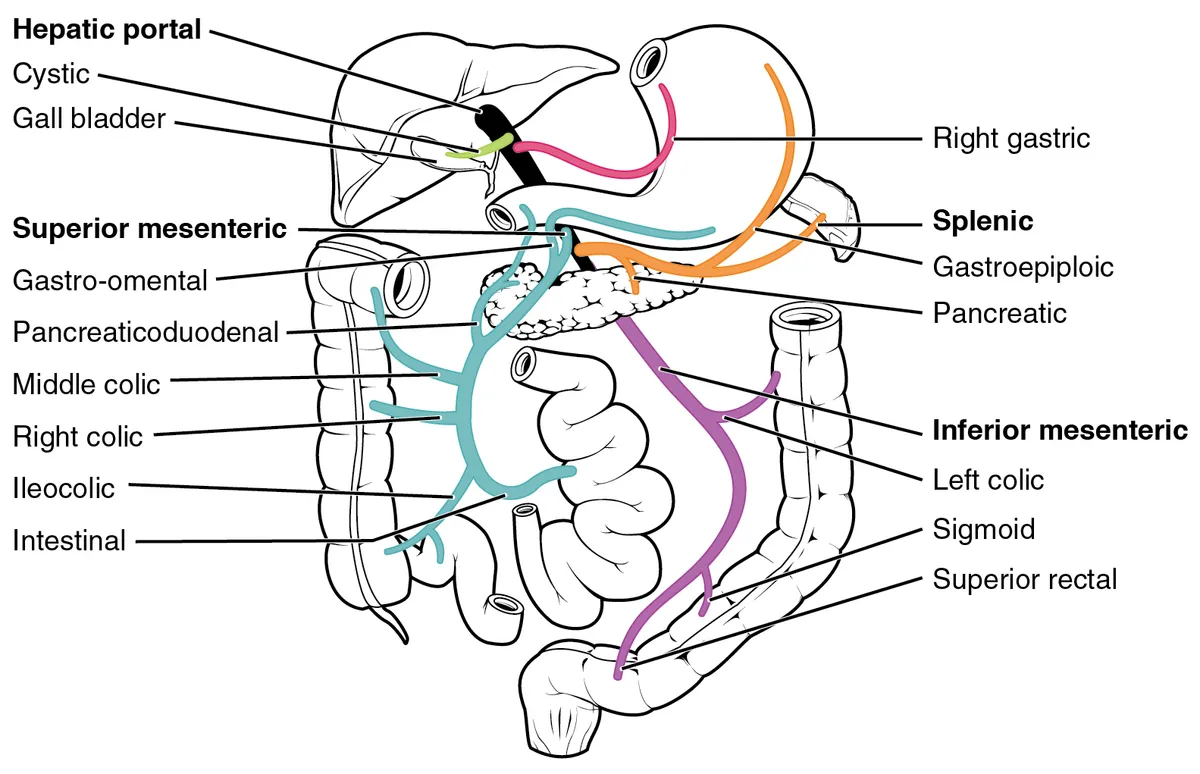
Formed behind the neck of the pancreas at L1–L2 by the union of the superior mesenteric and splenic veins, about 8 cm long, it runs in the free edge of the lesser omentum behind the bile duct and proper hepatic artery. It carries about 75% of the blood reaching the liver and divides at the porta hepatis into right and left branches.
- 02
Splenic vein
Vena splenica
Runs from the splenic hilum along the back of the body of the pancreas (receiving many small pancreatic veins), collecting the short gastric, left gastro-omental and usually the inferior mesenteric veins. Pancreatitis can cause splenic vein thrombosis and left-sided portal hypertension with gastric fundal varices.
- 03
Superior mesenteric vein
Vena mesenterica superior
Runs to the right of its artery in the root of the mesentery, draining the small intestine, caecum, ascending and transverse colon (ileocolic, right and middle colic veins), the right gastro-omental and pancreaticoduodenal veins. Its relation to the pancreatic head determines resectability of pancreatic tumours.
- 04
Inferior mesenteric vein
Vena mesenterica inferior
Begins as the superior rectal vein, ascends retroperitoneally to the left of its artery, receives the sigmoid and left colic veins, and usually ends in the splenic vein (or at the confluence, or in the superior mesenteric vein).
- 05
Portosystemic anastomoses
Anastomoses portocavales
Sites where portal and systemic veins meet: the lower oesophagus (left gastric to azygos), the rectum (superior to middle and inferior rectal), round the umbilicus (para-umbilical to epigastric) and retroperitoneally. In portal hypertension, usually from cirrhosis, they dilate into oesophageal varices, caput medusae and anorectal varices.
- 06
Intrahepatic branches
Ramus dexter et sinister venae portae
The short right branch divides into anterior and posterior branches for segments V–VIII; the longer left branch has transverse and umbilical parts supplying segments II–IV and is joined by the ligamentum teres. The portal tree runs with the hepatic artery and bile duct in the portal tracts and is the basis of segmental liver resection.
Position and relations
The portal vein forms behind the neck of the pancreas at about L2 from the union of the superior mesenteric and splenic veins (the inferior mesenteric usually drains into the splenic). It ascends behind the first part of the duodenum into the free edge of the lesser omentum (hepatoduodenal ligament) with the common bile duct and proper hepatic artery, and divides into right and left branches at the porta hepatis.
Form and structure
Within the liver, portal branches divide segmentally with the hepatic artery and bile ducts (the portal triad); portal and arterial blood mix in the sinusoids and drain to the central veins, hepatic veins and inferior vena cava. The portal system has no valves, so raised pressure is transmitted back to all its tributaries.
Vessels and nerves
In portal hypertension (usually from cirrhosis), blood returns to the systemic circulation through portosystemic anastomoses: oesophagogastric varices (risk of bleeding), paraumbilical veins (caput medusae), rectal and retroperitoneal veins, with splenomegaly and ascites.
Portal hypertension and collaterals
Portal hypertension is the main non-neoplastic complication of cirrhosis, with life-threatening consequences such as variceal bleeding, ascites and hepatic encephalopathy. It begins with increased resistance of the cirrhotic liver vasculature to portal flow and is aggravated by the hyperdynamic circulatory syndrome. A profibrogenic phenotype of sinusoidal cells, raising intrahepatic vascular tone and fibrosis, is considered the initiating step, and the altered stiffness of the cirrhotic liver feeds a vicious cycle.[1][2]
When the extrahepatic portal vein thromboses, the veins around the bile duct dilate and become tortuous to bridge the block, forming a portal cavernoma. They include the 3 o’clock and 9 o’clock marginal veins, the paracholedochal plexus of Petren, the epicholedochal plexus of Saint and the cholecystic veins. The cavernoma forms over a week to a year, may become a fibrotic hilar mass and can compress the bile ducts (portal biliopathy). Colour Doppler ultrasound makes the diagnosis easily: the portal vein is not seen, and multiple serpiginous channels occupy the hilum.[3]
In developing countries portal cavernoma affects children, after portal vein thrombosis caused by neonatal umbilical sepsis and dehydration. In adults the causes include hypercoagulable states, myeloproliferative disorders, Behçet’s disease, pancreatitis and pylephlebitis, and about 30% have no identifiable cause. A cavernoma rarely forms when thrombosis complicates cirrhosis, because stasis in the portal system prevents collaterals from developing.[3]
Full-text reading (StatPearls)
The StatPearls chapters on this topic, reproduced verbatim. Their CC BY-NC-ND 4.0 licence allows unmodified copies only, so they are not translated or shortened.
Anatomy, Abdomen and Pelvis, Portal Venous System (Hepatic Portal System)Harkins JM, Ahmad B · StatPearls 2023 · NBK554589 · 9k characters
Loading…
Harkins JM, Ahmad B. Anatomy, Abdomen and Pelvis, Portal Venous System (Hepatic Portal System). StatPearls [Internet]. Treasure Island (FL): StatPearls Publishing; 2023. NBK554589 · PMID 32119476 · CC BY-NC-ND 4.0. Reproduced verbatim, unmodified.
Organs in this system
References
Sources for the values and descriptions on this page, indexed in PubMed (US National Library of Medicine).
- [1]Pathophysiology and therapeutic options for cirrhotic portal hypertensionGuixé-Muntet S et al., The lancet. Gastroenterology & hepatology 2024PMID 38642564
- [2]The Pathophysiology of Portal HypertensionRamkissoon R et al., Clinics in liver disease 2024PMID 38945632
- [3]Portal biliopathyKhuroo MS et al., World journal of gastroenterology 2016PMID 27672292
- [4]Physiology, Cardiac OutputStatPearls (NCBI Bookshelf)PMID 29262215Supports: Heart rate, cardiac output
- [5]Physiology, CardiovascularStatPearls (NCBI Bookshelf)PMID 29630249