Pineal gland
Model
Layers
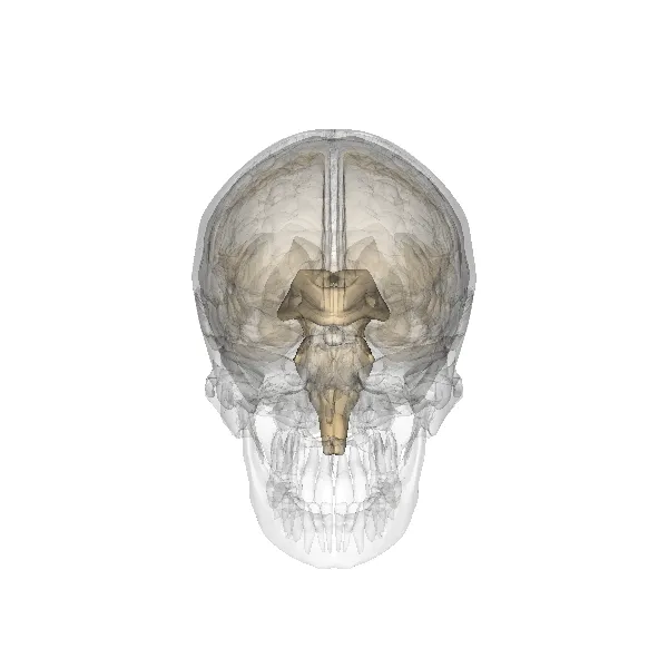
Z-Anatomy anatomical model based on BodyParts3D, licensed CC BY-SA 4.0.
Description
A small pine-cone-shaped gland about 8 mm long, attached to the roof of the third ventricle at the back of the diencephalon, between the superior colliculi. It secretes melatonin mainly at night, driven by light signals from the retina via the suprachiasmatic nucleus and the cervical sympathetic chain, helping to set circadian rhythm. It is often calcified in adults, serving as a midline landmark on imaging.
Parts
Each part has its own card; press “Show on the model” to fly to it and select it in the 3D model above.
- 01
Pinealocytes
Pinealocyti
About 95% of the cells, modified neurons that synthesise melatonin from serotonin, mainly at night. Secretion follows the day–night rhythm set by the suprachiasmatic nucleus; light suppresses it.
- 02
Interstitial (glial) cells
Cellulae interstitiales; Neuroglia
Astrocyte-like cells between the cords of pinealocytes and round the vessels, supporting the gland. The pineal is a circumventricular organ with fenestrated capillaries outside the blood–brain barrier.
- 03
Corpora arenacea (brain sand)
Corpora arenacea
Calcium phosphate and carbonate concretions that increase with age and are visible on CT in most adults. The calcified midline pineal was a classic radiological landmark: its shift suggests an intracranial mass.
- 04
Position and relations
Recessus pinealis; Commissura habenularis et posterior
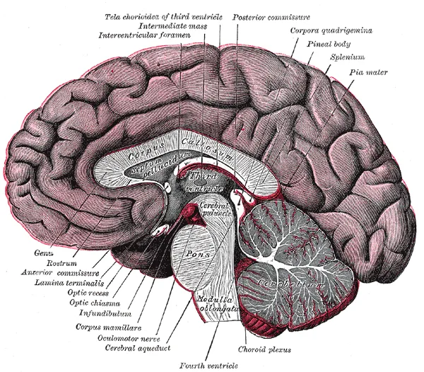
A pine-cone-shaped gland about 8 mm long on the posterior wall of the third ventricle, attached by a stalk with the habenular commissure above and the posterior commissure below, enclosing the pineal recess. It rests above the superior colliculi, so pineal tumours compress the tectum (Parinaud syndrome) and the aqueduct (hydrocephalus).
- 05
Neural control pathway
Innervatio sympathica glandulae pinealis
Light information passes from the retina (melanopsin-containing ganglion cells) to the suprachiasmatic nucleus, paraventricular nucleus, thoracic intermediolateral column and superior cervical ganglion, then by sympathetic fibres to the pineal; nocturnal noradrenaline stimulates melatonin synthesis. Cervical sympathetic lesions (Horner syndrome) can disturb this rhythm.
Position and relations
The pineal gland lies in the midline between the splenium of the corpus callosum above and the superior colliculi of the midbrain tectum below, its stalk attached to the posterior and habenular commissures. Tumours here may compress the tectum, causing upgaze palsy (Parinaud syndrome), and the cerebral aqueduct, causing hydrocephalus.
Form and structure
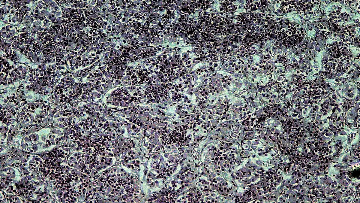
The parenchyma consists of pinealocytes and glial cells, divided into lobules by connective-tissue septa. With age, calcium concretions (brain sand) accumulate within it and are visible on CT in most adults.
Vessels and nerves
It is supplied by posterior choroidal branches (from the posterior cerebral artery) and, like other circumventricular organs, lacks a blood–brain barrier. Its innervation is postganglionic sympathetic fibres from the superior cervical ganglion, the final link in the light pathway from the retina to the gland.
Melatonin and biological rhythms
The pineal gland lies in the posterior cranial fossa behind the third ventricle and weighs under 0.2 g in adults; its main function is to secrete about 30 µg of melatonin a day. Melatonin is always produced at night, and the duration of secretion depends directly on night length; tied to the light–dark cycle, it synchronises behaviour and physiology with the environmental day and season.[1][2][3]
During each nightly episode, melatonin coordinates night-time physiology through immediate effects and primes daytime responses through prospective effects that appear only when it is absent. Likewise, the annual history of episode duration primes the neuroendocrine system for the coming season, and maternal melatonin programmes fetal behaviour and physiology for the light–dark cycle after birth.[2]
Melatonin is made not only by the pineal gland but also in the gut, skin and leukocytes, and by bacteria and plants, along the same tryptophan-derived pathway. Circulating melatonin is mostly 6-hydroxylated by hepatic P450 enzymes and excreted as 6-sulfatoxymelatonin; it acts through membrane and nuclear receptors to regulate sleep, stimulate immunity and provide antioxidant and neuroprotective effects.[4]
Clinical correlations
Melatonin secretion declines with age in parallel with poorer sleep, so melatonin is used for insomnia and circadian rhythm disorders (with bright-light therapy); evidence for this replacement approach remains limited and optimal dosing is undefined.[5][6]
Pineal calcification is very common and a radiological landmark; pineal region masses include pineal cysts, parenchymal tumours (pineocytoma, tumour of intermediate differentiation, pineoblastoma) and papillary tumours. They can compress the cerebral aqueduct (hydrocephalus) or tectum (Parinaud syndrome) and, in children, cause precocious puberty. The 5th WHO classification uses molecular features to guide treatment, from resection alone to multimodal therapy.[3][7]
Claims that the pineal gland secretes N,N-dimethyltryptamine (DMT) at birth, during dreaming or near death are not supported by evidence: the DMT detected in the brain is far too little to have psychoactive effects.[1]
Full-text reading (StatPearls)
The StatPearls chapters on this topic, reproduced verbatim. Their CC BY-NC-ND 4.0 licence allows unmodified copies only, so they are not translated or shortened.
Physiology, Pineal GlandIlahi S, Beriwal N, Ilahi TB · StatPearls 2023 · NBK525955 · 12k characters
Loading…
Ilahi S, Beriwal N, Ilahi TB. Physiology, Pineal Gland. StatPearls [Internet]. Treasure Island (FL): StatPearls Publishing; 2023. NBK525955 · PMID 30247830 · CC BY-NC-ND 4.0. Reproduced verbatim, unmodified.
Organs in this system
References
Sources for the values and descriptions on this page, indexed in PubMed (US National Library of Medicine).
- [1]N,N-dimethyltryptamine and the pineal gland: Separating fact from mythNichols DE, Journal of psychopharmacology 2018PMID 29095071
- [2]Melatonin as a Hormone: New Physiological and Clinical InsightsCipolla-Neto J et al., Endocrine reviews 2018PMID 30215696
- [3]Revisiting the pineal gland: a review of calcification, masses, precocious puberty, and melatonin functionsPatel S et al., The International journal of neuroscience 2020PMID 31714865
- [4]MelatoninHardeland R et al., The international journal of biochemistry & cell biology 2006PMID 16219483
- [5]Melatonin in sleep disordersPoza JJ et al., Neurologia 2022PMID 36064286
- [6]Circadian Rhythm Dysregulation and Restoration: The Role of MelatoninVasey C et al., Nutrients 2021PMID 34684482
- [7]SNO-EANO-EURACAN consensus on management of pineal parenchymal tumorsLiu APY et al., Neuro-oncology 2024PMID 39073785
- [8]Physiology, Pituitary HormonesStatPearls (NCBI Bookshelf)PMID 32491488Supports: Pituitary and endocrine axes
- [9]Physiology, Anterior PituitaryStatPearls (NCBI Bookshelf)PMID 29763073