Thyroid gland
Model
Layers
Z-Anatomy anatomical model based on BodyParts3D, licensed CC BY-SA 4.0.
Description
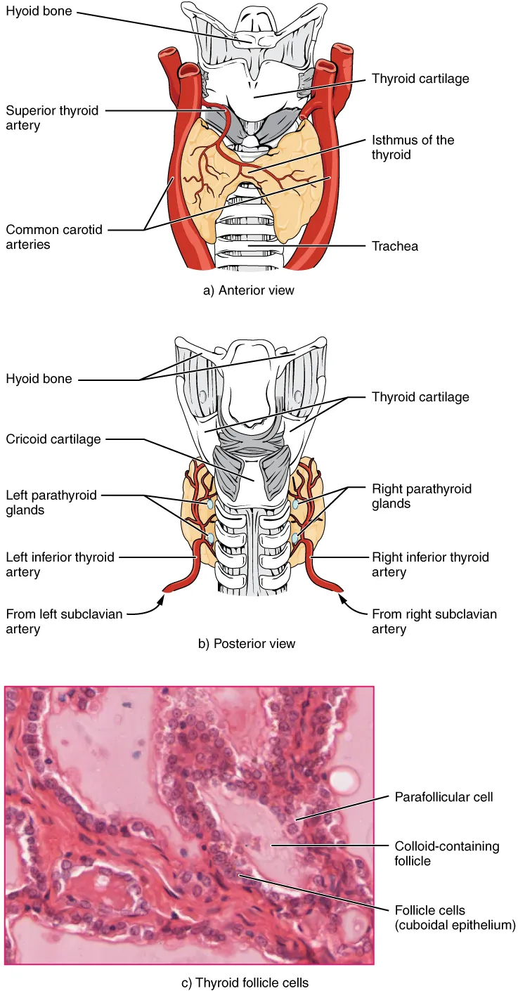
An H-shaped gland anterior to the trachea at the C5–T1 level, with two lobes joined by an isthmus. It secretes thyroxine (T4) and triiodothyronine (T3), which set the basal metabolic rate, as well as calcitonin.
Parts
Each part has its own card; press “Show on the model” to fly to it and select it in the 3D model above.
- 01
Right and left lobes
Lobus dexter et sinister glandulae thyroideae
Each pyramidal lobe, about 5 cm high, embraces the larynx and trachea from the thyroid cartilage to the fifth or sixth tracheal ring, with the carotid sheath posterolaterally. The gland weighs about 15–25 g and moves on swallowing because the pretracheal fascia binds it to the larynx.
- 02
Isthmus
Isthmus glandulae thyroideae
The band joining the lobes across the second to fourth tracheal rings; it is usually divided or retracted at tracheostomy. A thyroidea ima artery (in about 10% of people) may ascend in front of the trachea here.
- 03
Pyramidal lobe and thyroglossal duct
Lobus pyramidalis; Ductus thyroglossalis
About half of people have a pyramidal lobe rising from the isthmus, the lower remnant of the thyroglossal duct along which the gland descended from the foramen caecum of the tongue. A persistent duct forms a midline thyroglossal cyst that moves on tongue protrusion.
- 04
Thyroid follicles
Folliculi glandulae thyroideae
The functional unit: spheres 50–500 µm across lined by follicular cells (thyrocytes) round a colloid of thyroglobulin. Under TSH the cells trap iodide and synthesise and release T4 and T3; they are flat when resting and tall when overactive, as in Graves disease.
- 05
Parafollicular (C) cells
Cellulae parafolliculares
Pale cells between the follicles, of neural crest origin (via the ultimobranchial body), secreting calcitonin, which lowers blood calcium. Medullary thyroid carcinoma arises from them and may be part of MEN 2.
- 06
Vessels and nerve relations
Arteriae thyroideae; Nervus laryngeus recurrens
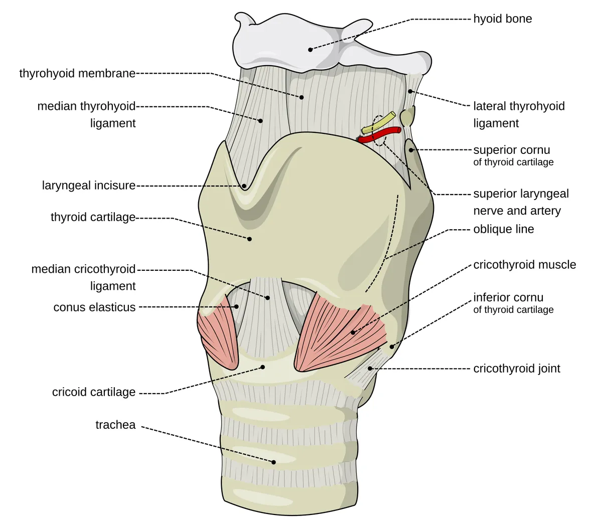
The superior thyroid artery (from the external carotid, with the external branch of the superior laryngeal nerve) and the inferior thyroid artery (from the thyrocervical trunk, crossing the recurrent laryngeal nerve). Thyroid surgery must preserve the recurrent nerves and the parathyroids on the posterior surface.
Position and relations
The thyroid lies in the lower anterior neck, in front of the trachea, at the level of C5–T1. It has two lateral lobes joined by an isthmus (over tracheal rings 2–4). Its posterior aspect relates to the carotid sheath, the parathyroid glands and the recurrent laryngeal nerves, which must be protected during thyroid surgery.
Form and structure
The structural unit is the follicle: a single layer of epithelial cells around a lumen filled with colloid (thyroglobulin). Between follicles lie parafollicular (C) cells that secrete calcitonin. The thyroid is the only endocrine gland that stores its hormone extracellularly, in amounts sufficient for several weeks.[1]
Vessels and nerves
The thyroid has a very rich blood supply from the superior thyroid artery (from the external carotid) and the inferior thyroid artery (from the thyrocervical trunk of the subclavian). Superior, middle and inferior thyroid veins form a plexus on its surface.
Physiology
Follicular cells take up iodide through the sodium/iodide symporter and incorporate it into thyroglobulin to form T4 and T3, under the control of pituitary TSH acting on the TSH receptor. Population TSH levels rise with age, so diagnosing subclinical hypothyroidism in older adults requires age-specific reference ranges.[1][2]
Surgical anatomy and development
Injury to the recurrent or superior laryngeal nerve or to the parathyroid glands during thyroid surgery can have lifelong consequences (hoarseness, airway compromise, hypocalcaemia). Surgeons therefore need a thorough knowledge of the anatomy and embryology of the thyroid, parathyroids and neighbouring structures to identify and preserve them.[3]
The thyroid produces hormone that acts on practically every organ system. Key molecules such as the TSH receptor and the sodium/iodide symporter (NIS) are well characterised, enabling new diagnostics and treatments. Developmental anomalies (thyroid dysgenesis: ectopy, athyreosis, hypoplasia) underlie most congenital hypothyroidism; ectopy is the commonest and remains unexplained.[1][4]
Clinical correlations
Thyroid nodules are very common and matter mainly for their malignant potential; serum TSH and ultrasound are the basis of evaluation, and fine-needle aspiration is the most accurate way to diagnose malignancy and select patients for surgery.[5]
Thyroid hormone has profound cardiac effects: hypothyroidism reduces contractility, raises vascular resistance and diastolic pressure and slows the heart; hyperthyroidism causes tachycardia, atrial fibrillation and high output. Thyrotoxic states are classified by increased or decreased glandular metabolic function, separating Graves’ disease and toxic nodules from thyroiditis.[6][7][8]
Full-text reading (StatPearls)
The StatPearls chapters on this topic, reproduced verbatim. Their CC BY-NC-ND 4.0 licence allows unmodified copies only, so they are not translated or shortened.
Histology, Thyroid GlandKhan YS, Farhana A · StatPearls 2025 · NBK551659 · 21k characters
Loading…
Khan YS, Farhana A. Histology, Thyroid Gland. StatPearls [Internet]. Treasure Island (FL): StatPearls Publishing; 2025. NBK551659 · PMID 31869123 · CC BY-NC-ND 4.0. Reproduced verbatim, unmodified.
Anatomy, Head and Neck, ThyroidAllen E, Fingeret A · StatPearls 2025 · NBK470452 · 14k characters
Loading…
Allen E, Fingeret A. Anatomy, Head and Neck, Thyroid. StatPearls [Internet]. Treasure Island (FL): StatPearls Publishing; 2025. NBK470452 · PMID 29262169 · CC BY-NC-ND 4.0. Reproduced verbatim, unmodified.
Anatomy, Head and Neck, Thyroid MusclesMnatsakanian A, Al Khalili Y · StatPearls 2023 · NBK541063 · 10k characters
Loading…
Mnatsakanian A, Al Khalili Y. Anatomy, Head and Neck, Thyroid Muscles. StatPearls [Internet]. Treasure Island (FL): StatPearls Publishing; 2023. NBK541063 · PMID 31082107 · CC BY-NC-ND 4.0. Reproduced verbatim, unmodified.
Anatomy, Head and Neck, Thyroid ArteriesWhite AM, Lasrado S · StatPearls 2023 · NBK560666 · 8k characters
Loading…
White AM, Lasrado S. Anatomy, Head and Neck, Thyroid Arteries. StatPearls [Internet]. Treasure Island (FL): StatPearls Publishing; 2023. NBK560666 · PMID 32809501 · CC BY-NC-ND 4.0. Reproduced verbatim, unmodified.
Anatomy, Head and Neck, Thyroid Thyroglossal DuctHussain A, Sajjad H · StatPearls 2023 · NBK554494 · 7k characters
Loading…
Hussain A, Sajjad H. Anatomy, Head and Neck, Thyroid Thyroglossal Duct. StatPearls [Internet]. Treasure Island (FL): StatPearls Publishing; 2023. NBK554494 · PMID 32119381 · CC BY-NC-ND 4.0. Reproduced verbatim, unmodified.
Organs in this system
References
Sources for the values and descriptions on this page, indexed in PubMed (US National Library of Medicine).
- [1]Thyroid physiologyStathatos N, The Medical clinics of North America 2012PMID 22443969
- [2]The aging thyroidTabatabaie V et al., Current opinion in endocrinology, diabetes, and obesity 2013PMID 23974775
- [3]Anatomy of thyroid and parathyroid glands and neurovascular relationsMohebati A et al., Clinical anatomy 2012PMID 21800365
- [4]Disorders of thyroid morphogenesisAbu-Khudir R et al., Best practice & research. Clinical endocrinology & metabolism 2017PMID 28648504
- [5]Thyroid nodulesPopoveniuc G et al., The Medical clinics of North America 2012PMID 22443979
- [6]Hypothyroidism and the HeartUdovcic M et al., Methodist DeBakey cardiovascular journal 2017PMID 28740582
- [7]Hyperthyroidism and the HeartOsuna PM et al., Methodist DeBakey cardiovascular journal 2017PMID 28740583
- [8]ThyrotoxicosisSeigel SC et al., The Medical clinics of North America 2012PMID 22443970
- [9]Physiology, Pituitary HormonesStatPearls (NCBI Bookshelf)PMID 32491488Supports: Pituitary and endocrine axes
- [10]Physiology, Anterior PituitaryStatPearls (NCBI Bookshelf)PMID 29763073