Urinary system
Model
Z-Anatomy anatomical model based on BodyParts3D, licensed CC BY-SA 4.0.
Overview
The urinary system comprises the two kidneys, two ureters, the urinary bladder and the urethra. The kidneys filter plasma, form urine and regulate extracellular fluid volume, electrolyte and acid–base balance, and blood pressure.
Functions
- 01Excretion of nitrogenous waste (urea, creatinine)
- 02Regulation of water, electrolyte and acid–base balance
- 03Endocrine function: renin, erythropoietin, calcitriol
The glomerular filtration barrier
The glomerular capillary wall has three layers: fenestrated endothelium, the glomerular basement membrane and the foot processes of podocytes. The basement membrane is built mainly from laminins, type IV collagen, nidogens and heparan sulfate proteoglycans; inherited defects of these components cause diseases such as Alport syndrome (COL4A3–5 genes).[1]
Loss of podocytes (absolute, or relative to glomerular volume) and effacement of their foot processes are a common mechanism of many glomerular diseases. When the filtration barrier is damaged, serum proteins reach the tubules and injure the tubulointerstitium, showing that glomeruli and tubules interact in the progression of chronic kidney disease.[2][3]
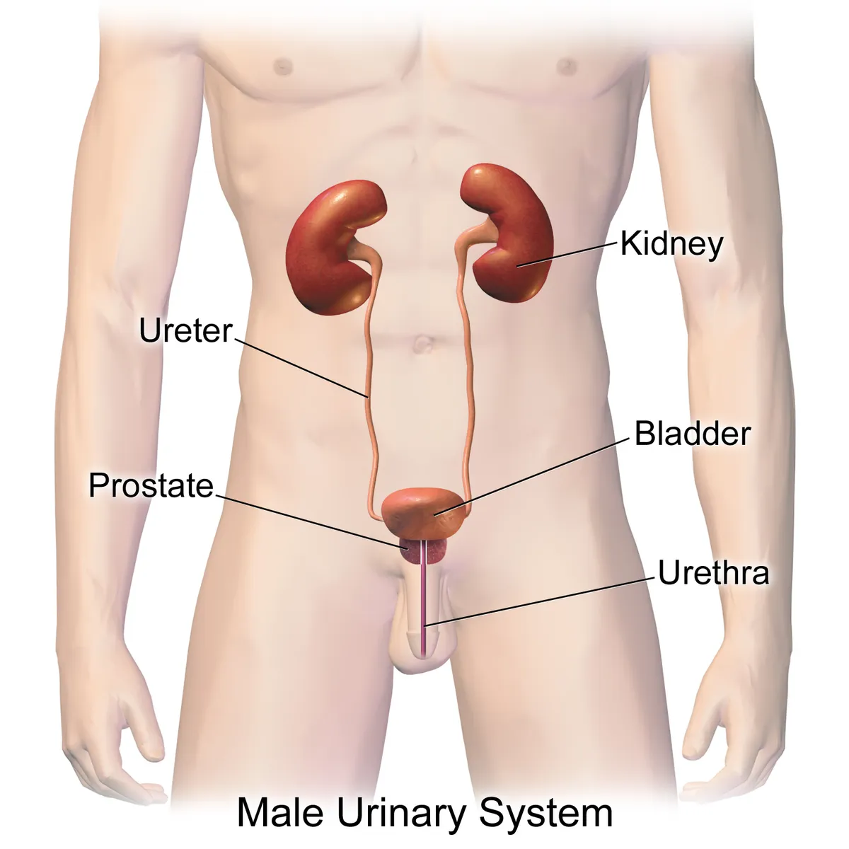
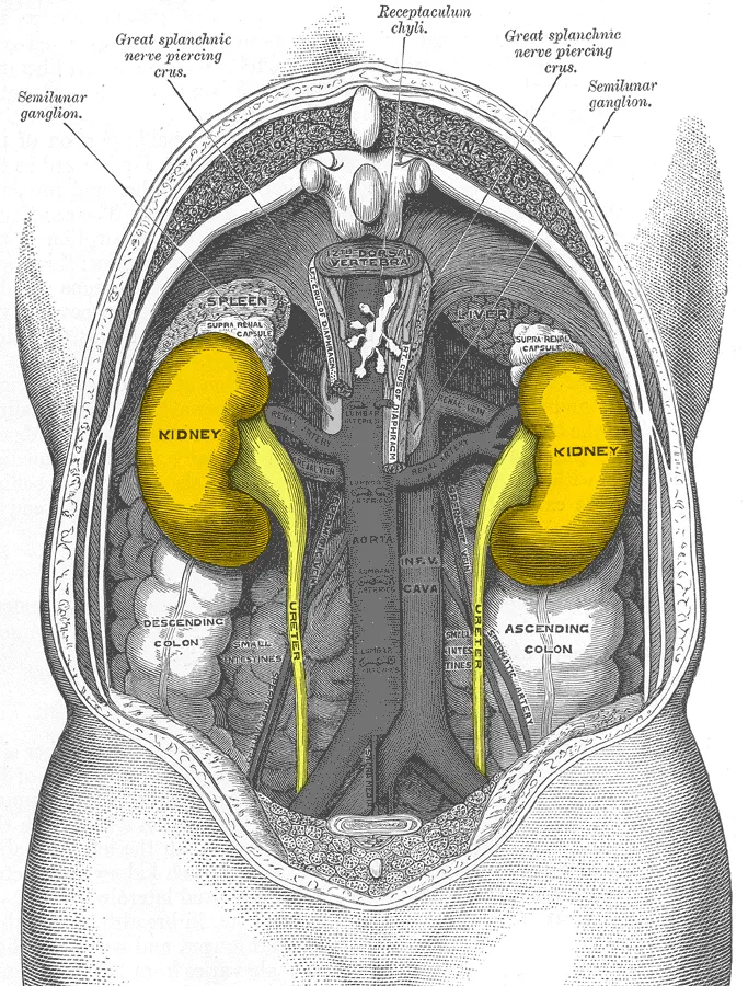
Position and relations of the kidneys
The kidneys are paired, bean-shaped retroperitoneal organs weighing on average 160 g in men and 135 g in women, 10–12 cm long, 5–7 cm wide and 3–5 cm thick, about the size of a fist; the left is usually slightly larger, some 10 g heavier. They lie between the transverse processes of T12 and L3, the right a little lower because of the liver. The long axis is tilted about 15–20° to the spine, parallel to psoas: the upper poles are more medial and posterior, the lower poles further apart, lateral and anterior. The upper pole rests on the diaphragm and is often crossed by the twelfth rib; the lower pole lies on psoas and quadratus lumborum, poorly shielded by the rib cage and so more exposed to trauma. Renal size correlates with body surface area, height and weight and declines with age.[4]
Anterior relations differ between the sides. The right kidney meets the liver across the hepatorenal recess (Morison pouch), the second part of the duodenum medially and the ascending colon below. The left kidney lies behind the descending colon, the tail of the pancreas (which may cover the hilum), the greater curvature of the stomach and the spleen, to which it is tied by the splenorenal ligament. A suprarenal gland caps each upper pole. At the hilum the structures lie, from front to back, renal vein, renal artery and renal pelvis.[4]
Renal circulation
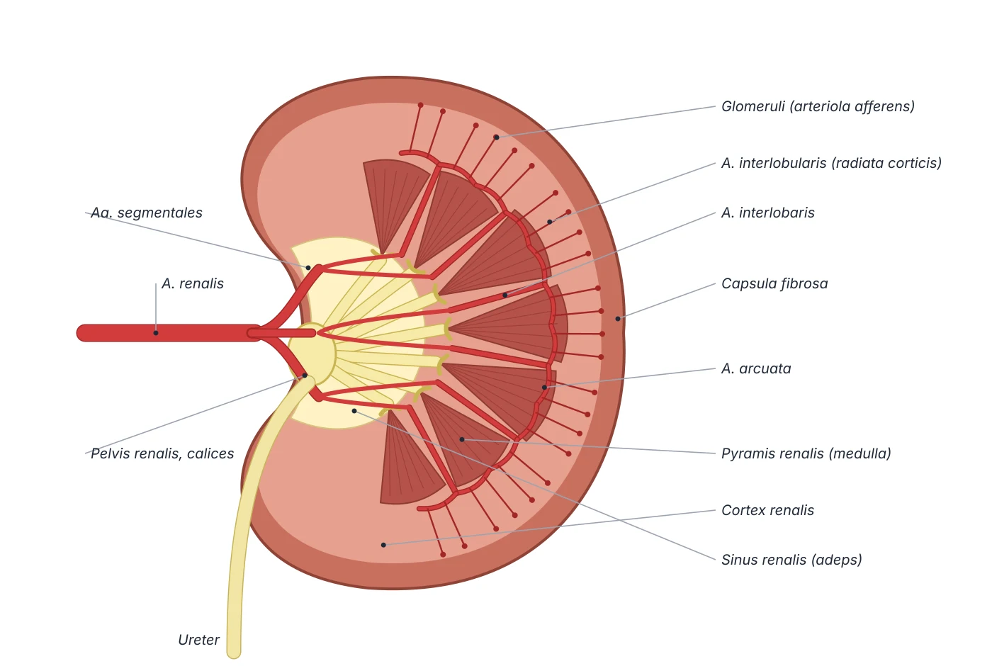
The kidneys receive about 20% of cardiac output, 1–1.1 L per minute. With a normal glomerular filtration rate of 120 mL/min, about 180 L of fluid is filtered each day while urine output is only about 1.5 L: more than 99% of the filtrate must be reabsorbed by the tubules and the peritubular capillary network. The renal arteries leave the aorta just below the superior mesenteric artery and enter the hilum at L2; the right is longer and passes behind the inferior vena cava. Each artery gives an anterior division (about 75% of flow, forming four segmental arteries) and a posterior division (the posterior segmental artery), which continue as interlobar, arcuate and interlobular arteries, afferent arterioles, glomeruli, efferent arterioles, peritubular capillaries and vasa recta.[4]
Segmental arteries are end arteries that do not anastomose within the parenchyma, so occlusion of one causes infarction of its segment. Accessory renal arteries occur in 20–30% of people, most often as an extrahilar branch to the upper pole; they are also end arteries, may cause ureteropelvic junction obstruction and must be identified before nephrectomy, transplantation or aortic aneurysm repair. The renal vein lies in front of the artery; the left renal vein is several centimetres longer and crosses in front of the aorta to reach the inferior vena cava at L2–L3, which is why the left kidney is usually chosen in living-donor nephrectomy. Sympathetic fibres (T8–L1) wrapped around the renal artery increase sodium retention, renin release and renal vasoconstriction; catheter-based renal denervation exploits this position to treat resistant hypertension.[4]
Development: pronephros, mesonephros, metanephros
The kidney develops from intermediate mesoderm in three successive generations. The pronephros, in the cervical region, appears briefly in week 4 and never functions in humans. The mesonephros, extending from the upper thoracic to the upper lumbar region, has about 40 pairs of excretory units with primitive glomeruli draining into the mesonephric (Wolffian) duct; it regresses in weeks 8–10, but in males the duct is kept as the epididymis, ductus deferens, seminal vesicle and ejaculatory duct. The metanephros, the permanent kidney, appears in week 5 through reciprocal induction between the ureteric bud (an outgrowth of the mesonephric duct) and the metanephric blastema. The bud dilates into the renal pelvis and ureter and branches into major and minor calyces and collecting ducts; the blastema forms the nephrons. Early splitting of the bud produces a bifid ureter.[4][5]
A molecular balance ensures a single ureteric bud on each side: GDNF–RET signalling drives bud outgrowth, BMP4 suppresses bud formation while lengthening the stalk, and Gremlin antagonises BMP4. The transcription factor WT1 regulates signalling from the blastema; WT1 mutations are associated with Wilms tumour and the Denys–Drash, Frasier and WAGR syndromes. Common developmental variants include the horseshoe kidney (about 1 in 400), whose lower poles fuse in front of the aorta so that ascent is arrested, the pelves face forward, accessory arteries are frequent and ureteropelvic obstruction, stones and infection are more common; the rarer fused pelvic (pancake) kidney, a single mass in the pelvis; and the retroaortic left renal vein.[5][4]
Ureters, bladder and urethra
The ureteric mucosa is lined by transitional epithelium (urothelium) on a thick fibroelastic lamina propria, with no submucosa and no glands, forming a watertight barrier. Its smooth muscle has inner longitudinal, middle circular and outer longitudinal layers; pacemaker cells in the minor calyces and renal pelvis launch peristaltic waves down the ureter. An outer adventitia carries the vessels, lymphatics and nerves. The ureter descends in front of psoas, crosses the pelvic brim at the bifurcation of the common iliac artery, is crossed anteriorly by the gonadal vessels and runs along the lateral pelvic wall to the posteroinferior bladder. It traverses the detrusor obliquely (intramural segment) before opening at the trigone, an oblique course that acts as an antireflux valve. Because the ureter contracts rhythmically, a normal contrast study never shows its whole length at once; a fully opacified column (columnisation) suggests obstruction.[5]
The bladder is an actively emptying reservoir. Parasympathetic fibres from S2–S4 in the pelvic splanchnic nerves contract the detrusor; sympathetic fibres from T10–L2 via the superior and inferior hypogastric plexuses support storage; the pudendal nerve (S2–S4, from Onuf’s nucleus in the sacral ventral horn) supplies the striated external urethral sphincter. Stretch receptors in the wall signal the central nervous system; sensation from the dome travels with the sympathetic pathway and from the lower bladder with the parasympathetic. The pontine micturition centre coordinates detrusor contraction with relaxation of both sphincters. The male urethra is long and has four parts: preprostatic (0.5–1.5 cm, within the bladder neck), prostatic (3–4 cm, the widest, with the urethral crest, seminal colliculus and prostatic duct openings), membranous and spongy. Its lining changes from urothelium proximally to pseudostratified columnar epithelium distally.[6][7]
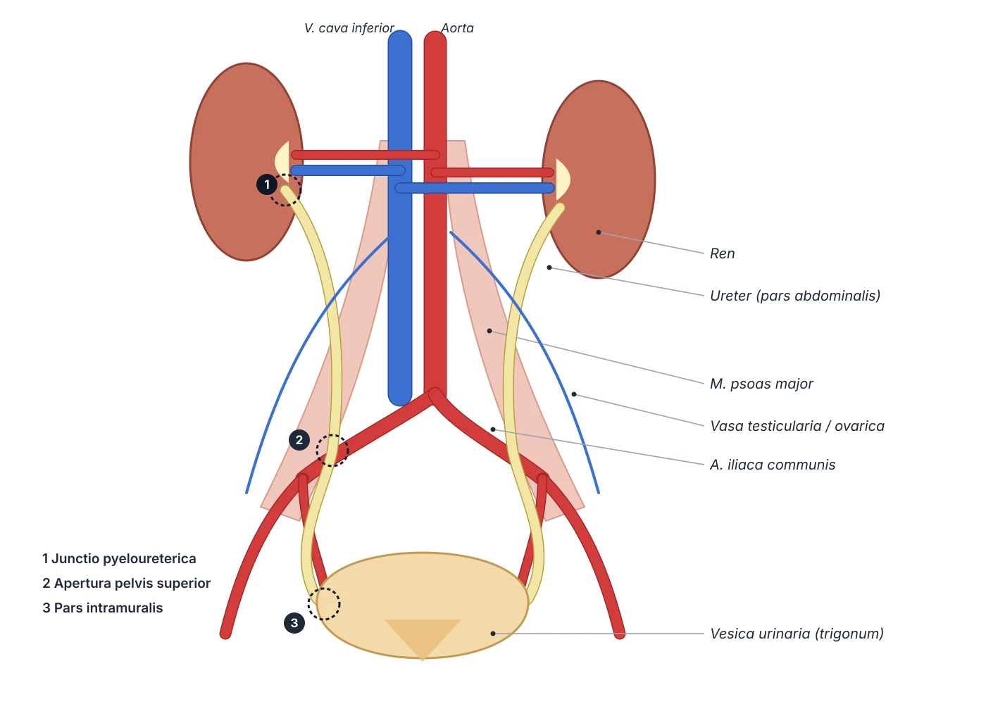
Ureteropelvic junction
- Location
- Renal pelvis funnels into the ureter at the hilum
- Significance
- Congenital obstruction; crossing lower-pole vessel
Pelvic brim
- Location
- Crossing the common iliac bifurcation
- Significance
- Stone impaction; pelvic surgical landmark
Ureterovesical junction
- Location
- Intramural course through the detrusor
- Significance
- Narrowest and aperistaltic: about 60% of stones lodge here
| Physiological narrowing | Location | Significance |
|---|---|---|
| Ureteropelvic junction | Renal pelvis funnels into the ureter at the hilum | Congenital obstruction; crossing lower-pole vessel |
| Pelvic brim | Crossing the common iliac bifurcation | Stone impaction; pelvic surgical landmark |
| Ureterovesical junction | Intramural course through the detrusor | Narrowest and aperistaltic: about 60% of stones lodge here |
Principal organs
Bean-shaped retroperitoneal organs lying either side of the vertebral column at the T12–L3 level. Each kidney contains about one million nephrons.
UreterUreterA muscular tube about 25–30 cm long that carries urine from the renal pelvis to the bladder by peristaltic waves. It has three physiological narrowings where kidney stones tend to lodge.
Urinary bladderVesica urinariaA hollow organ with a smooth-muscle wall (detrusor) lying in the lesser pelvis that stores urine; the first desire to void usually appears at about 150–200 mL.
UrethraUrethraThe tube that carries urine from the bladder neck to the exterior. The female urethra is short (about 4 cm); the male urethra is about 18–20 cm long and is a common path for urine and semen.
Reference values
- A normal glomerular filtration rate is about 125 mL/min, roughly 180 L of filtrate per day, of which all but 1–2 L is reabsorbed.
References
Sources for the values and descriptions on this page, indexed in PubMed (US National Library of Medicine).
- [1]Complexities of the glomerular basement membraneNaylor RW et al., Nature reviews. Nephrology 2021PMID 32839582
- [2]Design-based stereological methods for estimating numbers of glomerular podocytesPuelles VG et al., Annals of anatomy = Anatomischer Anzeiger 2014PMID 23845787
- [3]Crosstalk between glomeruli and tubulesFogo AB et al., Nature reviews. Nephrology 2025PMID 39643696
- [4]Anatomy, Abdomen and Pelvis: KidneysGarza FA et al., StatPearls 2025PMID 29494007
- [5]Anatomy, Abdomen and Pelvis UreterLescay HA et al., StatPearls 2024PMID 30422575
- [6]Anatomy, Abdomen and Pelvis: BladderShermadou ES et al., StatPearls 2023PMID 30285360
- [7]Histology, Male UrethraStoddard N et al., StatPearls 2023PMID 31194395
- [8]Normal reference values for glomerular filtration rate: what do we really know?Delanaye P et al., 2012PMID 22802582Supports: Glomerular filtration rate