Connective tissue, cartilage and bone
Model
Hover or tap to see names, click to read a description; click a name in the legend to fly to it. Procedurally built to histological proportions; colours follow illustration conventions.
Description
Tissue with relatively few cells scattered in abundant extracellular matrix (fibres and ground substance). The properties of the matrix set the function: soft and hydrated in loose connective tissue, tough in tendons and ligaments, compression-resistant in cartilage and rigid with calcium salts in bone.
Extracellular matrix and ground substance
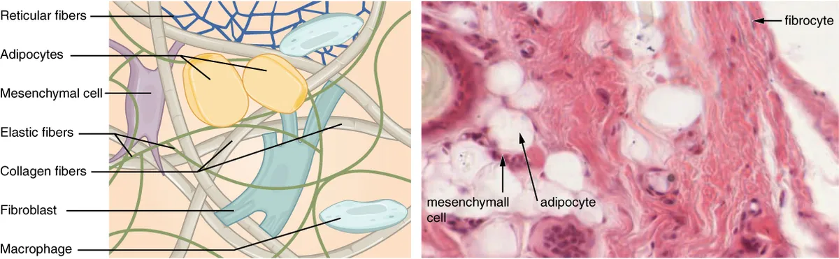
Unlike epithelium, connective tissue has few cells scattered through an abundant extracellular matrix, and it is the matrix that sets the tissue’s mechanical properties. The matrix consists of fibres (collagen, elastic, reticular) embedded in an amorphous ground substance. Ground substance is a hydrated gel of glycosaminoglycans such as hyaluronan, chondroitin sulfate and keratan sulfate; apart from hyaluronan they are attached to core proteins as proteoglycans such as aggrecan and decorin. Their dense negative charge attracts cations and water, generating a swelling pressure that resists compression, and it provides the medium through which nutrients and signals diffuse from capillaries to cells.[1][2]
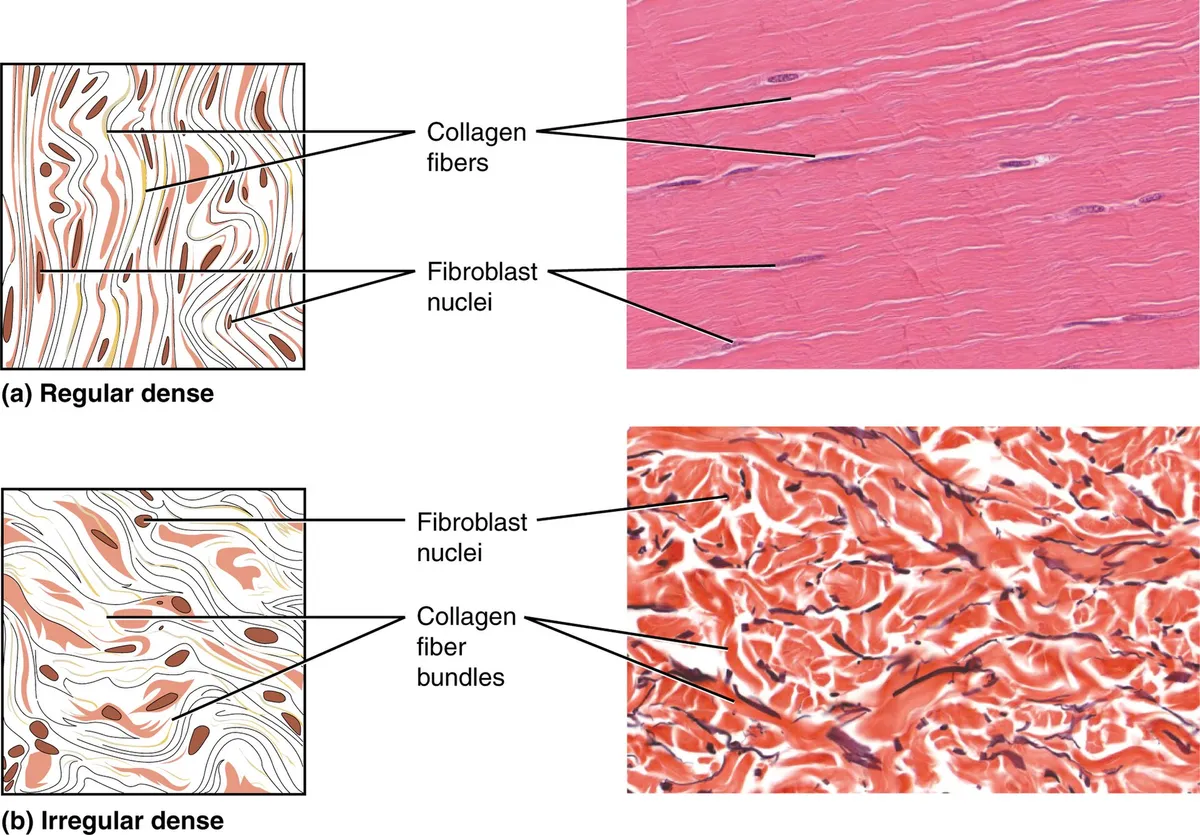
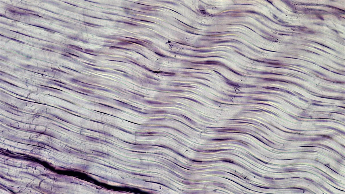
Connective tissue proper is divided into loose (areolar) and dense connective tissue. Loose tissue lies beneath epithelia and around vessels; it is rich in ground substance, its fibres are sparse and loosely arranged, and it is full of immune cells, the stage on which inflammation and oedema play out. Dense irregular tissue (reticular dermis, organ capsules) has collagen bundles running in all directions to resist forces from many sides; dense regular tissue (tendon, ligament, aponeurosis) has parallel bundles resisting tension along one axis. The specialised connective tissues are adipose tissue, cartilage, bone, blood and the reticular tissue of haematopoietic organs.[1][2]
Collagen, elastic and reticular fibres
Collagen makes up about a quarter of body protein and comes in nearly 30 types. Each molecule is a triple helix of three α chains with the repeating sequence Gly-X-Y, X and Y often being proline and hydroxyproline. Synthesis begins in the rough ER: proline and lysine are hydroxylated by vitamin C-dependent enzymes, three chains wind into procollagen and are secreted; outside the cell peptidases trim both ends, tropocollagen molecules self-assemble in a staggered array that gives the 67 nm banding, and lysyl oxidase cross-links them. Lack of vitamin C weakens collagen, causing the bleeding gums and poor wound healing of scurvy.[3][4]
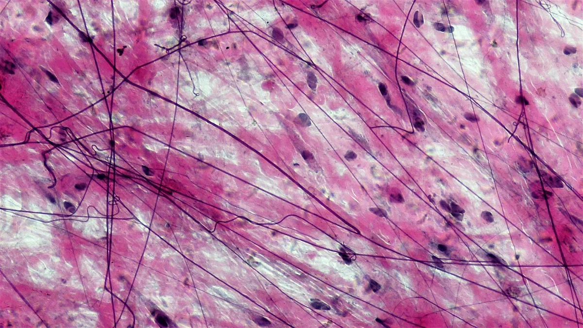
Collagen I forms the thick tension-resisting fibres of tendon, skin and bone; mutations in COL1A1 or COL1A2 cause osteogenesis imperfecta with brittle bones, blue sclerae and deafness. Collagen III forms the fine reticular fibres that support vessels, gut and haematopoietic organs; COL3A1 mutations cause vascular Ehlers–Danlos syndrome with a risk of arterial rupture. Elastic fibres have a core of elastin cross-linked by desmosine within a sheath of fibrillin microfibrils, stretching to about one and a half times their length and recoiling; they abound in large arteries, lung and skin. Fibrillin-1 mutations cause Marfan syndrome with aortic root dilation.[5][6][7][8]
Connective tissue cells
Fibroblasts are the main resident cells: spindle-shaped with a flat nucleus, and with prominent rough ER and Golgi when active, they synthesise and remodel collagen, elastin, proteoglycans and the metalloproteinases that degrade matrix. After injury, under the influence of TGF-β, fibroblasts become myofibroblasts containing α-smooth-muscle actin that pull wound edges together and lay down scar. If this signal does not switch off, scarring spreads and becomes fibrosis, as in pulmonary fibrosis, cirrhosis or keloids.[9][10]
Loose connective tissue is also an immune outpost. Resident macrophages, many of embryonic origin and self-maintaining, engulf debris and bacteria and present antigen; blood monocytes reinforce them in inflammation. Mast cells sit next to vessels, their cytoplasm packed with heparin- and histamine-filled granules; when surface IgE meets an allergen they degranulate within seconds, causing the vasodilation, oedema and itch of allergy and, in severe cases, anaphylaxis. Plasma cells secrete antibody, and eosinophils are common in the connective tissue of gut and lung.[11][12][13]
White and brown adipose tissue
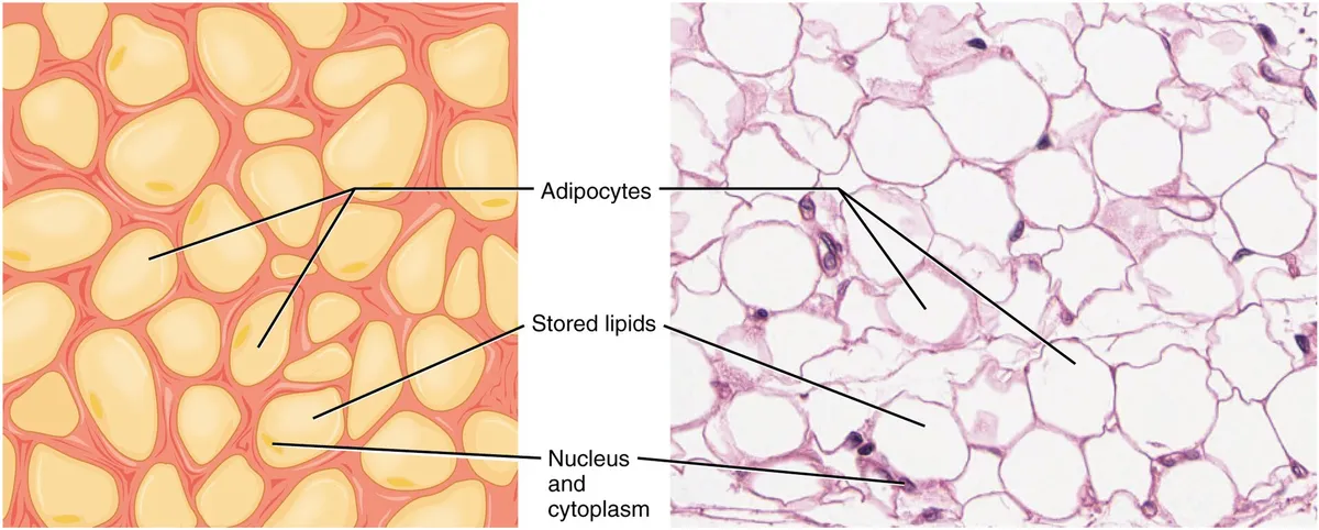
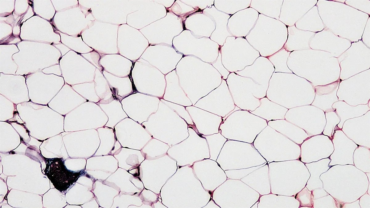
White adipocytes contain one large lipid droplet occupying over 90% of the cell, squeezing nucleus and cytoplasm into a thin “signet ring” rim. White fat stores energy as triglyceride, insulates, cushions and acts as an endocrine organ secreting leptin, a satiety signal, and adiponectin, which enhances insulin sensitivity. In obesity, adipocytes enlarge, become hypoxic and attract macrophages, creating a low-grade chronic inflammation that contributes to insulin resistance.[14][15]
Brown adipocytes contain many small lipid droplets and are crowded with mitochondria rich in uncoupling protein 1 (UCP1); UCP1 lets protons flow back across the inner membrane without making ATP, turning respiratory energy into heat. Brown fat is plentiful in newborns (between the shoulder blades, around the kidneys) to keep them warm; adults retain depots in the neck and above the clavicles, activated by cold through sympathetic nerves. “Beige” fat consists of thermogenic cells that appear within white fat during prolonged cold; besides UCP1 they use other heat-producing cycles, such as creatine and calcium cycling.[14][15][16][17]
Cartilage
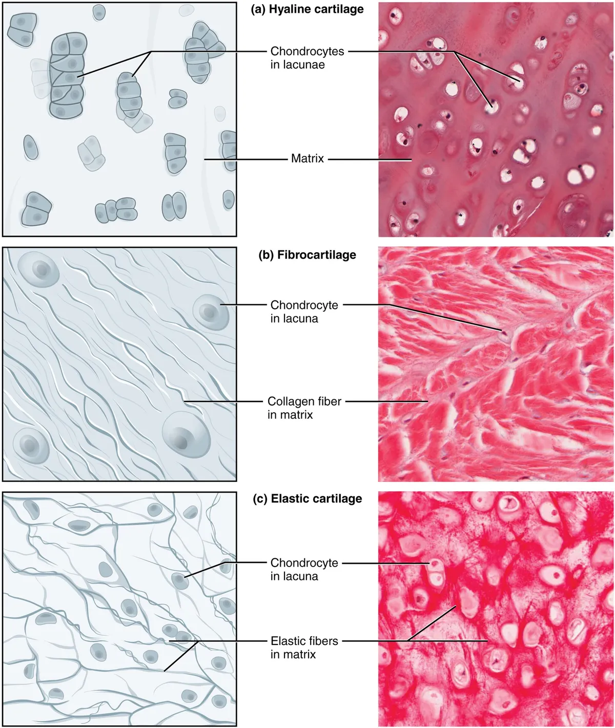
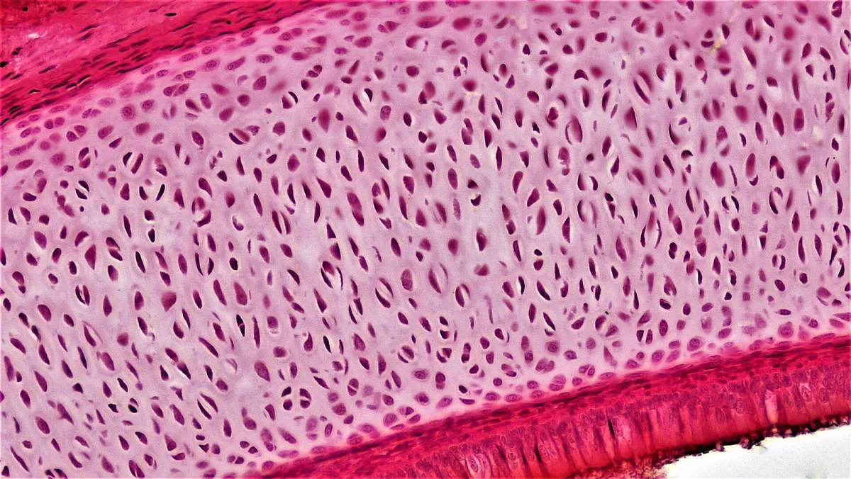
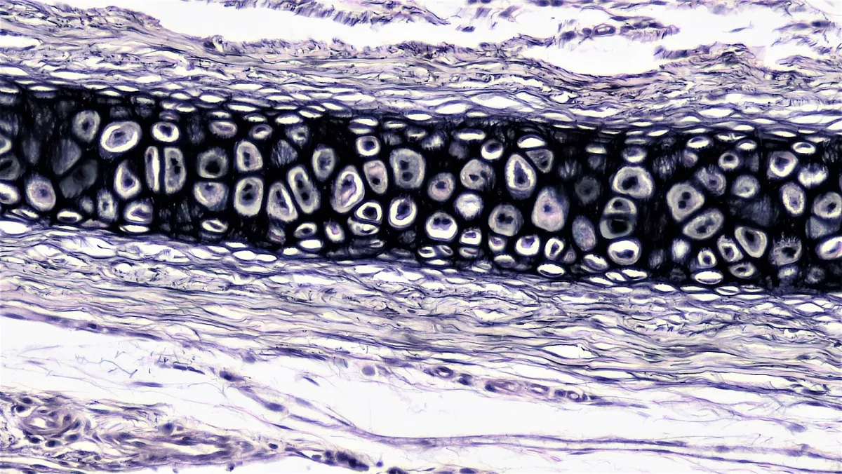
Cartilage is a firm yet resilient connective tissue with no blood vessels, nerves or lymphatics; chondrocytes live in lacunae and are nourished by diffusion, so cartilage heals poorly. Hyaline cartilage has a fine collagen II network and aggrecan-rich matrix and is found in articular surfaces, costal cartilage and the trachea, and as the template for endochondral ossification of long bones. Elastic cartilage adds elastic fibres for flexibility (auricle, epiglottis); fibrocartilage has abundant collagen I bundles and little matrix, resisting tension and compression in intervertebral discs, menisci and the pubic symphysis.[18][19]
Articular cartilage is zoned: a superficial zone with flattened cells and fibres parallel to the surface, a middle zone with oblique fibres, a deep zone with columns of cells and perpendicular fibres, then a tidemark separating it from calcified cartilage. Under load, water is squeezed out of the matrix and drawn back in, reducing friction and pumping nutrients to the cells. Osteoarthritis is a disease of the whole joint: with age, chondrocytes accumulate glycation products, respond less to growth factors and secrete degrading enzymes such as ADAMTS-5 and MMP-13, so the cartilage wears away and the subchondral bone remodels.[20][21][22]
Bone tissue
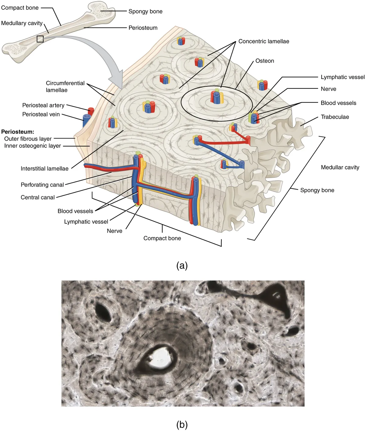
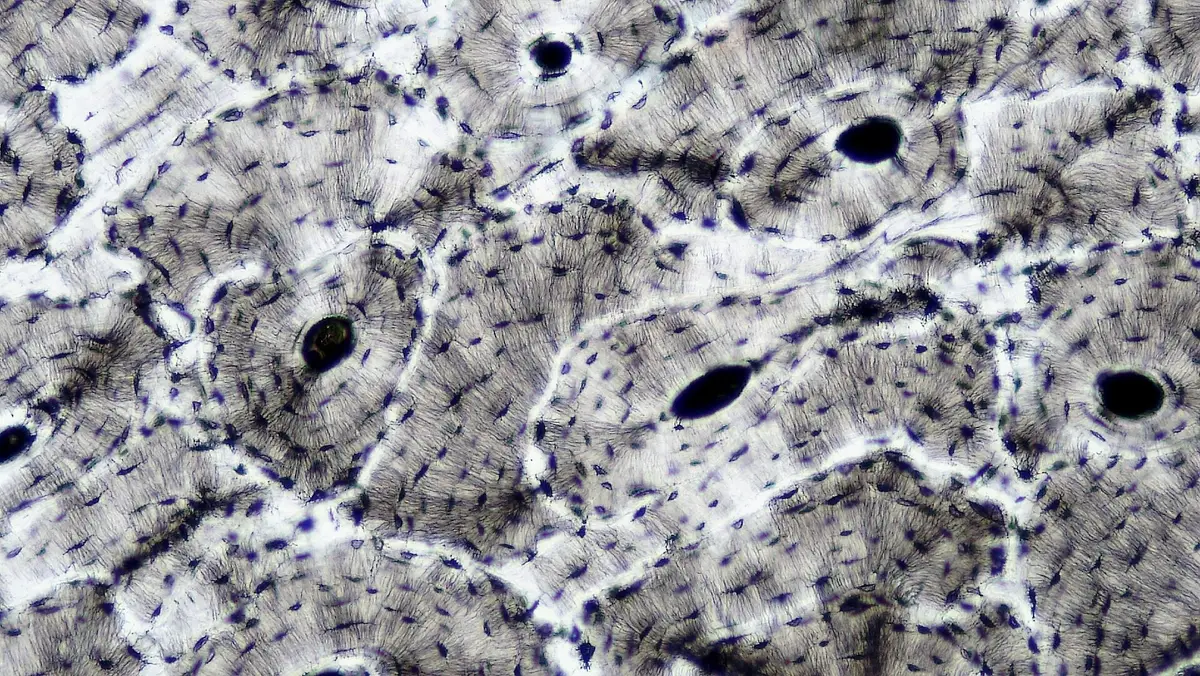
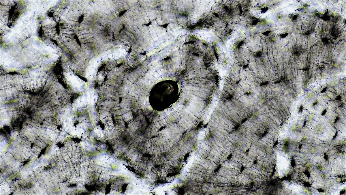
Bone is connective tissue with a mineralised matrix: about 60% of its weight is hydroxyapatite crystals deposited on a collagen I scaffold, the rest being non-collagenous proteins and water. Collagen gives toughness, mineral gives stiffness. The compact bone of the cortex is built from cylindrical osteons: concentric lamellae around a Haversian canal carrying vessels and nerves, linked to one another and to the periosteum by transverse Volkmann canals; interstitial lamellae fill the gaps between osteons and circumferential lamellae run around the outer and inner surfaces. Spongy bone at the bone ends is a lattice of trabeculae aligned with stress lines, with marrow in between.[23][5]
Three cell types share the work. Osteoblasts, of mesenchymal origin, secrete osteoid and mineralise it; once buried they become osteocytes, more than 90% of bone cells, which sit in lacunae and connect through cytoplasmic processes in canaliculi. Osteocytes sense mechanical load and secrete sclerostin and RANKL to steer the other two. Osteoclasts are multinucleated cells of the monocyte lineage that seal onto bone and secrete acid and cathepsin K through a ruffled border to resorb it. About 10% of the skeleton is replaced each year by remodelling units; when resorption outpaces formation, as after menopause, bone thins into osteoporosis.[23][24][25][26]
Full-text reading (StatPearls)
The StatPearls chapters on this topic, reproduced verbatim. Their CC BY-NC-ND 4.0 licence allows unmodified copies only, so they are not translated or shortened.
Anatomy, Connective TissueKamrani P, Marston G, Arbor TC, Jan A · StatPearls 2023 · NBK538534 · 15k characters
Loading…
Kamrani P, Marston G, Arbor TC, Jan A. Anatomy, Connective Tissue. StatPearls [Internet]. Treasure Island (FL): StatPearls Publishing; 2023. NBK538534 · PMID 30860769 · CC BY-NC-ND 4.0. Reproduced verbatim, unmodified.
Biochemistry, Collagen SynthesisRahimi N, Launico MV · StatPearls 2026 · NBK507709 · 13k characters
Loading…
Rahimi N, Launico MV. Biochemistry, Collagen Synthesis. StatPearls [Internet]. Treasure Island (FL): StatPearls Publishing; 2026. NBK507709 · PMID 29939531 · CC BY-NC-ND 4.0. Reproduced verbatim, unmodified.
Anatomy, Head and Neck: Larynx Arytenoid CartilageAndaloro C, Sharma P, La Mantia I · StatPearls 2023 · NBK513252 · 13k characters
Loading…
Andaloro C, Sharma P, La Mantia I. Anatomy, Head and Neck: Larynx Arytenoid Cartilage. StatPearls [Internet]. Treasure Island (FL): StatPearls Publishing; 2023. NBK513252 · PMID 30020624 · CC BY-NC-ND 4.0. Reproduced verbatim, unmodified.
Anatomy, Head and Neck, Cricoid CartilageMathews S, Jain S · StatPearls 2023 · NBK539821 · 12k characters
Loading…
Mathews S, Jain S. Anatomy, Head and Neck, Cricoid Cartilage. StatPearls [Internet]. Treasure Island (FL): StatPearls Publishing; 2023. NBK539821 · PMID 30969643 · CC BY-NC-ND 4.0. Reproduced verbatim, unmodified.
Histology, FibroblastDick MK, Miao JH, Limaiem F · StatPearls 2023 · NBK541065 · 12k characters
Loading…
Dick MK, Miao JH, Limaiem F. Histology, Fibroblast. StatPearls [Internet]. Treasure Island (FL): StatPearls Publishing; 2023. NBK541065 · PMID 31082109 · CC BY-NC-ND 4.0. Reproduced verbatim, unmodified.
Anatomy, Head and Neck: Larynx CartilageFlynn W, Vickerton P · StatPearls 2023 · NBK553185 · 11k characters
Loading…
Flynn W, Vickerton P. Anatomy, Head and Neck: Larynx Cartilage. StatPearls [Internet]. Treasure Island (FL): StatPearls Publishing; 2023. NBK553185 · PMID 31985978 · CC BY-NC-ND 4.0. Reproduced verbatim, unmodified.
Histology, Mast CellsFong M, Crane JS · StatPearls 2023 · NBK499904 · 9k characters
Loading…
Fong M, Crane JS. Histology, Mast Cells. StatPearls [Internet]. Treasure Island (FL): StatPearls Publishing; 2023. NBK499904 · PMID 29763079 · CC BY-NC-ND 4.0. Reproduced verbatim, unmodified.
Physiology, Connective TissueNezwek TA, Varacallo MA · StatPearls 2022 · NBK542226 · 9k characters
Loading…
Nezwek TA, Varacallo MA. Physiology, Connective Tissue. StatPearls [Internet]. Treasure Island (FL): StatPearls Publishing; 2022. NBK542226 · PMID 31194383 · CC BY-NC-ND 4.0. Reproduced verbatim, unmodified.
Anatomy, CartilageChang LR, Marston G, Martin A · StatPearls 2022 · NBK532964 · 8k characters
Loading…
Chang LR, Marston G, Martin A. Anatomy, Cartilage. StatPearls [Internet]. Treasure Island (FL): StatPearls Publishing; 2022. NBK532964 · PMID 30422559 · CC BY-NC-ND 4.0. Reproduced verbatim, unmodified.
Organs in this system
References
Sources for the values and descriptions on this page, indexed in PubMed (US National Library of Medicine).
- [1]Anatomy, Connective TissueKamrani P et al., StatPearls 2023PMID 30860769
- [2]Physiology, Connective TissueNezwek TA et al., StatPearls 2022PMID 31194383
- [3]Biochemistry, Collagen SynthesisRahimi N et al., StatPearls 2026PMID 29939531
- [4]Collagen Fibril Assembly and FunctionHolmes DF et al., Current topics in developmental biology 2018PMID 29853175
- [5]Type 1 collagen: Synthesis, structure and key functions in bone mineralizationSelvaraj V et al., Differentiation; research in biological diversity 2024PMID 38437764
- [6]Osteogenesis imperfectaMarini JC et al., Nature reviews. Disease primers 2017PMID 28820180
- [7]Type III collagen (COL3A1): Gene and protein structure, tissue distribution, and associated diseasesKuivaniemi H et al., Gene 2019PMID 31075413
- [8]Elastin architectureVindin H et al., Matrix biology 2019PMID 31301399
- [9]Histology, FibroblastDick MK et al., StatPearls 2023PMID 31082109
- [10]Concise review: how do red blood cells born, live, and die?Corrons JLV et al., Annals of hematology 2021PMID 34342697
- [11]Monocytes and macrophages: developmental pathways and tissue homeostasisGinhoux F et al., Nature reviews. Immunology 2014PMID 24854589
- [12]Histology, Mast CellsFong M et al., StatPearls 2023PMID 29763079
- [13]IgE, mast cells, basophils, and eosinophilsStone KD et al., The Journal of allergy and clinical immunology 2010PMID 20176269
- [14]Brown and beige fat: development, function and therapeutic potentialHarms M et al., Nature medicine 2013PMID 24100998
- [15]Brown Adipose Tissue-A Translational PerspectiveCarpentier AC et al., Endocrine reviews 2023PMID 35640259
- [16]UCP1-independent thermogenesisRoesler A et al., The Biochemical journal 2020PMID 32059055
- [17]Control of brown and beige fat developmentWang W et al., Nature reviews. Molecular cell biology 2016PMID 27552974
- [18]Anatomy, CartilageChang LR et al., StatPearls 2022PMID 30422559
- [19]Basic Science of Articular CartilageCarballo CB et al., Clinics in sports medicine 2017PMID 28577703
- [20]Restoring articular cartilage: insights from structure, composition and developmentPueyo Moliner A et al., Nature reviews. Rheumatology 2025PMID 40155694
- [21]Cartilage Homeostasis and OsteoarthritisFujii Y et al., International journal of molecular sciences 2022PMID 35682994
- [22]Ageing and the pathogenesis of osteoarthritisLoeser RF et al., Nature reviews. Rheumatology 2016PMID 27192932
- [23]Biology of Bone Tissue: Structure, Function, and Factors That Influence Bone CellsFlorencio-Silva R et al., BioMed research international 2015PMID 26247020
- [24]The biology of normal bone remodellingKatsimbri P, European journal of cancer care 2017PMID 28786518
- [25]Osteoblast-Osteoclast Communication and Bone HomeostasisKim JM et al., Cells 2020PMID 32927921
- [26]Physiological Bone Remodeling: Systemic Regulation and Growth Factor InvolvementSiddiqui JA et al., Physiology 2016PMID 27053737