Pectoralis major
Model
Layers
Z-Anatomy anatomical model based on BodyParts3D, licensed CC BY-SA 4.0.
Description
A broad fan-shaped muscle of the anterior chest wall forming the anterior axillary fold. It adducts, medially rotates and flexes the arm and is an accessory muscle of respiration.
Parts
Each part has its own card; press “Show on the model” to fly to it and select it in the 3D model above.
- 01
Clavicular head
Pars clavicularis musculi pectoralis majoris
Arises from the medial half of the anterior border of the clavicle, separated from the sternocostal head by a small groove. It flexes the arm from extension and adducts it; it is separated from deltoid by the deltopectoral groove, which carries the cephalic vein.
- 02
Sternocostal head
Pars sternocostalis musculi pectoralis majoris
The largest part, arising from the front of the sternum and the first six costal cartilages. It powerfully adducts and medially rotates the arm and extends it from a flexed position, as in climbing.
- 03
Abdominal part
Pars abdominalis musculi pectoralis majoris
The small lowest slip, arising from the anterior layer of the rectus sheath, helping to form the anterior axillary fold with the lower sternocostal fibres.
- 04
Twisted insertion
Insertio
The fibres converge into a flat tendon on the lateral lip of the intertubercular groove of the humerus. The tendon is twisted: clavicular fibres insert low and in front, the lowest sternocostal fibres high and behind, which rounds the anterior axillary fold. Tendon rupture typically occurs during heavy bench pressing.
- 05
Nerve and blood supply
Innervatio et vascularisatio
The lateral pectoral nerve (C5–C7) supplies the clavicular head and the medial pectoral nerve (C8–T1), piercing pectoralis minor, the sternocostal head. Blood comes mainly from the pectoral branch of the thoraco-acromial artery, the pedicle on which a pectoralis major flap is rotated for head and neck reconstruction.
- 06
Pectoralis minor (deep layer)
Musculus pectoralis minor
A triangular muscle beneath pectoralis major, from ribs 3–5 to the coracoid process, drawing the scapula forwards and down. It divides the axillary artery into three parts and is the landmark for the levels of axillary nodes in breast cancer surgery.
Position and relations
It has three parts: clavicular from the medial half of the clavicle, sternocostal from the sternum and upper six costal cartilages, and abdominal from the rectus sheath. The fibres twist as they insert on the lateral lip of the intertubercular groove, the lower fibres inserting higher, forming the rounded anterior axillary fold. The breast lies on the pectoral fascia.
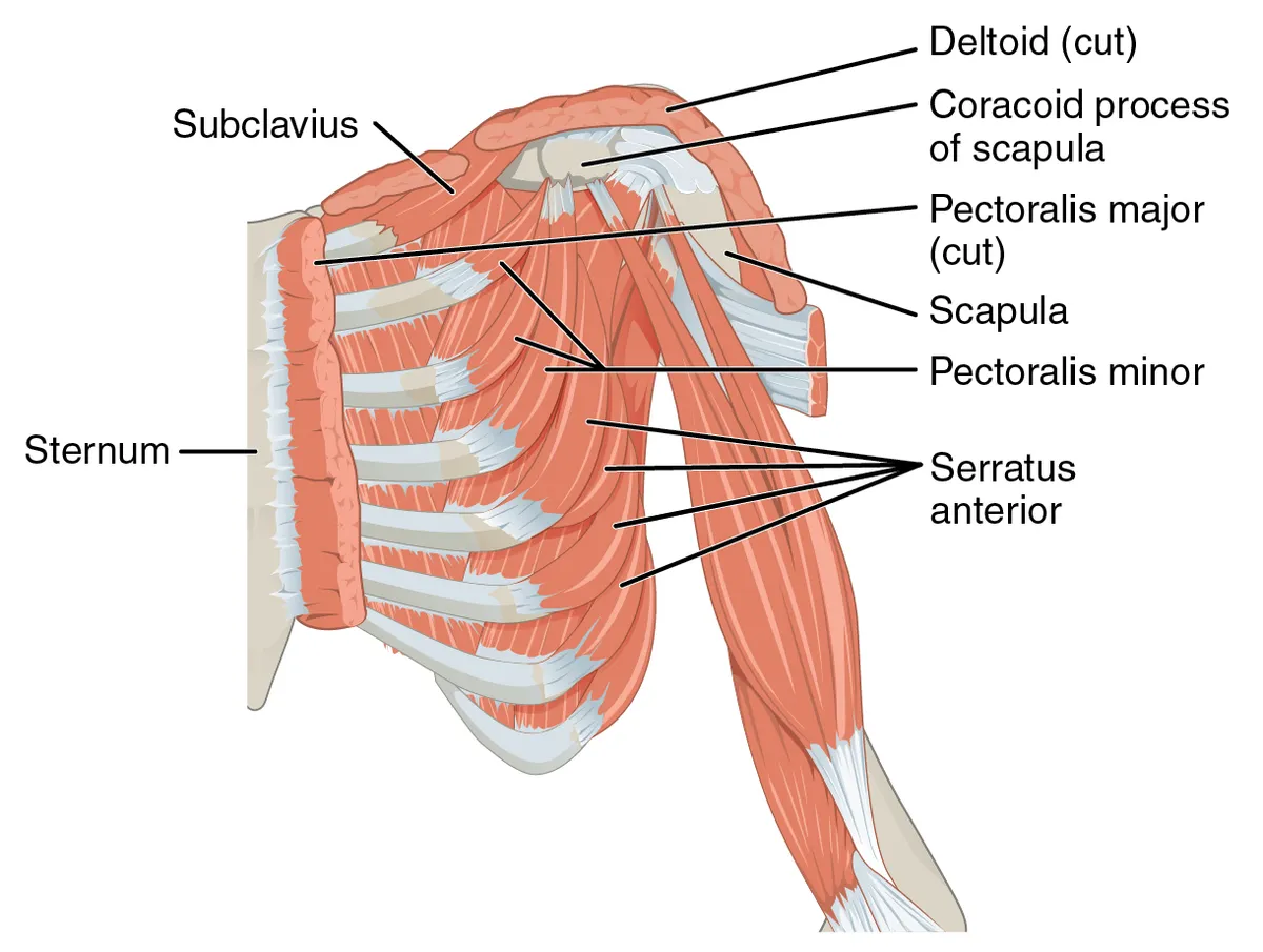
Form and structure
The clavicular head flexes the arm and the sternocostal head adducts it and extends it from flexion; with the arm fixed (leaning on a table) the muscle lifts the rib cage in forced inspiration. Tendon avulsion is typical in bench-press lifters; congenital absence of the sternocostal head characterises Poland syndrome.
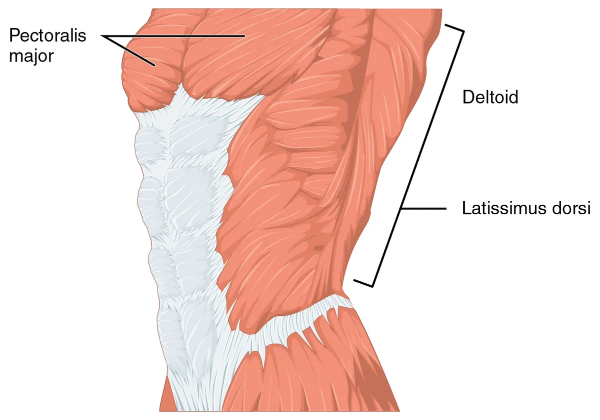
Vessels and nerves
The lateral and medial pectoral nerves (brachial plexus, C5–T1) supply it; blood comes from the pectoral branch of the thoracoacromial artery and perforators of the internal thoracic artery. The pedicled pectoralis major flap is a classic workhorse in head-and-neck and chest-wall reconstruction.
Heads and pectoral nerves
The pectoralis major is the most superficial and largest muscle of the anterior chest wall. Thick and fan-shaped, it lies beneath the breast and forms the anterior wall of the axilla. It has two heads: the clavicular head arises from the anterior medial half of the clavicle, and the sternocostal head from the anterior sternum, the first seven costal cartilages, the sternal end of the sixth rib and the external oblique aponeurosis, in 2 to 7 distinct segments. It inserts on the lateral lip of the intertubercular sulcus of the humerus.[1]
A meta-analysis found that the lateral pectoral nerve most often arises by two branches from the anterior divisions of the upper and middle trunks (33.8%) or as a single root from the lateral cord (23.4%), and the medial pectoral nerve from the medial cord (49.3%) or the anterior division of the lower trunk (43.8%). The two are usually linked just distal to the thoraco-acromial artery by the ansa pectoralis, and in 50–100% of cases at least some medial pectoral branches pierce pectoralis minor. The lateral nerve supplies the upper pectoralis major; the medial nerve supplies its lower parts and pectoralis minor.[2]
The pectoral nerves can be injured by direct trauma, hypertrophic muscle compression or iatrogenically in breast surgery, axillary dissection or pectoralis major transfer, and they also serve as donors in nerve transfers. Pectoralis major injuries are uncommon but increasing; the muscle’s complex, variable anatomy hampers standard classification, and MRI diagnosis helps avoid surgical delay.[2][3]
Full-text reading (StatPearls)
The StatPearls chapters on this topic, reproduced verbatim. Their CC BY-NC-ND 4.0 licence allows unmodified copies only, so they are not translated or shortened.
Anatomy, Thorax, Pectoralis Major MajorSolari F, Burns B · StatPearls 2023 · NBK525991 · 7k characters
Loading…
Solari F, Burns B. Anatomy, Thorax, Pectoralis Major Major. StatPearls [Internet]. Treasure Island (FL): StatPearls Publishing; 2023. NBK525991 · PMID 30252247 · CC BY-NC-ND 4.0. Reproduced verbatim, unmodified.
Organs in this system
References
Sources for the values and descriptions on this page, indexed in PubMed (US National Library of Medicine).
- [1]Anatomy, Thorax, Pectoralis Major MajorSolari F et al., StatPearls 2023PMID 30252247
- [2]Surgical anatomy of the pectoral nerves and the pectoral musculaturePorzionato A et al., Clinical anatomy 2012PMID 22125052
- [3]Magnetic resonance imaging of pectoralis major injuries: a radiologist's essential guideLake MG et al., Emergency radiology 2025PMID 40772985
- [4]Anatomy, Bony Pelvis and Lower Limb, Gluteus Maximus MuscleStatPearls (NCBI Bookshelf)PMID 30855781Supports: Gluteus maximus