Breast (mammary gland)
Model
Hover or tap to see names, click to read a description; click a name in the legend to fly to it. Procedurally built to histological proportions; colours follow illustration conventions.
Description
An exocrine gland in the subcutaneous tissue of the anterior chest wall, usually from the second to the sixth rib, with an axillary tail extending towards the axilla. The glandular tissue has 15–20 lobes, each draining through a lactiferous duct with a sinus before opening on the nipple; suspensory (Cooper’s) ligaments tie the skin to the pectoral fascia. Lymph drains mainly to the axillary nodes.
Parts
Each part has its own card; press “Show on the model” to fly to it and select it in the 3D model above.
- 01
Lobes and lobules
Lobi et lobuli glandulae mammariae
The breast consists of 15–20 lobes arranged like spokes around the nipple, each a branched tubulo-alveolar gland. The functional unit is the terminal duct lobular unit (TDLU), where most breast cancers arise. Glandular tissue is most abundant in the upper outer quadrant and extends into the axilla as the axillary tail (of Spence).
- 02
Lactiferous ducts
Ductus lactiferi
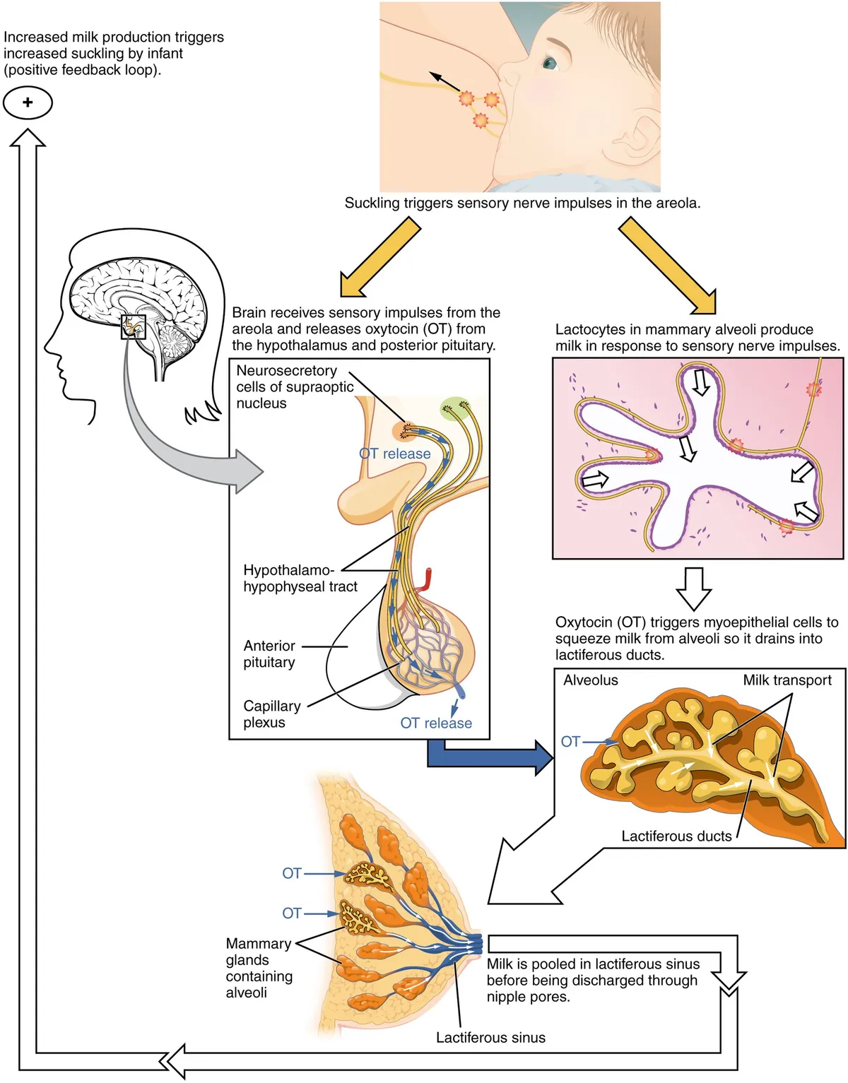
Each lobe has a main lactiferous duct opening on the nipple; just beneath the areola the duct widens into a lactiferous sinus. Ducts are lined by a double layer of luminal and myoepithelial cells; the myoepithelial cells contract under oxytocin to eject milk.
- 03
Nipple and areola
Papilla mammaria et areola mammae
The nipple usually lies at the fourth intercostal space in men; it carries the duct openings and many smooth muscle fibres that make it erect. The pigmented areola contains Montgomery’s (areolar) glands, which lubricate and protect it during breastfeeding.
- 04
Suspensory ligaments (Cooper’s)
Ligamenta suspensoria mammaria
Fibrous septa running from the deep fascia through the breast to the dermis; they support the gland and divide the breast fat into compartments. When a tumour infiltrates and shortens them, the skin dimples or takes a peau d’orange appearance.
- 05
Retromammary space and bed
Spatium retromammarium
The breast bed extends from the second to sixth ribs and from the sternal edge to the midaxillary line, over pectoralis major, serratus anterior and part of the external oblique. The loose retromammary space lets the gland slide over pectoralis major; a tumour fixed to the muscle loses this mobility.
- 06
Blood supply and lymphatic drainage
Vasa et lymphatica mammae
Arteries come from perforating branches of the internal thoracic artery (about 60%), the lateral thoracic artery and the intercostal arteries. About 75% of the lymph, especially from the lateral quadrants, drains to the axillary nodes; most of the rest, chiefly from the medial quadrants, drains to parasternal nodes along the internal thoracic artery.
Position and relations
The breast lies on the fascia of pectoralis major and part of serratus anterior, separated by a loose retromammary space that lets it move. Its axillary tail (of Spence) passes through the axillary fascia. Clinically it is divided into four quadrants around the nipple; the upper outer quadrant holds the most glandular tissue and is the commonest site of cancer.
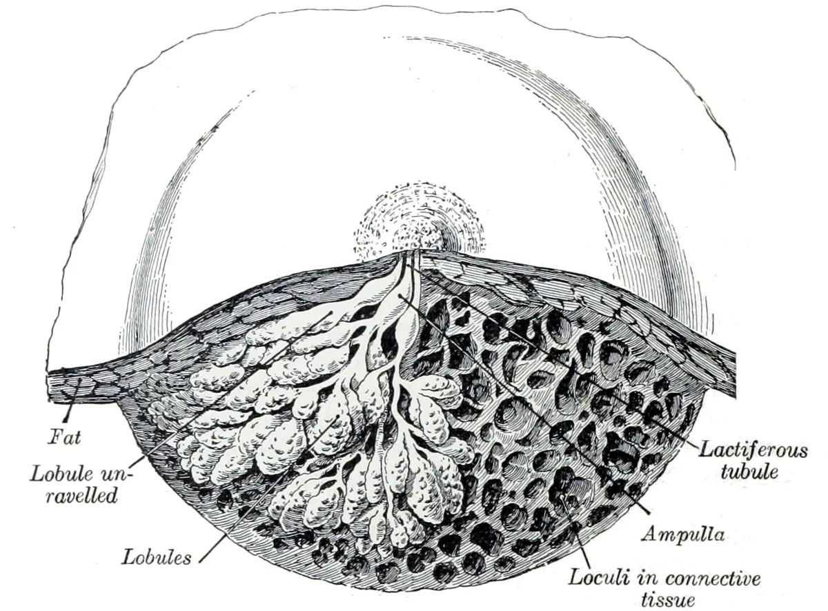
Form and structure
Each lobe contains many lobules, whose functional unit, the terminal duct lobular unit, is where most breast cancers arise. The glandular tissue is embedded in fat and fibrous tissue; its proportion rises sharply in pregnancy and lactation and declines after menopause. The areola carries Montgomery glands that secrete a lubricant.
Vessels and nerves
Arteries come from the mammary branches of the internal thoracic artery, the lateral thoracic artery, the pectoral branches of the thoraco-acromial artery and the intercostal arteries. About 75% of lymph drains to the axillary nodes and the rest to the parasternal nodes; the axillary sentinel node underlies sentinel-node biopsy in breast cancer surgery. Sensation comes from cutaneous branches of the fourth to sixth intercostal nerves.
Development
The mammary gland begins in the embryonic milk lines: placodes form, invaginate into flask-shaped buds and branch into a miniature bilayered ductal tree (luminal and myoepithelial cells). The signalling pathways that drive this stage are highly conserved and are reactivated in breast cancer.[1]
The mammary gland reaches maturity only during pregnancy and lactation. Puberty lays the groundwork; the incomplete breast is mostly adipose tissue with lobes that drain into lactiferous ducts, the lactiferous sinus and the nipple–areola complex. After weaning the gland regresses to a resting state and can repeat the cycle of expansion and regression with each pregnancy, a plasticity that implies tight hormonal control.[2][3]
Physiology of lactation
In the first trimester oestrogen elongates and branches the ductal system into the fat pad and stimulates pituitary prolactin release. By about week 20 the gland can make milk components, but high oestrogen and progesterone still inhibit secretion, so only colostrum is produced. The areola darkens, the breast enlarges and the Montgomery glands become prominent.[3]
Delivery of the placenta causes abrupt progesterone withdrawal and initiates lactation, which is maintained by prolactin (milk synthesis) and oxytocin (ejection through myoepithelial contraction), both driven by suckling. The gland is also endocrine: it secretes parathyroid hormone-related peptide (PTHrP), which mobilises maternal skeletal calcium for milk. After weaning, local cytokines and falling prolactin trigger large-scale cell loss and involution.[4]
The calcium demand of milk is met mainly by temporary maternal bone resorption (driven by PTHrP and low oestrogen) rather than by increased intestinal absorption; bone density recovers after weaning.[5]
Full-text reading (StatPearls)
The StatPearls chapters on this topic, reproduced verbatim. Their CC BY-NC-ND 4.0 licence allows unmodified copies only, so they are not translated or shortened.
Anatomy, Thorax, BreastRivard AB, Peterson DC · StatPearls 2023 · NBK519575 · 9k characters
Loading…
Rivard AB, Peterson DC. Anatomy, Thorax, Breast. StatPearls [Internet]. Treasure Island (FL): StatPearls Publishing; 2023. NBK519575 · PMID 30137859 · CC BY-NC-ND 4.0. Reproduced verbatim, unmodified.
Organs in this system
References
Sources for the values and descriptions on this page, indexed in PubMed (US National Library of Medicine).
- [1]Embryonic mammary gland developmentSpina E et al., Seminars in cell & developmental biology 2021PMID 33472760
- [2]Anatomy of the human mammary gland: Current status of knowledgeHassiotou F et al., Clinical anatomy 2013PMID 22997014
- [3]Anatomy and Physiology of the Breast during Pregnancy and LactationAlex A et al., Advances in experimental medicine and biology 2020PMID 32816256
- [4]Hormonal regulation of mammary gland development and lactationHannan FM et al., Nature reviews. Endocrinology 2023PMID 36192506
- [5]Maternal Mineral and Bone Metabolism During Pregnancy, Lactation, and Post-Weaning RecoveryKovacs CS, Physiological reviews 2016PMID 26887676