Femur
Model
Hover or tap to see names, click to read a description; click a name in the legend to fly to it. Procedurally built to histological proportions; colours follow illustration conventions.
Description
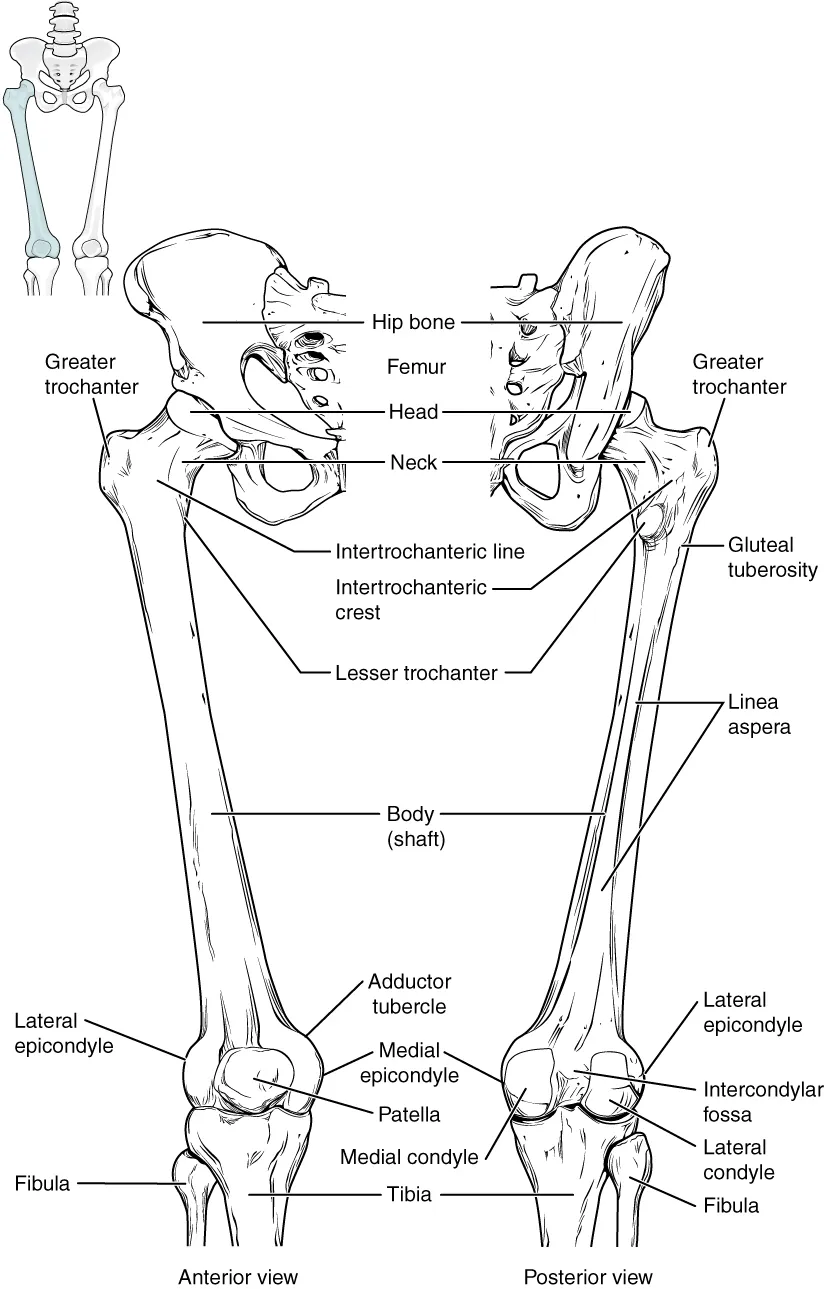
The longest and strongest bone, about one quarter of body height; it articulates with the hip bone at the hip joint and with the tibia at the knee.
Parts
Each part has its own card; press “Show on the model” to fly to it and select it in the 3D model above.
- 01
Femoral head
Caput femoris
About two thirds of a sphere fitting snugly into the acetabulum to form the stable hip joint. A central pit, the fovea, anchors the ligament of the head; its artery supplies only a small part of the head in adults, most blood coming from the medial circumflex femoral artery along the neck.
- 02
Femoral neck
Collum femoris
It joins head to shaft at a neck–shaft angle of about 125° in adults with about 10–15° of anteversion. Femoral neck fractures are very common in the elderly; displaced fractures tear the retinacular arteries supplying the head and lead to avascular necrosis, so they are often treated by joint replacement.
- 03
Greater and lesser trochanters
Trochanter major et minor
The greater trochanter, palpable on the side of the hip, receives gluteus medius and minimus and the lateral rotators; the lesser trochanter on the posteromedial side receives iliopsoas. The intertrochanteric line in front and crest behind join them and mark the capsular attachment.
- 04
Shaft
Corpus femoris
The longest and strongest shaft in the body, bowed forwards and slanting medially downwards to bring the knees together. Its posterior surface bears the linea aspera, the attachment of the adductors, the vasti and the short head of biceps femoris. A shaft fracture can lose 1–1.5 litres of blood into the thigh.
- 05
Medial and lateral condyles
Condyli femoris
Two large knuckles at the lower end that meet the tibial plateau through the menisci and join in front as the patellar surface. The intercondylar fossa behind anchors the cruciate ligaments. The longer medial condyle contributes to the “screw-home” locking of the knee in full extension.
Form and structure
The femur is the longest and strongest bone in the body. Its upper end has the head, articulating with the acetabulum, a neck forming an angle of about 125° with the shaft, and the greater and lesser trochanters for muscle attachment. The shaft bears the linea aspera posteriorly; the lower end has two condyles articulating with the tibia and a patellar surface.
Vessels and nerves
The femoral head is supplied mainly by branches of the medial circumflex femoral artery running along the neck. A femoral neck fracture, common in older people with osteoporosis, can tear these vessels and cause avascular necrosis of the head.[1]
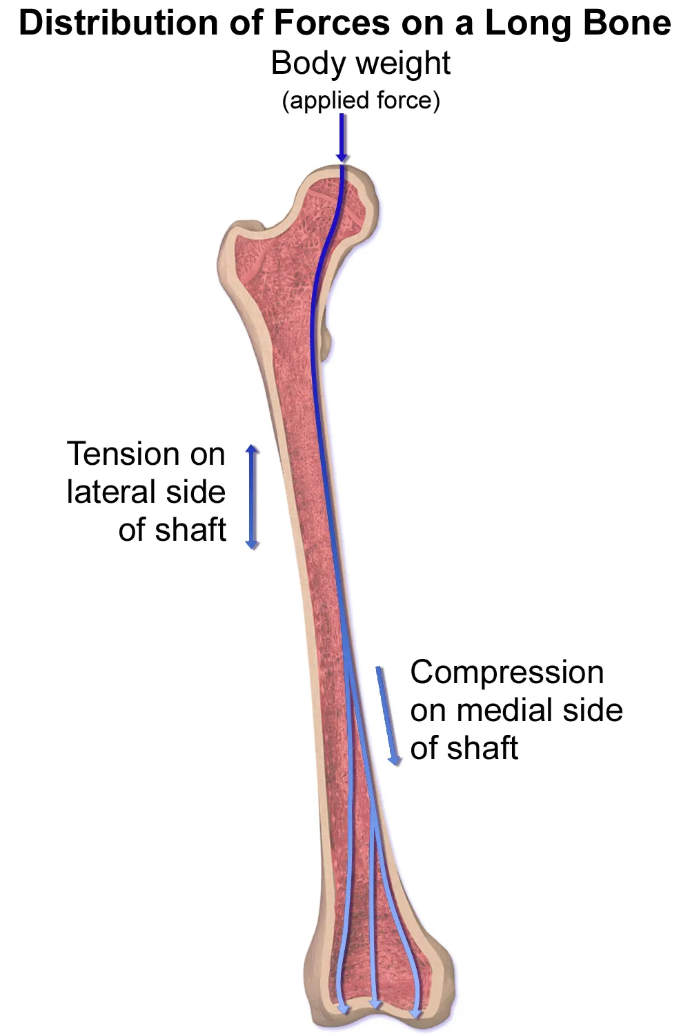
Growth of a long bone
The femur is a typical long bone formed by endochondral ossification: cartilage at the epiphyses and metaphyses proliferates and then mineralises into new bone, lengthening the bone at the growth plate and widening it radially. After maturity, the bone keeps being remodelled to maintain strength; after a fracture, bone formation rises sharply to heal it.[2][3]
The knee joint
The distal femur articulates with the tibial plateau and patella to form the knee. The two menisci are fibrocartilaginous crescents anchored to bone by roots and ligaments; their composition and multilayered structure convert compressive into hoop tensile forces, improve congruity and absorb shock under load. The medial meniscus has more attachments at its horns and body than the lateral, so it is less mobile and more prone to tears.[4][5]
Only the peripheral meniscus is vascularised (the red–red zone); the inner part is nearly avascular, so tears there heal poorly, and the meniscal roots are key landmarks for root repair and transplantation. The discoid lateral meniscus is a congenital variant prone to tearing and is more common in Asian populations.[5][6]
The anterolateral ligament, described by Segond in 1879 and “rediscovered” in 2013, limits internal rotation and lateral meniscal extrusion; it contributes to the pivot-shift phenomenon, so anterolateral stabilisation alongside ACL reconstruction is considered to reduce postoperative instability.[7]
Pain around the knee may arise from many structures: iliotibial tract syndrome, pes anserinus bursitis, Baker’s cyst, popliteus tenosynovitis and the infrapatellar (Hoffa) fat pad. Patellofemoral pain syndrome is the commonest cause of anterior knee pain under 60, worse with loaded flexion (squatting, descending stairs), and is treated mainly by strengthening hip, trunk and knee muscles.[8][9][10]
The hip joint
Femoral anteversion is the angle between the femoral neck and shaft, reflecting femoral torsion. It varies by up to 30° among healthy adults, increases substantially during gestation and then decreases steadily until maturity. Differences alter muscle moment arms and joint loading, affect gait and may contribute to osteoarthritis.[11]
The hip capsule is reinforced by three ligaments (iliofemoral, ischiofemoral, pubofemoral), which dominate mobility and stability; the zona orbicularis resists distraction and acts as an aperture mechanism against edge-loading at extreme flexion and extension. The ligament of the head of the femur, long dismissed, may add stability, and the acetabular labrum deepens the socket; labral tears are a mechanical cause of hip pain.[12][13][14]
Femoroacetabular impingement is early, painful contact between the femoral head–neck and the acetabular rim: pincer type from acetabular over-coverage, cam type from an aspherical head–neck junction; it damages labrum and cartilage and can lead to hip osteoarthritis. Osteonecrosis of the femoral head, staged by the ARCO system, is another cause of joint destruction.[15][16][17]
Hip capsule: ligaments, zona orbicularis and stability
Human ligaments are mostly type I collagen (85%), with types III, V, VI, XI and XIV making up the rest; in the hip, more type III collagen in the capsule is associated with instability and in cartilage with progressive degeneration. The iliofemoral ligament has lateral (superior) and medial (inferior) arms that share an origin on the anterior inferior iliac spine and fan out to the intertrochanteric line, forming the inverted-Y ligament of Bigelow, which reinforces the capsule in external rotation and extension. The ischiofemoral ligament runs from the ischium behind and below the acetabular rim to the posterior intertrochanteric line, resisting internal rotation in neutral and in flexion–adduction–internal rotation (FADIR). The pubofemoral ligament, from the superior pubic ramus, blends with the medial iliofemoral and inferior ischiofemoral fibres and limits excessive abduction and external rotation in extension.[12]
The triangular ligamentum teres joins the acetabular notch to the fovea of the femoral head; it carries small vessels and nerves to the head and contributes to proprioception and stability, a function that may decline with age. The circular fibres of the zona orbicularis form a collar that closes around the femoral neck like an aperture: in extension its posteroinferior part tightens to medialise and secure the head anteriorly, in deep flexion its anteroinferior part secures it posteriorly. It may also circulate synovial fluid between the central and peripheral compartments.[12]
Tensile testing showed the iliofemoral ligament larger in cross-section and stiffer than the posterior capsule, so the anterior capsule is stronger. On MRI in femoroacetabular impingement, the anterosuperior (iliofemoral) capsule was thickest, about 6 mm versus 3.5–4.2 mm in normal hips; in another study capsules in cam impingement and chondrolabral disease were thicker than in controls (6.8 ± 1.6 vs 5.3 ± 2.3 mm), without correlation with the alpha angle. Stepwise resection studies identified the lateral iliofemoral arm as the main restraint to external rotation and the pubofemoral ligament as restraining external rotation and abduction in extension; the labrum and ligamentum teres act as secondary restraints, and the iliocapsularis muscle over the iliofemoral ligament adds stability, especially in dysplastic hips.[12]
Full-text reading (StatPearls)
The StatPearls chapters on this topic, reproduced verbatim. Their CC BY-NC-ND 4.0 licence allows unmodified copies only, so they are not translated or shortened.
Anatomy, Bony Pelvis and Lower Limb: FemurChang A, Breeland G, Black AC, Hubbard JB · StatPearls 2023 · NBK532982 · 26k characters
Loading…
Chang A, Breeland G, Black AC, Hubbard JB. Anatomy, Bony Pelvis and Lower Limb: Femur. StatPearls [Internet]. Treasure Island (FL): StatPearls Publishing; 2023. NBK532982 · PMID 30422577 · CC BY-NC-ND 4.0. Reproduced verbatim, unmodified.
Organs in this system
References
Sources for the values and descriptions on this page, indexed in PubMed (US National Library of Medicine).
- [1]Biology of Bone Tissue: Structure, Function, and Factors That Influence Bone CellsFlorencio-Silva R et al., BioMed research international 2015PMID 26247020
- [2]The biology of normal bone remodellingKatsimbri P, European journal of cancer care 2017PMID 28786518
- [3]Type H blood vessels in bone modeling and remodelingPeng Y et al., Theranostics 2020PMID 31903130
- [4]Meniscus Form and FunctionMarkes AR et al., Clinics in sports medicine 2020PMID 31767101
- [5]Meniscus Anatomy and Basic ScienceGee SM et al., Sports medicine and arthroscopy review 2021PMID 34398117
- [6]Discoid lateral meniscus: current conceptsTapasvi S et al., Journal of ISAKOS 2021PMID 33833041
- [7]The anterolateral ligament (ALL) and its role in rotational extra-articular stability of the knee joint: a review of anatomy and surgical conceptsRoessler PP et al., Archives of orthopaedic and trauma surgery 2016PMID 26714471
- [8]Clinical anatomy of the kneeSaavedra MÁ et al., Reumatologia clinica 2012PMID 23219082
- [9]Evaluation and treatment of disorders of the infrapatellar fat padDragoo JL et al., Sports medicine 2012PMID 22149697
- [10]Patellofemoral Pain SyndromeGaitonde DY et al., American family physician 2019PMID 30633480
- [11]Femoral anteversion: significance and measurementScorcelletti M et al., Journal of anatomy 2020PMID 32579722
- [12]Hip Joint Capsular Anatomy, Mechanics, and Surgical ManagementNg KCG et al., The Journal of bone and joint surgery. American volume 2019PMID 31800428
- [13]Ligament of the head of femur: A comprehensive review of its anatomy, embryology, and potential functionPerumal V et al., Clinical anatomy 2016PMID 26480296
- [14]Acetabular labrumBlankenbaker DG et al., Magnetic resonance imaging clinics of North America 2013PMID 23168180
- [15]Femoroacetabular impingementAnderson SE et al., European journal of radiology 2012PMID 21924849
- [16]Hip Osteoarthritis: Etiopathogenesis and Implications for ManagementMurphy NJ et al., Advances in therapy 2016PMID 27671326
- [17]Osteonecrosis of the Femoral Head: an Updated Review of ARCO on Pathogenesis, Staging and TreatmentHines JT et al., Journal of Korean medical science 2021PMID 34155839
- [18]Anatomy, BonesStatPearls (NCBI Bookshelf)PMID 30725884Supports: Number of bones
- [19]Calcium metabolism in health and diseasePeacock M, 2010PMID 20089499Supports: Skeletal calcium store