Venae cavae
Model
Layers
Z-Anatomy anatomical model based on BodyParts3D, licensed CC BY-SA 4.0.
Description
The two largest veins, returning systemic blood to the right atrium. The superior vena cava (about 7 cm) forms behind the right first costal cartilage from the two brachiocephalic veins and receives the azygos vein. The inferior vena cava begins at L5 from the two common iliac veins, ascends to the right of the aorta, pierces the diaphragm at T8 and enters the right atrium.
Parts
Each part has its own card; press “Show on the model” to fly to it and select it in the 3D model above.
- 01
Superior vena cava
Vena cava superior
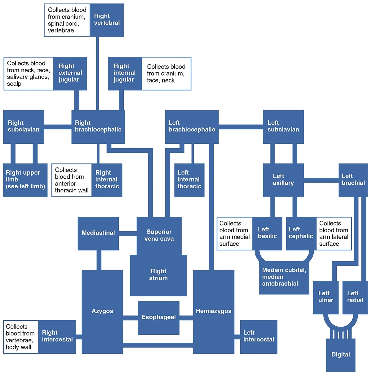
Formed by the two brachiocephalic veins behind the first right costal cartilage, about 7 cm long and valveless, it enters the right atrium at the third costal cartilage. It receives the azygos vein just before entering the pericardium; compression by mediastinal or lung tumours causes superior vena cava syndrome (swelling of face, neck and arms).
- 02
Brachiocephalic veins
Venae brachiocephalicae
Each is formed behind the sternoclavicular joint by the internal jugular and subclavian veins; the thoracic duct ends at the left venous angle. The left vein is twice as long as the right and runs obliquely behind the manubrium, receiving the inferior thyroid and internal thoracic veins.
- 03
Azygos system
Systema azygos
The azygos (right), hemiazygos and accessory hemiazygos (left) veins drain the thoracic and posterior walls, the azygos arching over the right lung root into the superior vena cava. It is a key caval–caval collateral when the inferior vena cava is obstructed.
- 04
Inferior vena cava – abdominal part
Pars abdominalis venae cavae inferioris
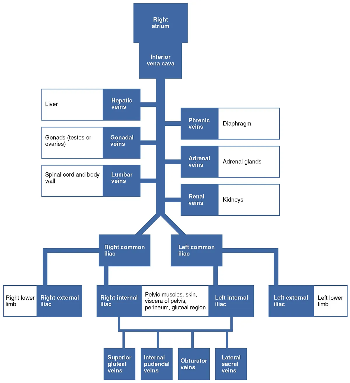
Formed by the two common iliac veins at L5, right of the aorta, it ascends behind the duodenum, head of pancreas and liver (lying in a groove on the bare area). It receives lumbar, right gonadal, renal, right suprarenal, inferior phrenic and three hepatic veins; the left renal vein crosses in front of the aorta to reach it.
- 05
Inferior vena cava – thoracic part
Pars thoracica venae cavae inferioris
Passes through the caval opening in the central tendon of the diaphragm at T8, runs a short course of about 2 cm and enters the lower right atrium, guarded by a rudimentary valve (Eustachian valve) that in the fetus directed blood through the foramen ovale.
Position and relations
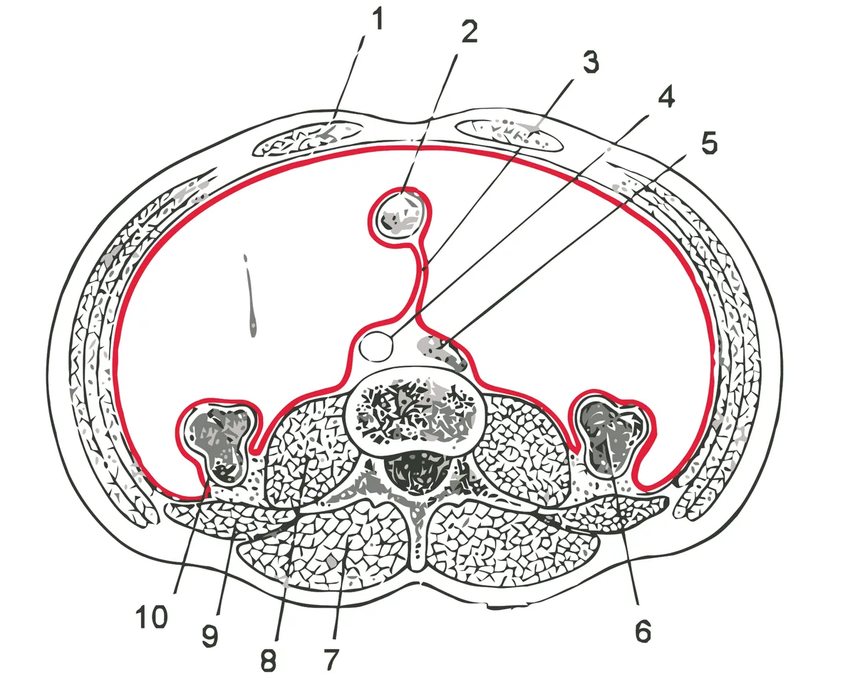
The superior vena cava lies in the superior and middle mediastinum to the right of the ascending aorta, its lower half within the pericardium; tumours of the right lung apex or mediastinum can compress it and cause superior vena cava syndrome (swelling of the face, neck and upper limbs). The inferior vena cava is retroperitoneal, to the right of the abdominal aorta, and runs in a groove on the back of the liver before passing through the caval opening of the diaphragm.
Form and structure
The superior vena cava has no valves. The inferior vena cava receives the lumbar, right gonadal, renal, right suprarenal and inferior phrenic veins and, just below the diaphragm, the hepatic veins; its atrial opening has a rudimentary valve (Eustachian valve), which in the fetus directs blood towards the foramen ovale.
Vessels and nerves
The two venae cavae are linked through the azygos system and the vertebral venous plexus, providing collateral routes if either is blocked. The inferior vena cava develops from several embryonic venous segments, so variants such as a duplicated or left-sided inferior vena cava are not rare and matter in retroperitoneal surgery.
Development and variants
Both venae cavae arise from the embryonic cardinal venous system, so variants occur when embryonic segments regress or persist abnormally. A persistent left superior vena cava results when the left superior cardinal vein below the brachiocephalic vein fails to regress; it usually drains into the coronary sinus, which appears dilated on echocardiography, may accompany atrial septal defect, bicuspid aortic valve or coarctation, and complicates pacemaker or catheter placement from the left subclavian approach.[1]
In a left-sided inferior vena cava, the right common iliac vein crosses in front of the spine to join the left, the IVC ascends to the left of the abdominal aorta, receives the left renal vein and then crosses to the right. In the complete form the hepatic segment is absent and blood returns through the azygos vein. Most cases are asymptomatic but matter in retroperitoneal surgery and vascular interventions. A duplicated IVC is another rare variant, usually found incidentally on CT.[2][3]
Clinical correlations
Inferior vena cava agenesis is rare but found in almost 5% of people under 30 with unprovoked deep vein thrombosis, typically young men with proximal (often bilateral) thrombosis after major exertion; venous return relies on collaterals. Ultrasound usually misses it, so CT or MRI is needed; treatment is prolonged anticoagulation and compression stockings.[4]
The inferior vena cava may be invaded by tumour thrombus from renal cell or hepatocellular carcinoma, and rarely gives rise to leiomyosarcoma, the commonest primary malignancy of large veins; resection with caval reconstruction is the main treatment.[5][6]
Full-text reading (StatPearls)
The StatPearls chapters on this topic, reproduced verbatim. Their CC BY-NC-ND 4.0 licence allows unmodified copies only, so they are not translated or shortened.
Anatomy, Thorax, Superior Vena CavaWhite HJ, Soos MP · StatPearls 2023 · NBK545255 · 11k characters
Loading…
White HJ, Soos MP. Anatomy, Thorax, Superior Vena Cava. StatPearls [Internet]. Treasure Island (FL): StatPearls Publishing; 2023. NBK545255 · PMID 31424839 · CC BY-NC-ND 4.0. Reproduced verbatim, unmodified.
Anatomy, Abdomen and Pelvis: Inferior Vena CavaTucker WD, Shrestha R, Burns B · StatPearls 2023 · NBK482353 · 10k characters
Loading…
Tucker WD, Shrestha R, Burns B. Anatomy, Abdomen and Pelvis: Inferior Vena Cava. StatPearls [Internet]. Treasure Island (FL): StatPearls Publishing; 2023. NBK482353 · PMID 29493975 · CC BY-NC-ND 4.0. Reproduced verbatim, unmodified.
Organs in this system
References
Sources for the values and descriptions on this page, indexed in PubMed (US National Library of Medicine).
- [1]Persistent left superior vena cava: a case report and review of literatureGoyal SK et al., Cardiovascular ultrasound 2008PMID 18847480
- [2]Left-Sided Inferior Vena Cava: A Retrospective Study and Literature ReviewDeng Y et al., Annals of vascular surgery 2025PMID 39341555
- [3]Duplication of Inferior Vena Cava, Possible Cause of Internal Hernia? Literature Review and Rare Case PresentationKwizera C et al., Chirurgia 2020PMID 33138904
- [4]Inferior vena cava agenesis and deep vein thrombosis: 10 patients and review of the literatureLambert M et al., Vascular medicine 2010PMID 21183652
- [5]Histopathology and outcomes of non-metastatic renal cell carcinoma with inferior vena cava tumor thrombus: systematic review and meta-analysisLasorsa F et al., European journal of surgical oncology 2025PMID 41046750
- [6]Leiomyosarcoma of the Inferior Vena Cava: A Case Series and Review of the LiteratureAlkhalili E et al., Annals of vascular surgery 2016PMID 26802297
- [7]Physiology, Cardiac OutputStatPearls (NCBI Bookshelf)PMID 29262215Supports: Heart rate, cardiac output
- [8]Physiology, CardiovascularStatPearls (NCBI Bookshelf)PMID 29630249