Urinary bladder
Model
Hover or tap to see names, click to read a description; click a name in the legend to fly to it. Procedurally built to histological proportions; colours follow illustration conventions.
Description
A hollow organ with a smooth-muscle wall (detrusor) lying in the lesser pelvis that stores urine; the first desire to void usually appears at about 150–200 mL.
Parts
Each part has its own card; press “Show on the model” to fly to it and select it in the 3D model above.
- 01
Apex and body
Apex et corpus vesicae
The apex points towards the pubic symphysis and is joined to the umbilicus by the median umbilical ligament (the urachal remnant); the body lies between apex and fundus. Empty, the bladder lies wholly in the pelvis; full (400–600 mL) it rises above the pubis and can be punctured suprapubically without entering the peritoneum.
- 02
Fundus (base)
Fundus vesicae
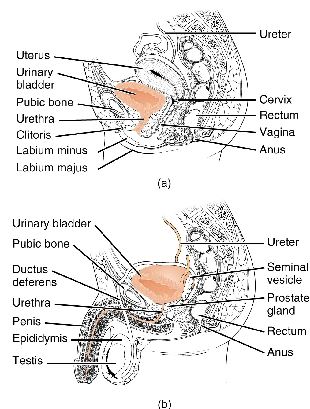
The triangular posteroinferior surface. In men it is related to the seminal glands, the ampullae of the ductus deferens and the rectum (across Denonvilliers fascia); in women to the cervix and anterior vaginal wall, so anterior vaginal prolapse carries the bladder down (cystocele).
- 03
Trigone
Trigonum vesicae
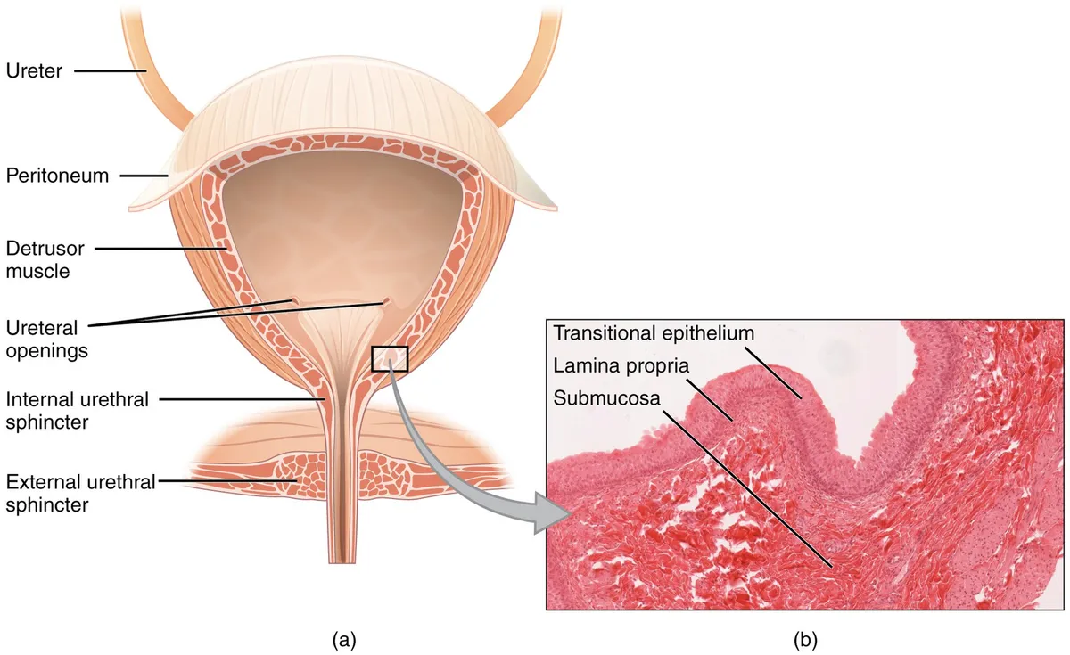
A smooth triangular area at the base between the two ureteric orifices and the internal urethral orifice, where the mucosa is firmly bound to the muscle and remains unfolded even when the bladder is empty. The interureteric ridge joining the ureteric orifices guides their identification at cystoscopy.
- 04
Detrusor muscle
Musculus detrusor vesicae
Three interlacing layers of smooth muscle that contract as a whole during voiding under S2–S4 parasympathetic drive (muscarinic M3 receptors) and relax during filling under sympathetic (β3) influence. Overactive bladder reflects uninhibited contractions of this muscle.
- 05
Bladder neck
Cervix vesicae; Ostium urethrae internum
The lowest part, continuous with the urethra; in men it rests on the base of the prostate and has an internal sphincter (smooth muscle, sympathetic α1) that closes during ejaculation to prevent retrograde flow. In women it rests on levator ani and is supported by the pubovesical ligaments.
Position and relations
The empty bladder lies in the lesser pelvis behind the pubic bones; when full, it rises above the symphysis into the abdomen. In males its posterior surface relates to the seminal vesicles and rectum; in females, to the uterus and vagina. The bladder neck continues as the urethra.
Form and structure
The bladder wall consists of smooth muscle (the detrusor) and a mucosa of transitional epithelium (urothelium) that thins as the bladder fills. The trigone is a smooth area of mucosa between the two ureteric orifices and the internal urethral orifice. Normal capacity is about 400–500 mL.
Vessels and nerves
Parasympathetic fibres (pelvic splanchnic nerves, S2–S4) contract the detrusor during voiding; sympathetic fibres (T11–L2) relax it and contract the internal sphincter to store urine. The external sphincter is striated and under voluntary control via the pudendal nerve; the pontine micturition centre coordinates these reflexes.[1]
Bladder sensation
Filling sensations progress in a continuum: first sensation, first desire to void, then strong desire. Functional MRI shows that these sensations map mainly to the insula, shifting anteriorly as they intensify. Urgency in overactive bladder syndrome is not part of this normal continuum and is accompanied by abnormal brain responses.[1]
Neural control of micturition
The lower urinary tract alternates between storing and voiding urine. A hierarchical neural system in the brain, spinal cord and peripheral autonomic ganglia coordinates the bladder smooth muscle (detrusor) with the smooth and striated muscle of the urethral outlet. Spinal storage mechanisms are governed by rostral brainstem circuitry that initiates reflex voiding, and the forebrain decides when voiding occurs voluntarily.[2]
Barrington’s nucleus (the pontine micturition centre) projects to the sacral parasympathetic nucleus, where preganglionic neurons supplying the bladder lie; it raises bladder pressure rapidly and strongly and sustains it during voiding.[3]
Infants and young children void reflexively until about 3–5 years, after which micturition becomes voluntary. In adults, neurological disease or injury can bring back involuntary reflex voiding, causing incontinence and neurogenic voiding dysfunction, a major problem in people with neurological impairment.[4][5]
Full-text reading (StatPearls)
The StatPearls chapters on this topic, reproduced verbatim. Their CC BY-NC-ND 4.0 licence allows unmodified copies only, so they are not translated or shortened.
Anatomy, Abdomen and Pelvis: BladderShermadou ES, Rahman S, Leslie SW · StatPearls 2023 · NBK531465 · 12k characters
Loading…
Shermadou ES, Rahman S, Leslie SW. Anatomy, Abdomen and Pelvis: Bladder. StatPearls [Internet]. Treasure Island (FL): StatPearls Publishing; 2023. NBK531465 · PMID 30285360 · CC BY-NC-ND 4.0. Reproduced verbatim, unmodified.
Histology, BladderBolla SR, Odeluga N, Amraei R, Jetti R · StatPearls 2023 · NBK540963 · 10k characters
Loading…
Bolla SR, Odeluga N, Amraei R, Jetti R. Histology, Bladder. StatPearls [Internet]. Treasure Island (FL): StatPearls Publishing; 2023. NBK540963 · PMID 31082007 · CC BY-NC-ND 4.0. Reproduced verbatim, unmodified.
Anatomy, Abdomen and Pelvis: Bladder Detrusor MuscleSam P, Nassereddin A, LaGrange CA · StatPearls 2023 · NBK482181 · 8k characters
Loading…
Sam P, Nassereddin A, LaGrange CA. Anatomy, Abdomen and Pelvis: Bladder Detrusor Muscle. StatPearls [Internet]. Treasure Island (FL): StatPearls Publishing; 2023. NBK482181 · PMID 29489195 · CC BY-NC-ND 4.0. Reproduced verbatim, unmodified.
Organs in this system
References
Sources for the values and descriptions on this page, indexed in PubMed (US National Library of Medicine).
- [1]Imaging bladder sensationsGriffiths D, Neurourology and urodynamics 2007PMID 17654564
- [2]Neural control of the lower urinary tractde Groat WC et al., Comprehensive physiology 2015PMID 25589273
- [3]Role of Barrington's nucleus in micturitionSasaki M, The Journal of comparative neurology 2005PMID 16255005
- [4]The neural control of micturitionFowler CJ et al., Nature reviews. Neuroscience 2008PMID 18490916
- [5]Neurogenic voiding dysfunctionGeorgopoulos P et al., Current opinion in urology 2017PMID 28212120
- [6]Normal reference values for glomerular filtration rate: what do we really know?Delanaye P et al., 2012PMID 22802582Supports: Glomerular filtration rate