Cranial nerves
Model
Layers
Z-Anatomy anatomical model based on BodyParts3D, licensed CC BY-SA 4.0.
Description
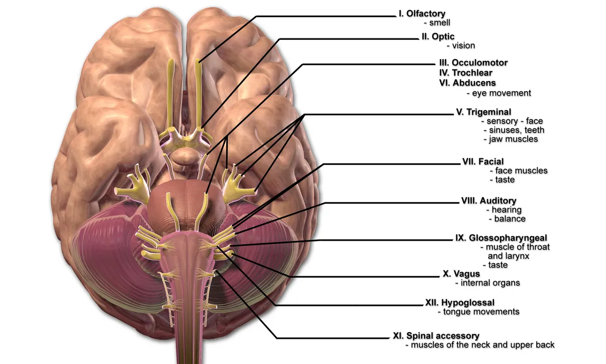
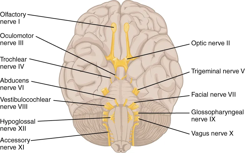
Twelve pairs of nerves that arise directly from the brain, numbered I–XII from front to back. They carry the special senses (smell, vision, hearing, taste), supply the muscles of the eye, face, tongue, pharynx and larynx, and convey parasympathetic fibres (III, VII, IX, X).
Parts
Each part has its own card; press “Show on the model” to fly to it and select it in the 3D model above.
- 01
Olfactory nerve (I)
Nervus olfactorius (I)
About 20 bundles of unmyelinated axons of olfactory receptor cells that pass from the roof of the nasal cavity through the cribriform plate to the olfactory bulb. Anterior skull-base fractures can tear them, causing anosmia and CSF rhinorrhoea.
- 02
Optic nerve (II)
Nervus opticus (II)
In fact a central white-matter tract of about 1.2 million retinal ganglion cell axons, wrapped in meninges. It runs through the optic canal to the optic chiasm, where fibres from the nasal hemiretinae cross.
- 03
Oculomotor nerve (III)
Nervus oculomotorius (III)
Emerges in the interpeduncular fossa, runs in the lateral wall of the cavernous sinus and enters the orbit through the superior orbital fissure. It supplies levator palpebrae superioris, the superior, medial and inferior recti and inferior oblique, and via the ciliary ganglion carries parasympathetic fibres for pupillary constriction and accommodation.
- 04
Trochlear nerve (IV)
Nervus trochlearis (IV)
The thinnest cranial nerve and the only one to leave the back of the brainstem (below the inferior colliculus), after crossing. It has the longest intracranial course and supplies only the superior oblique; palsy causes vertical diplopia, worse when looking down, as when descending stairs.
- 05
Trigeminal nerve (V)
Nervus trigeminus (V)
The largest cranial nerve, leaving the anterolateral pons as a large sensory and a small motor root; its ganglion lies in Meckel’s cave. The ophthalmic (V1), maxillary (V2) and mandibular (V3) divisions supply sensation to the face, nasal and oral mucosa and teeth; V3 also supplies the muscles of mastication.
- 06
Abducens nerve (VI)
Nervus abducens (VI)
Leaves at the pontomedullary junction, runs a long course over the clivus and through the cavernous sinus beside the internal carotid, and supplies only the lateral rectus. Its long course makes it vulnerable in raised intracranial pressure, causing esotropia and horizontal diplopia.
- 07
Facial nerve (VII)
Nervus facialis (VII)
Enters the internal acoustic meatus with VIII, runs in the facial canal of the temporal bone (geniculate ganglion) and leaves at the stylomastoid foramen to branch within the parotid. It supplies the muscles of facial expression and stapedius; the chorda tympani carries taste from the anterior two-thirds of the tongue and secretomotor fibres to the submandibular and sublingual glands, and the greater petrosal nerve serves the lacrimal gland.
- 08
Vestibulocochlear nerve (VIII)
Nervus vestibulocochlearis (VIII)
A special sensory nerve passing from the inner ear through the internal acoustic meatus to the cerebellopontine angle. Its cochlear part carries hearing from the organ of Corti; its vestibular part carries balance from the utricle, saccule and semicircular canals. A vestibular schwannoma typically causes unilateral sensorineural deafness.
- 09
Glossopharyngeal nerve (IX)
Nervus glossopharyngeus (IX)
Leaves the skull through the jugular foramen. It carries general sensation and taste from the posterior third of the tongue, sensation from the pharynx (afferent limb of the gag reflex) and from the carotid sinus and body, supplies stylopharyngeus, and sends parasympathetic fibres via the lesser petrosal nerve and otic ganglion to the parotid.
- 10
Vagus nerve (X)
Nervus vagus (X)
Leaves through the jugular foramen, descends in the carotid sheath and continues into thorax and abdomen. It supplies the muscles of pharynx, soft palate and larynx (via the recurrent laryngeal nerve) and carries about 75% of the body’s parasympathetic fibres to the heart, lungs and gut as far as the distal transverse colon.
- 11
Accessory nerve (XI)
Nervus accessorius (XI)
Its spinal root arises from the accessory nucleus in the anterior horn of C1–C5, ascends through the foramen magnum and leaves the skull through the jugular foramen. It supplies sternocleidomastoid and trapezius and is vulnerable in the posterior triangle during node biopsy, causing a drooping shoulder.
- 12
Hypoglossal nerve (XII)
Nervus hypoglossus (XII)
Emerges from the preolivary sulcus of the medulla, leaves the skull through the hypoglossal canal and curves forward below the angle of the jaw. It supplies all tongue muscles except palatoglossus; unilateral palsy makes the protruded tongue deviate towards the weak side.
The twelve cranial nerves
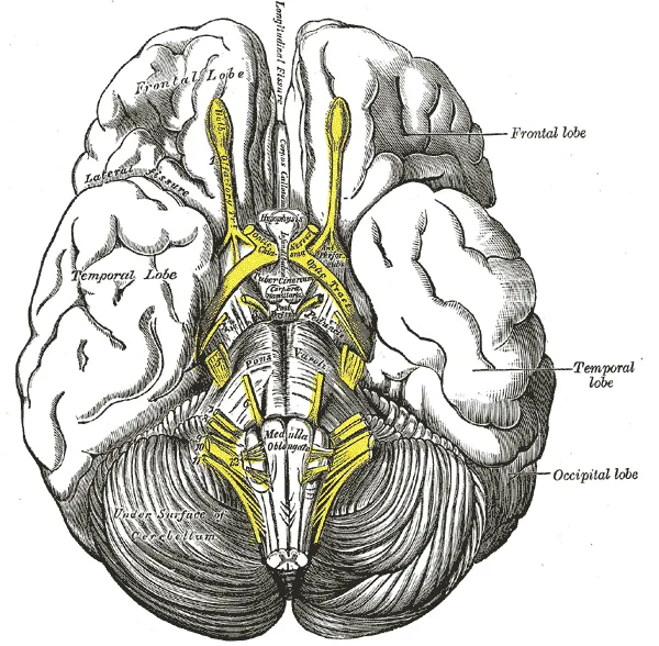
The cranial nerves are numbered in the order in which they emerge from the base of the brain, front to back. The first two (olfactory, optic) are really extensions of the brain; III and IV arise from the midbrain, V to VIII from the pons and pontomedullary junction, and IX to XII from the medulla. All except the accessory nerve arise entirely within the cranial vault and leave through foramina in the skull base.[1]
I
- Nerve
- Olfactory (n. olfactorius)
- Fibre type
- Sensory
- Main distribution
- Smell
II
- Nerve
- Optic (n. opticus)
- Fibre type
- Sensory
- Main distribution
- Vision
III
- Nerve
- Oculomotor (n. oculomotorius)
- Fibre type
- Motor, parasympathetic
- Main distribution
- Most extra-ocular muscles, levator palpebrae; pupil constriction, accommodation
IV
- Nerve
- Trochlear (n. trochlearis)
- Fibre type
- Motor
- Main distribution
- Superior oblique
V
- Nerve
- Trigeminal (n. trigeminus)
- Fibre type
- Mixed
- Main distribution
- Sensation of face, cornea, mouth; muscles of mastication
VI
- Nerve
- Abducens (n. abducens)
- Fibre type
- Motor
- Main distribution
- Lateral rectus
VII
- Nerve
- Facial (n. facialis)
- Fibre type
- Mixed
- Main distribution
- Muscles of facial expression; taste, anterior two-thirds of tongue; lacrimal, submandibular, sublingual glands
VIII
- Nerve
- Vestibulocochlear (n. vestibulocochlearis)
- Fibre type
- Sensory
- Main distribution
- Hearing, balance
IX
- Nerve
- Glossopharyngeal (n. glossopharyngeus)
- Fibre type
- Mixed
- Main distribution
- Taste and sensation, posterior third of tongue and pharynx; parotid gland; carotid sinus
X
- Nerve
- Vagus (n. vagus)
- Fibre type
- Mixed
- Main distribution
- Pharynx, larynx; parasympathetic supply of heart, lungs and gut
XI
- Nerve
- Accessory (n. accessorius)
- Fibre type
- Motor
- Main distribution
- Sternocleidomastoid, trapezius
XII
- Nerve
- Hypoglossal (n. hypoglossus)
- Fibre type
- Motor
- Main distribution
- Muscles of the tongue
| Nerve | Fibre type | Main distribution | |
|---|---|---|---|
| I | Olfactory (n. olfactorius) | Sensory | Smell |
| II | Optic (n. opticus) | Sensory | Vision |
| III | Oculomotor (n. oculomotorius) | Motor, parasympathetic | Most extra-ocular muscles, levator palpebrae; pupil constriction, accommodation |
| IV | Trochlear (n. trochlearis) | Motor | Superior oblique |
| V | Trigeminal (n. trigeminus) | Mixed | Sensation of face, cornea, mouth; muscles of mastication |
| VI | Abducens (n. abducens) | Motor | Lateral rectus |
| VII | Facial (n. facialis) | Mixed | Muscles of facial expression; taste, anterior two-thirds of tongue; lacrimal, submandibular, sublingual glands |
| VIII | Vestibulocochlear (n. vestibulocochlearis) | Sensory | Hearing, balance |
| IX | Glossopharyngeal (n. glossopharyngeus) | Mixed | Taste and sensation, posterior third of tongue and pharynx; parotid gland; carotid sinus |
| X | Vagus (n. vagus) | Mixed | Pharynx, larynx; parasympathetic supply of heart, lungs and gut |
| XI | Accessory (n. accessorius) | Motor | Sternocleidomastoid, trapezius |
| XII | Hypoglossal (n. hypoglossus) | Motor | Muscles of the tongue |
Nuclei and course
The nuclei of nerves III to XII lie in the brainstem, arranged in functional columns: somatic motor nuclei near the midline (III, IV, VI, XII), branchial motor nuclei (nucleus ambiguus, motor nuclei of V and VII), parasympathetic nuclei (Edinger–Westphal, superior and inferior salivatory, dorsal vagal) and, laterally, sensory nuclei (solitary nucleus, trigeminal complex, vestibular and cochlear nuclei). The level of a brainstem lesion can therefore be deduced from which cranial nerves are affected.
The main exits are the cribriform plate (I), optic canal (II), superior orbital fissure (III, IV, V1, VI), foramen rotundum (V2), foramen ovale (V3), internal acoustic meatus (VII, VIII), jugular foramen (IX, X, XI) and hypoglossal canal (XII). MRI is the modality of choice for the nerves themselves, while CT shows the bony foramina.[1]
Clinical correlations
Peripheral facial (Bell’s) palsy weakens the whole half of the face on the same side, including the forehead, whereas a central lesion such as a stroke usually spares the forehead. An oculomotor palsy from a posterior communicating artery aneurysm typically includes a dilated pupil, because the parasympathetic fibres lie on the surface of the nerve. Trigeminal neuralgia causes brief, shock-like pain in one trigeminal division.
Functional organisation
All twelve cranial nerves have important functions and, except for the accessory nerve (XI), all arise solely within the cranial vault. MRI is the modality of choice for the nerves themselves, while CT is important for the bony foramina they traverse.[1]
The visual pathway starts as retinal ganglion cell axons forming the optic nerve (II). At the chiasm, fibres from the nasal retina cross to join the temporal fibres of the other eye, so each occipital cortex receives the opposite half of the visual field of both eyes. Fibres continue as the optic tract, synapse in the lateral geniculate nucleus and run via the optic radiations to the visual cortex, supplied mainly by the posterior cerebral artery; the central-vision area at the occipital pole may also receive middle cerebral branches. Chiasmal lesions (such as pituitary adenoma) typically cause bitemporal hemianopia.[2][3]
The hypoglossal nerve (XII) leaves the brainstem in the preolivary sulcus, crosses the premedullary cistern and exits the skull through the hypoglossal canal. It is purely motor, supplying all intrinsic tongue muscles, three extrinsic muscles (styloglossus, hyoglossus, genioglossus) and the geniohyoid; unilateral palsy makes the protruded tongue deviate towards the weak side. Neoplasia is the most common cause of palsy.[4]
Clinical correlations
Trigeminal neuralgia may be essential or secondary. The trigeminal pathway comprises the cervical cord, brainstem, root and the three peripheral branches V1, V2 and V3. Neurovascular compression is the main cause of the essential form and requires two conditions: contact at the root entry zone (2–6 mm from its emergence from the pons) and perpendicular to the root.[5]
After leaving the stylomastoid foramen, the facial nerve (VII) enters the parotid gland and divides there into its branches to the muscles of facial expression, so parotid surgery must preserve its trunk and branches. The parotid itself receives secretomotor parasympathetic fibres from the glossopharyngeal nerve via the otic ganglion.[6]
Full-text reading (StatPearls)
The StatPearls chapters on this topic, reproduced verbatim. Their CC BY-NC-ND 4.0 licence allows unmodified copies only, so they are not translated or shortened.
Neuroanatomy, Cranial NerveSonne J, Omole AE, Lopez-Ojeda W · StatPearls 2025 · NBK470353 · 18k characters
Loading…
Sonne J, Omole AE, Lopez-Ojeda W. Neuroanatomy, Cranial Nerve. StatPearls [Internet]. Treasure Island (FL): StatPearls Publishing; 2025. NBK470353 · PMID 29261885 · CC BY-NC-ND 4.0. Reproduced verbatim, unmodified.
Neuroanatomy, Cranial Nerve 11 (Accessory)Bordoni B, Varacallo MA · StatPearls 2025 · NBK507722 · 16k characters
Loading…
Bordoni B, Varacallo MA. Neuroanatomy, Cranial Nerve 11 (Accessory). StatPearls [Internet]. Treasure Island (FL): StatPearls Publishing; 2025. NBK507722 · PMID 29939544 · CC BY-NC-ND 4.0. Reproduced verbatim, unmodified.
Neuroanatomy, Cranial Nerve 1 (Olfactory)Helwany M, Bordoni B · StatPearls 2023 · NBK556051 · 16k characters
Loading…
Helwany M, Bordoni B. Neuroanatomy, Cranial Nerve 1 (Olfactory). StatPearls [Internet]. Treasure Island (FL): StatPearls Publishing; 2023. NBK556051 · PMID 32310511 · CC BY-NC-ND 4.0. Reproduced verbatim, unmodified.
Neuroanatomy, Cranial Nerve 2 (Optic)Smith AM, Czyz CN · StatPearls 2022 · NBK507907 · 10k characters
Loading…
Smith AM, Czyz CN. Neuroanatomy, Cranial Nerve 2 (Optic). StatPearls [Internet]. Treasure Island (FL): StatPearls Publishing; 2022. NBK507907 · PMID 29939684 · CC BY-NC-ND 4.0. Reproduced verbatim, unmodified.
Neuroanatomy, Cranial Nerve 4 (Trochlear)Kim SY, Motlagh M, Naqvi IA · StatPearls 2023 · NBK537244 · 9k characters
Loading…
Kim SY, Motlagh M, Naqvi IA. Neuroanatomy, Cranial Nerve 4 (Trochlear). StatPearls [Internet]. Treasure Island (FL): StatPearls Publishing; 2023. NBK537244 · PMID 30725929 · CC BY-NC-ND 4.0. Reproduced verbatim, unmodified.
Neuroanatomy, Cranial Nerve 6 (Abducens)Nguyen V, Reddy V, Varacallo MA · StatPearls 2022 · NBK430711 · 8k characters
Loading…
Nguyen V, Reddy V, Varacallo MA. Neuroanatomy, Cranial Nerve 6 (Abducens). StatPearls [Internet]. Treasure Island (FL): StatPearls Publishing; 2022. NBK430711 · PMID 28613463 · CC BY-NC-ND 4.0. Reproduced verbatim, unmodified.
Organs in this system
References
Sources for the values and descriptions on this page, indexed in PubMed (US National Library of Medicine).
- [1]Cranial Nerve AnatomyTraylor KS et al., Neuroimaging clinics of North America 2022PMID 35843663
- [2]Anatomy of the visual pathwaysDe Moraes CG, Journal of glaucoma 2013PMID 23733119
- [3]The optic chiasmKidd D, Clinical anatomy 2014PMID 24824063
- [4]The Hypoglossal Nervede Sousa Costa R et al., Seminars in ultrasound, CT, and MR 2023PMID 37055141
- [5]Trigeminal neuralgiaLeclercq D et al., Diagnostic and interventional imaging 2013PMID 24007773
- [6]Facial Nerve and Parotid Gland AnatomyKochhar A et al., Otolaryngologic clinics of North America 2016PMID 27040583
- [7]The human brain in numbers: a linearly scaled-up primate brainHerculano-Houzel S, 2009PMID 19915731Supports: Neuron count
- [8]Determinants of conduction velocity in myelinated nerve fibersWaxman SG, 1980PMID 6245357Supports: Conduction velocity
- [9]Quantitative imaging of energy expenditure in human brainPMID 22487547Supports: Cerebral energy expenditure