Knee and popliteal fossa
Model
Layers
Z-Anatomy anatomical model based on BodyParts3D, licensed CC BY-SA 4.0.
Overview
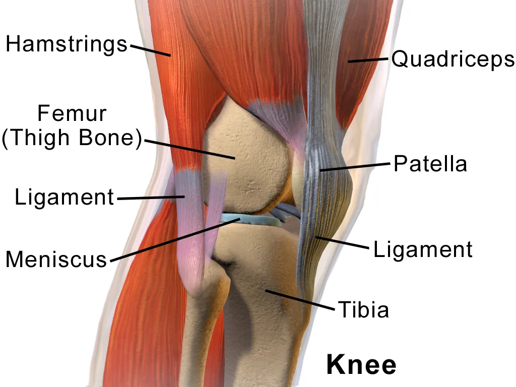
A regional model of the (right) knee and popliteal fossa: the lower femur, patella, upper tibia and fibula, the two menisci, the anterior and posterior cruciate and collateral ligaments, the bursae around the knee, quadriceps and the patellar ligament, the hamstrings and the two heads of gastrocnemius that bound the popliteal fossa, the popliteal artery and vein, and the tibial and common fibular nerves.
Boundaries and subdivisions
The knee is the largest joint in the body, comprising the tibiofemoral and patellofemoral joints within one capsule; the patella, the largest sesamoid bone, sits within the quadriceps tendon. The popliteal fossa is the diamond-shaped hollow behind the knee, bounded by four muscle bellies: biceps femoris above and laterally, semitendinosus and semimembranosus above and medially, and the medial and lateral heads of gastrocnemius below. Its floor is the popliteal surface of the femur, the knee capsule (oblique popliteal ligament) and popliteus; its roof is the popliteal fascia, pierced by the small saphenous vein.[1][2][3]
Layers from superficial to deep
In front of the knee, the quadriceps tendon, patella and patellar ligament form the extensor mechanism, flanked by the patellar retinacula; several bursae (suprapatellar, communicating with the joint, prepatellar, superficial and deep infrapatellar) reduce friction. Inside the joint the C-shaped medial and nearly circular lateral menisci deepen the tibial surfaces; the anterior cruciate ligament runs from the anterior intercondylar area of the tibia to the lateral femoral condyle and prevents anterior tibial translation, while the posterior cruciate prevents posterior translation. The tibial collateral ligament is firmly attached to the medial meniscus and resists valgus stress; the fibular collateral ligament is separate from the lateral meniscus.[1][4][5][6][7]
Vessels, nerves and lymphatics
In the popliteal fossa, from superficial to deep, lie the tibial nerve, popliteal vein and popliteal artery; the artery is deepest, against the popliteal surface of the femur and the capsule. The sciatic nerve usually divides into the tibial and common fibular nerves a few centimetres above the fossa; the common fibular nerve follows the medial border of biceps femoris and then winds around the neck of the fibula. The popliteal artery gives five genicular arteries forming an anastomosis around the joint and the sural branches to gastrocnemius, then divides into the anterior and posterior tibial arteries at the lower border of popliteus. The popliteal nodes receive lymph travelling with the small saphenous vein.[3][8][9]
Embryology
The knee begins as a mesenchymal condensation at 4 weeks and is identifiable as a joint from 6 weeks. The anterior cruciate ligament appears as a mesenchymal condensation at 6–7 weeks and the posterior cruciate at about 8 weeks; both are well oriented by 9 weeks. The fibular collateral ligament forms independently of the joint capsule at about 8 weeks, whereas the tibial collateral ligament begins at 9 weeks as a condensation of the developing capsule, which explains why the tibial collateral ligament is attached to the medial meniscus and the fibular one is separate from the lateral meniscus. The vascular system is mesodermal from about day 17, and the popliteal artery forms during the complex development of the lower limb bud.[4][5][6][9]
Anatomical variants
In a digital subtraction angiography study of 621 patients (1,242 limbs), 89.2% had the textbook popliteal branching; variants included a hypoplastic or aplastic posterior tibial artery (5.1%), hypoplastic or aplastic anterior tibial artery (1.7%), high origin of the anterior tibial artery (1.2%), hypoplasia of both tibial arteries (0.8%), high origin of the posterior tibial artery (0.4%) and an anterior tibioperoneal trunk (0.1%). When one limb had a variant, the other limb also did in 28% of cases. Female anterior cruciate ligaments have lower load at failure, stiffness and elastic modulus than male ones, and women rupture the ACL 2 to 7 times more often than men of the same age. The size and position of the ACL’s femoral and tibial footprints also vary slightly.[9][4]
Clinical correlations
A twisting injury with the foot planted can cause the “unhappy triad”: tears of the anterior cruciate ligament, tibial collateral ligament and medial meniscus (the latter two being attached). The anterior drawer and Lachman tests assess the anterior cruciate ligament, the posterior drawer the posterior cruciate. A popliteal (Baker) cyst is a synovial bulge into the fossa and may rupture, mimicking deep vein thrombosis. Knee dislocation can injure the popliteal artery, which is tethered against the bone; distal pulses must be checked after every knee dislocation.[1][4][5][6]
Surgical and procedural considerations
The aim of anterior cruciate ligament reconstruction is to restore stability while preserving function; correct tunnel placement requires an understanding of the attachments, the orientation of the two bundles and their behaviour in flexion and extension. Grafts may be autografts (hamstring tendons, bone–patellar tendon–bone, quadriceps tendon), allografts or synthetic. Management of posterior cruciate injuries remains debated, with a shift from non-operative to surgical treatment and ongoing discussion of single- versus double-bundle reconstruction. Most isolated tibial collateral ligament tears heal well with bracing and rehabilitation, even in athletes; surgery is reserved for combined injuries or persistent instability. Posterior approaches to the knee demand knowledge of popliteal artery variants, and endovascular repair of popliteal aneurysms carries few complications.[4][5][6][9]
Key structures
Fossa poplitea
- Name
- Popliteal fossa
- Position, role
- Bounded by four muscle bellies; popliteal vessels, tibial and common fibular nerves
Lig. cruciatum anterius
- Name
- Anterior cruciate ligament
- Position, role
- Prevents anterior tibial translation
Lig. cruciatum posterius
- Name
- Posterior cruciate ligament
- Position, role
- Prevents posterior tibial translation
Meniscus medialis
- Name
- Medial meniscus
- Position, role
- C-shaped; attached to the tibial collateral ligament
Meniscus lateralis
- Name
- Lateral meniscus
- Position, role
- Nearly circular, more mobile
Patella
- Name
- Patella
- Position, role
- Largest sesamoid bone, within the quadriceps tendon
A. poplitea
- Name
- Popliteal artery
- Position, role
- Deepest structure in the fossa; five genicular arteries
| Terminologia Anatomica | Name | Position, role |
|---|---|---|
| Fossa poplitea | Popliteal fossa | Bounded by four muscle bellies; popliteal vessels, tibial and common fibular nerves |
| Lig. cruciatum anterius | Anterior cruciate ligament | Prevents anterior tibial translation |
| Lig. cruciatum posterius | Posterior cruciate ligament | Prevents posterior tibial translation |
| Meniscus medialis | Medial meniscus | C-shaped; attached to the tibial collateral ligament |
| Meniscus lateralis | Lateral meniscus | Nearly circular, more mobile |
| Patella | Patella | Largest sesamoid bone, within the quadriceps tendon |
| A. poplitea | Popliteal artery | Deepest structure in the fossa; five genicular arteries |
What each layer contains
SkinCutis6
- Popliteal fossa
- Anterior region of leg
- Anterior region of knee
- Posterior region of leg
- Posterior region of thigh
- Posterior region of knee
Muscles, fasciae and tendon sheathsMusculi, fasciae et vaginae tendinum38
- Subtendinous prepatellar bursa
- Adductor magnus
- Anserine bursa
- Deep infrapatellar bursa
- Semimembranosus bursa
- Subcutaneous infrapatellar bursa
- Subcutaneous prepatellar bursa
- Subcutaneous bursa of tuberosity of tibia
- Inferior subtendinous bursa of biceps femoris muscle
- Lateral subtendinous bursa of gastrocnemius muscle
- Medial subtendinous bursa of gastrocnemius muscle
- Subtendinous bursa of sartorius muscle
- Suprapatellar bursa
- Short head of biceps femoris
- Lateral head of gastrocnemius
- Long head of biceps femoris
- Medial head of gastrocnemius
- Extensor digitorum longus
- Fibularis longus muscle
- Gracilis muscle
- Plantaris muscle
- Popliteus muscle
- Semimembranosus muscle
- Semitendinosus muscle
- Soleus muscle
- Tibialis anterior muscle
- Tibialis posterior muscle
- Vastus intermedius muscle
- Vastus lateralis muscle
- Vastus medialis muscle
- Lateral patellar retinaculum
- Medial patellar retinaculum
- Anterior intermuscular septum of leg
- Lateral femoral intermuscular septum
- Medial femoral intermuscular septum
- Posterior intermuscular septum of leg
- Transverse intermuscular septum of leg
- Subfacial prepatellar bursa
Bones and cartilageOssa et cartilagines4
- Fibula
- Femur
- Patella
- Tibia
Joints and ligamentsJuncturae et ligamenta22
- Articular capsule of superior tibiofibular joint
- Articular capsule of knee joint
- Infrapatellar fat pad
- Anterior ligament of fibular head
- Fibular collateral ligament
- Anterior cruciate ligament
- Posterior cruciate ligament
- Meniscopatellar ligament
- Anterior meniscotibial ligament (Lateral meniscus)
- Anterior meniscotibial ligament (Medial meniscus)
- Posterior meniscotibial ligament (Lateral meniscus)
- Posterior meniscotibial ligament (Medial meniscus)
- Popliteofibular ligament
- Arcuate popliteal ligament
- Oblique popliteal ligament
- Posterior ligament of fibular head
- Transverse ligament of knee
- Interosseous membrane of leg
- Lateral meniscus
- Medial meniscus
- Deep part of tibial collateral ligament
- Superficial part of tibial collateral ligament
ArteriesArteriae9
- Inferior lateral genicular artery
- Inferior medial genicular artery
- Popliteal artery
- Superior lateral genicular artery
- Superior medial genicular artery
- Anterior tibial artery
- Middle genicular artery
- Descending branch of lateral circumflex femoral artery
- Patellar anastomosis
VeinsVenae4
- Popliteal vein
- Small saphenous vein
- Genicular veins
- Anterior tibial veins
NervesNervi8
- Medial sural cutaneous nerve
- Common fibular nerve
- Deep fibular nerve
- Superficial fibular nerve
- Sural nerve
- Tibial nerve
- Sural communicating branch of common fibular nerve
- Infrapatellar branch of saphenous nerve
LymphaticsSystema lymphoideum5
- (Fibular node)
- (Anterior tibial node)
- (Posterior tibial node)
- Deep popliteal nodes
- Superficial popliteal nodes
Full-text reading (StatPearls)
The StatPearls chapters on this topic, reproduced verbatim. Their CC BY-NC-ND 4.0 licence allows unmodified copies only, so they are not translated or shortened.
Anatomy, Bony Pelvis and Lower Limb: Knee Posterior Cruciate LigamentCox CF, Graefe SB, Black AC, Bordoni B · StatPearls 2023 · NBK535416 · 16k characters
Loading…
Cox CF, Graefe SB, Black AC, Bordoni B. Anatomy, Bony Pelvis and Lower Limb: Knee Posterior Cruciate Ligament. StatPearls [Internet]. Treasure Island (FL): StatPearls Publishing; 2023. NBK535416 · PMID 30571037 · CC BY-NC-ND 4.0. Reproduced verbatim, unmodified.
Anatomy, Bony Pelvis and Lower Limb, Knee Medial Collateral LigamentMabrouk A, Hubbard JB · StatPearls 2025 · NBK507780 · 16k characters
Loading…
Mabrouk A, Hubbard JB. Anatomy, Bony Pelvis and Lower Limb, Knee Medial Collateral Ligament. StatPearls [Internet]. Treasure Island (FL): StatPearls Publishing; 2025. NBK507780 · PMID 29939557 · CC BY-NC-ND 4.0. Reproduced verbatim, unmodified.
Anatomy, Bony Pelvis and Lower Limb, Knee Anterior Cruciate LigamentYoo H, Marappa-Ganeshan R · StatPearls 2023 · NBK559233 · 12k characters
Loading…
Yoo H, Marappa-Ganeshan R. Anatomy, Bony Pelvis and Lower Limb, Knee Anterior Cruciate Ligament. StatPearls [Internet]. Treasure Island (FL): StatPearls Publishing; 2023. NBK559233 · PMID 32644659 · CC BY-NC-ND 4.0. Reproduced verbatim, unmodified.
Anatomy, Bony Pelvis and Lower Limb: Popliteal ArteryBowers Z, Nassereddin A, Sinkler MA, Bordoni B · StatPearls 2023 · NBK537125 · 10k characters
Loading…
Bowers Z, Nassereddin A, Sinkler MA, Bordoni B. Anatomy, Bony Pelvis and Lower Limb: Popliteal Artery. StatPearls [Internet]. Treasure Island (FL): StatPearls Publishing; 2023. NBK537125 · PMID 30725810 · CC BY-NC-ND 4.0. Reproduced verbatim, unmodified.
Regional anatomy
References
Sources for the values and descriptions on this page, indexed in PubMed (US National Library of Medicine).
- [1]Anatomy, Bony Pelvis and Lower Limb, KneeGupton M et al., StatPearls 2023PMID 29763193
- [2]Anatomy, Bony Pelvis and Lower Limb, Knee PatellaCox CF et al., StatPearls 2023PMID 30137819
- [3]Anatomy, Bony Pelvis and Lower Limb: Tibial NerveDesai SS et al., StatPearls 2026PMID 30725713
- [4]Anatomy, Bony Pelvis and Lower Limb, Knee Anterior Cruciate LigamentYoo H et al., StatPearls 2023PMID 32644659
- [5]Anatomy, Bony Pelvis and Lower Limb: Knee Posterior Cruciate LigamentCox CF et al., StatPearls 2023PMID 30571037
- [6]Anatomy, Bony Pelvis and Lower Limb, Knee Medial Collateral LigamentMabrouk A et al., StatPearls 2025PMID 29939557
- [7]Anatomy, Bony Pelvis and Lower Limb, Knee Lateral MeniscusCox CF et al., StatPearls 2023PMID 30137778
- [8]Anatomy, Bony Pelvis and Lower Limb: Gastrocnemius MuscleBordoni B et al., StatPearls 2026PMID 30422541
- [9]Anatomy, Bony Pelvis and Lower Limb: Popliteal ArteryBowers Z et al., StatPearls 2023PMID 30725810