Eye
Model
Hover or tap to see names, click to read a description; click a name in the legend to fly to it. Procedurally built to histological proportions; colours follow illustration conventions.
Description
The organ of vision: the eyeball within the orbit and its accessory structures, namely six extra-ocular muscles, the eyelids, conjunctiva and lacrimal apparatus. The eyeball, about 24 mm across, has three coats (fibrous, vascular and neural) enclosing the transparent media: aqueous humour, lens and vitreous body.
Parts
Each part has its own card; press “Show on the model” to fly to it and select it in the 3D model above.
- 01
Cornea
Cornea
The transparent, avascular front of the fibrous coat, about 0.5 mm thick centrally, with five layers: epithelium, Bowman layer, stroma, Descemet membrane and endothelium. It provides about two-thirds of the eye’s refractive power (some 43 dioptres) and is richly innervated by the long ciliary nerves.
- 02
Sclera
Sclera
The tough white fibrous coat over the posterior five-sixths of the globe, giving attachment to the extraocular muscles. The optic nerve pierces it posteriorly at the lamina cribrosa; it meets the cornea at the limbus, where Schlemm’s canal drains the aqueous humour.
- 03
Iris and ciliary body
Iris; Corpus ciliare
The front of the uveal tract. The iris has a sphincter pupillae (parasympathetic, nerve III) and dilator pupillae (sympathetic) that regulate light entry. The ciliary body contains the ciliary muscle, which accommodates the lens through the zonule, and ciliary processes that secrete aqueous humour.
- 04
Choroid
Choroidea
The pigmented vascular layer between sclera and retina, the posterior part of the uvea. Its choriocapillaris nourishes the outer retina (photoreceptors) across Bruch’s membrane and has one of the highest blood flows per gram of any tissue.
- 05
Lens and ciliary zonule
Lens; Zonula ciliaris
A transparent, avascular biconvex lens suspended from the ciliary body by the zonule. When the ciliary muscle contracts, the zonule relaxes and the lens rounds up for near vision; its declining elasticity with age causes presbyopia, and opacity causes cataract.
- 06
Anterior and posterior chambers
Camera anterior et posterior bulbi; Humor aquosus
The posterior chamber lies between iris and lens, the anterior chamber between cornea and iris. Aqueous humour secreted by the ciliary body flows through the pupil into the anterior chamber and drains at the iridocorneal angle through the trabecular meshwork into Schlemm’s canal; impaired outflow raises intraocular pressure (glaucoma).
- 07
Vitreous body
Corpus vitreum
A clear gel of about 4 mL, four-fifths of the globe’s volume, made of 99% water with collagen fibrils and hyaluronic acid. It is firmly attached at the vitreous base and optic disc; age-related liquefaction and detachment can tear the retina.
- 08
Retina
Retina
The inner neural coat of ten layers, holding rods and cones, bipolar cells and ganglion cells. The macula and its central fovea contain only cones and give the sharpest vision; the optic disc (blind spot), about 3 mm nasal to it, is where ganglion cell axons form the optic nerve.
- 09
Lacrimal apparatus
Apparatus lacrimalis
The lacrimal gland in the upper outer orbit secretes tears (parasympathetic supply via VII); they drain through the puncta, canaliculi, lacrimal sac and nasolacrimal duct into the inferior meatus. Congenital nasolacrimal duct obstruction is common in infants and causes constant watering.
Position and relations
The eyeball occupies the front of the orbit, cushioned by orbital fat and slung in the fascial sheath (Tenon’s capsule). The orbit is a bony pyramid formed by seven bones; at its apex lie the optic canal (nerve II, ophthalmic artery) and the superior orbital fissure (III, IV, V1, VI). Its medial wall and floor are thin and fracture easily.
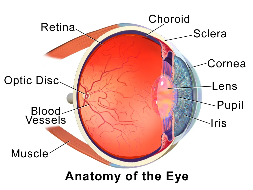
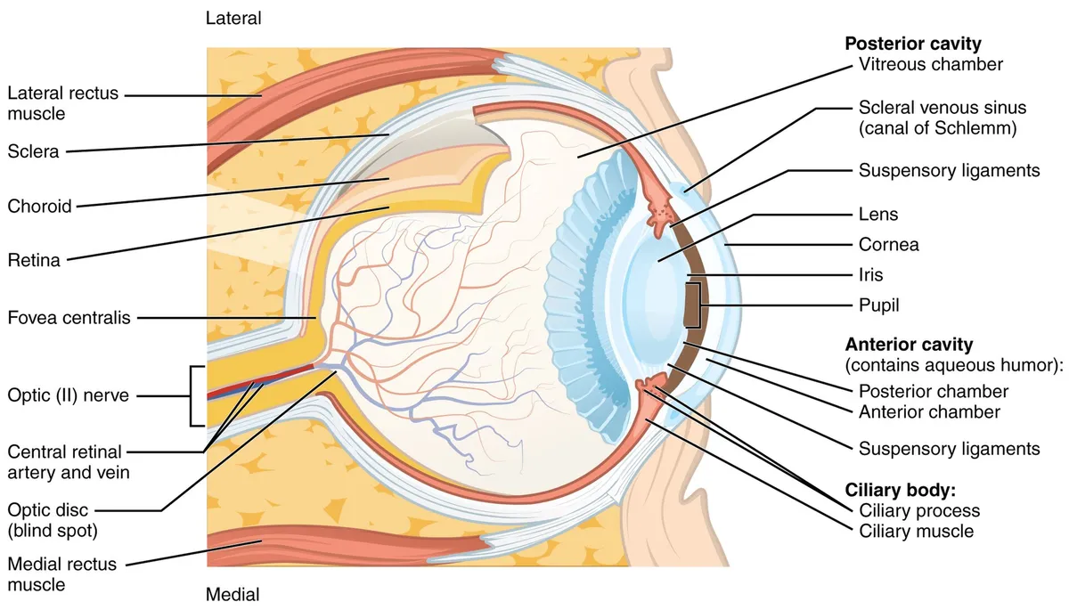
Coats and refractive media
The outer fibrous coat comprises the opaque white sclera behind and the transparent cornea in front, the most powerful refracting surface. The middle vascular coat (uvea) comprises the choroid, the ciliary body (which secretes aqueous humour and contains the ciliary muscle for accommodation) and the iris around the pupil. The inner coat is the retina, with rods and cones; the macula and its fovea give the sharpest vision, while the optic disc (blind spot) is where the fibres converge into the optic nerve.
Aqueous humour flows from the posterior chamber through the pupil to the anterior chamber and drains through the trabecular meshwork into Schlemm’s canal; obstruction raises intra-ocular pressure (glaucoma). The lens hangs on the zonular fibres; when the ciliary muscle contracts the zonules slacken and the lens rounds up for near vision. The gel-like vitreous body fills about four-fifths of the globe.
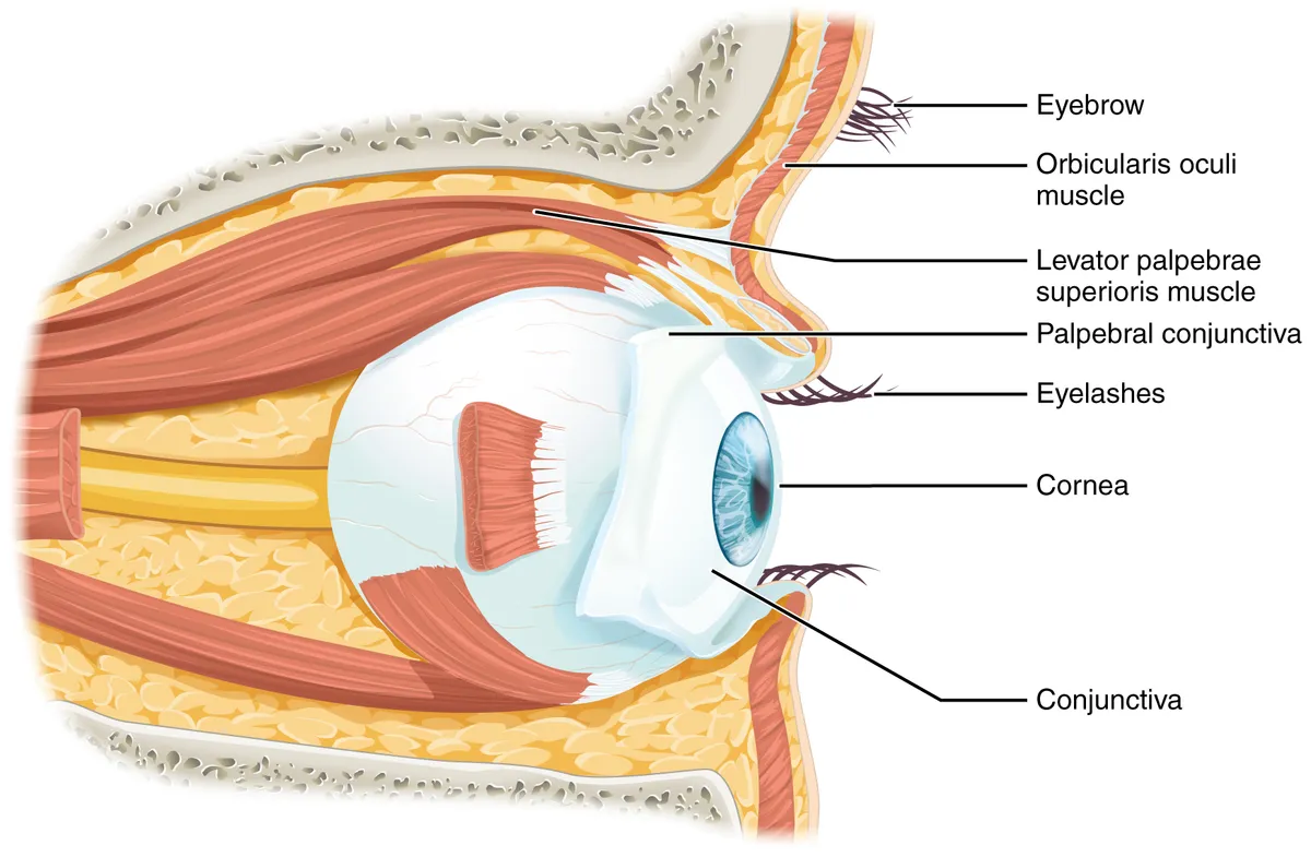
Extra-ocular muscles and lacrimal apparatus
Four recti arise from the common tendinous ring at the orbital apex, and two obliques rotate and elevate or depress the globe. The lacrimal gland in the upper lateral orbit secretes tears, which drain through the puncta and canaliculi into the lacrimal sac and down the nasolacrimal duct to the inferior meatus.
M. rectus superior
- Main action
- Elevation, adduction, intorsion
- Nerve
- III
M. rectus inferior
- Main action
- Depression, adduction, extorsion
- Nerve
- III
M. rectus medialis
- Main action
- Adduction
- Nerve
- III
M. rectus lateralis
- Main action
- Abduction
- Nerve
- VI
M. obliquus superior
- Main action
- Intorsion, depression in adduction
- Nerve
- IV
M. obliquus inferior
- Main action
- Extorsion, elevation in adduction
- Nerve
- III
| Muscle | Main action | Nerve |
|---|---|---|
| M. rectus superior | Elevation, adduction, intorsion | III |
| M. rectus inferior | Depression, adduction, extorsion | III |
| M. rectus medialis | Adduction | III |
| M. rectus lateralis | Abduction | VI |
| M. obliquus superior | Intorsion, depression in adduction | IV |
| M. obliquus inferior | Extorsion, elevation in adduction | III |
Visual pathway
Fibres from the nasal half of each retina cross in the optic chiasm, so each optic tract carries the opposite half of the visual field. The tract ends in the lateral geniculate body of the thalamus, from which the optic radiation runs to the visual cortex around the calcarine sulcus. A pituitary tumour pressing on the chiasm therefore causes bitemporal hemianopia, while a lesion behind the chiasm causes homonymous hemianopia.
The retina
The mammalian retina contains more than 60 neuronal types arranged in three processing stages. The first splits rod and cone output into about 12 parallel streams (via bipolar cell types); the second connects these streams to specific ganglion cell types; the third combines bipolar and amacrine activity into roughly 20 encodings of the visual world sent to the brain. The retina is thus a signal processor, not merely a film.[1]
The human retina has three glial types: microglia and two macroglia, astrocytes and Müller cells, which support photoreceptors and neurons metabolically. The fovea, the site of sharpest vision, lacks astrocytes and microglia; its Müller cells form the Müller cell cone that stabilises the foveal structure and improves light transmission to the photoreceptors.[2]
The blood–retinal barrier has an inner part (tight junctions between retinal capillary endothelial cells) and an outer part (tight junctions between retinal pigment epithelial cells). It regulates ion, protein and water flux, keeping the eye an immune-privileged site; its breakdown underlies diabetic retinopathy and age-related macular degeneration.[3]
Full-text reading (StatPearls)
The StatPearls chapters on this topic, reproduced verbatim. Their CC BY-NC-ND 4.0 licence allows unmodified copies only, so they are not translated or shortened.
Anatomy, Head and Neck, Eye Ciliary MusclesSuresh B, Ali T · StatPearls 2025 · NBK482132 · 16k characters
Loading…
Suresh B, Ali T. Anatomy, Head and Neck, Eye Ciliary Muscles. StatPearls [Internet]. Treasure Island (FL): StatPearls Publishing; 2025. NBK482132 · PMID 29489160 · CC BY-NC-ND 4.0. Reproduced verbatim, unmodified.
Anatomy, Head and Neck, Eye Corrugator MuscleYu M, Wang SM · StatPearls 2023 · NBK542280 · 13k characters
Loading…
Yu M, Wang SM. Anatomy, Head and Neck, Eye Corrugator Muscle. StatPearls [Internet]. Treasure Island (FL): StatPearls Publishing; 2023. NBK542280 · PMID 31194420 · CC BY-NC-ND 4.0. Reproduced verbatim, unmodified.
Histology, EyePradeep T, Mehra D, Le PH · StatPearls 2023 · NBK544343 · 12k characters
Loading…
Pradeep T, Mehra D, Le PH. Histology, Eye. StatPearls [Internet]. Treasure Island (FL): StatPearls Publishing; 2023. NBK544343 · PMID 31335063 · CC BY-NC-ND 4.0. Reproduced verbatim, unmodified.
Anatomy, Head and Neck, Eye CorneaLudwig PE, Lopez MJ, Sevensma KE · StatPearls 2023 · NBK470340 · 10k characters
Loading…
Ludwig PE, Lopez MJ, Sevensma KE. Anatomy, Head and Neck, Eye Cornea. StatPearls [Internet]. Treasure Island (FL): StatPearls Publishing; 2023. NBK470340 · PMID 29262108 · CC BY-NC-ND 4.0. Reproduced verbatim, unmodified.
Anatomy, Head and Neck, Eye ConjunctivaShumway CL, Motlagh M, Wade M · StatPearls 2023 · NBK519502 · 8k characters
Loading…
Shumway CL, Motlagh M, Wade M. Anatomy, Head and Neck, Eye Conjunctiva. StatPearls [Internet]. Treasure Island (FL): StatPearls Publishing; 2023. NBK519502 · PMID 30137787 · CC BY-NC-ND 4.0. Reproduced verbatim, unmodified.
Anatomy, Head and Neck, Eye FoveaRehman I, Mahabadi N, Motlagh M, Ali T · StatPearls 2023 · NBK482301 · 8k characters
Loading…
Rehman I, Mahabadi N, Motlagh M, Ali T. Anatomy, Head and Neck, Eye Fovea. StatPearls [Internet]. Treasure Island (FL): StatPearls Publishing; 2023. NBK482301 · PMID 29493926 · CC BY-NC-ND 4.0. Reproduced verbatim, unmodified.
Anatomy, Head and Neck: Eye, Superior Tarsal Muscle (Müller Muscle)Dave T, Sabeti S, Patel BC · StatPearls 2024 · NBK540964 · 8k characters
Loading…
Dave T, Sabeti S, Patel BC. Anatomy, Head and Neck: Eye, Superior Tarsal Muscle (Müller Muscle). StatPearls [Internet]. Treasure Island (FL): StatPearls Publishing; 2024. NBK540964 · PMID 31082008 · CC BY-NC-ND 4.0. Reproduced verbatim, unmodified.
Anatomy, Head and Neck: Eye Medial Rectus MusclesShumway CL, Motlagh M, Launico MV, Wade M · StatPearls 2023 · NBK519026 · 7k characters
Loading…
Shumway CL, Motlagh M, Launico MV, Wade M. Anatomy, Head and Neck: Eye Medial Rectus Muscles. StatPearls [Internet]. Treasure Island (FL): StatPearls Publishing; 2023. NBK519026 · PMID 30085568 · CC BY-NC-ND 4.0. Reproduced verbatim, unmodified.
Anatomy, Head and Neck: Eye Lacrimal DuctDucker L, Rivera RY · StatPearls 2023 · NBK531487 · 7k characters
Loading…
Ducker L, Rivera RY. Anatomy, Head and Neck: Eye Lacrimal Duct. StatPearls [Internet]. Treasure Island (FL): StatPearls Publishing; 2023. NBK531487 · PMID 30285382 · CC BY-NC-ND 4.0. Reproduced verbatim, unmodified.
Anatomy, Head and Neck: Eye Lateral Rectus MuscleFernández Cabrera A, Suárez-Quintanilla J · StatPearls 2023 · NBK539721 · 6k characters
Loading…
Fernández Cabrera A, Suárez-Quintanilla J. Anatomy, Head and Neck: Eye Lateral Rectus Muscle. StatPearls [Internet]. Treasure Island (FL): StatPearls Publishing; 2023. NBK539721 · PMID 30969543 · CC BY-NC-ND 4.0. Reproduced verbatim, unmodified.
Organs in this system
References
Sources for the values and descriptions on this page, indexed in PubMed (US National Library of Medicine).
- [1]The neuronal organization of the retinaMasland RH, Neuron 2012PMID 23083731
- [2]Glia of the human retinaReichenbach A et al., Glia 2020PMID 31793693
- [3]Blood-retinal barrierCunha-Vaz J et al., European journal of ophthalmology 2011PMID 23264323
- [4]The human brain in numbers: a linearly scaled-up primate brainHerculano-Houzel S, 2009PMID 19915731Supports: Neuron count
- [5]Determinants of conduction velocity in myelinated nerve fibersWaxman SG, 1980PMID 6245357Supports: Conduction velocity
- [6]Quantitative imaging of energy expenditure in human brainPMID 22487547Supports: Cerebral energy expenditure