Pulmonary trunk
Model
Layers
Z-Anatomy anatomical model based on BodyParts3D, licensed CC BY-SA 4.0.
Description
A short arterial trunk (about 5 cm) leaving the conus arteriosus of the right ventricle, running upwards and backwards to divide below the aortic arch, at T5–T6, into the right and left pulmonary arteries. After birth it is the only artery carrying deoxygenated blood; in the fetus the ductus arteriosus links it to the aortic arch.
Parts
Each part has its own card; press “Show on the model” to fly to it and select it in the 3D model above.
- 01
Pulmonary valve and root
Valva trunci pulmonalis
Sits at the top of the right ventricular infundibulum behind the left third costal cartilage, with three semilunar cusps (anterior, right, left). It is best heard in the left second intercostal space; pulmonary stenosis is one component of tetralogy of Fallot.
- 02
Pulmonary trunk
Truncus pulmonalis
A trunk about 5 cm long and 3 cm wide running up and back to the left of the ascending aorta, sharing its pericardial sleeve. It carries deoxygenated blood at a normal pressure of about 25/10 mmHg; a diameter over 29 mm on CT suggests pulmonary hypertension.
- 03
Bifurcation
Bifurcatio trunci pulmonalis
At about T5–T6, beneath the aortic arch and in front of the tracheal bifurcation, the trunk divides into the right and left pulmonary arteries. A large saddle embolus astride the bifurcation is a severe form of pulmonary embolism.
- 04
Right and left pulmonary arteries
Arteria pulmonalis dextra et sinistra
The right artery is longer and runs behind the ascending aorta and superior vena cava to the right hilum; the shorter left artery arches over the left main bronchus. Within the lungs they branch alongside the bronchial tree down to lobules and alveolar capillaries.
- 05
Ligamentum arteriosum
Ligamentum arteriosum
A fibrous band joining the origin of the left pulmonary artery to the underside of the aortic arch, the remnant of the fetal ductus arteriosus. The duct normally closes functionally within 1–2 days of birth; a patent duct gives a left-to-right shunt with a continuous murmur.
Position and relations
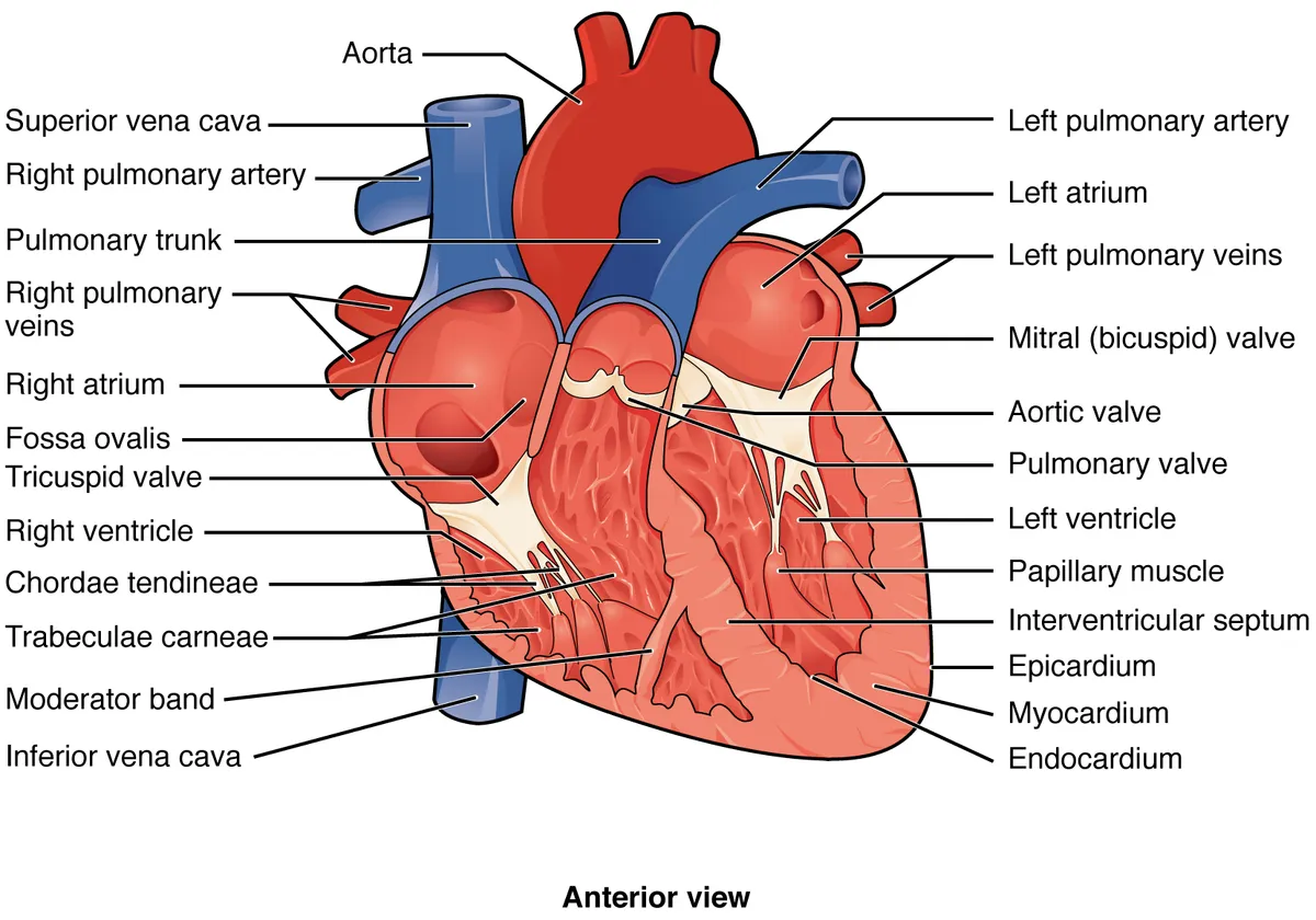
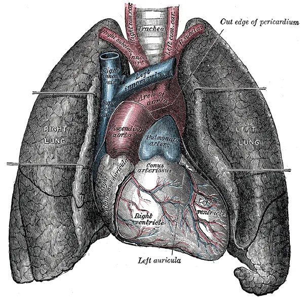
The pulmonary trunk lies within the pericardium with the ascending aorta, at first in front of it and then spiralling to its left. Its bifurcation lies under the aortic arch, where the ligamentum arteriosum (remnant of the ductus arteriosus) joins the two vessels; the left recurrent laryngeal nerve hooks around the arch just behind this ligament.
Form and structure
Its orifice carries the pulmonary valve with three semilunar cusps. Its wall is thinner than the aorta’s because the pulmonary circulation is a low-pressure system (normal mean pulmonary arterial pressure about 15 mmHg). The right pulmonary artery is longer and passes under the aortic arch behind the ascending aorta; the left is shorter and passes in front of the descending aorta.
Vessels and nerves
In the fetus, most blood leaving the pulmonary trunk passes through the ductus arteriosus into the aorta, because the lungs are not yet ventilated. After birth the ductus closes within days; if it persists, it causes a left-to-right shunt. The wall of the trunk is nourished by small branches of the coronary and bronchial arteries.
The pulmonary circulation
The pulmonary trunk carries deoxygenated blood from the right ventricle through the pulmonary arteries to the alveolar capillaries for gas exchange. The pulmonary circulation receives the whole cardiac output at a low driving pressure, so its thin-walled, low-resistance vessels differ markedly from the systemic circulation. It is not static: regional perfusion changes with posture, exercise and over time.[1]
Hypoxic pulmonary vasoconstriction (HPV) is a fundamental difference between the two circulations: when alveoli are hypoxic, intrapulmonary arteries constrict and divert blood to better-ventilated segments, optimising ventilation–perfusion matching. A mitochondrial sensor in pulmonary artery smooth muscle changes reactive oxygen species, inhibits potassium channels, depolarises the cell, opens voltage-gated calcium channels and causes constriction; sustained hypoxia activates rho kinase and HIF-1α, leading to vascular remodelling and pulmonary hypertension.[2]
In the fetus, the unventilated lung uses HPV to divert blood through the ductus arteriosus to the systemic circulation. In adults, focal HPV preserves oxygenation in pneumonia or atelectasis and reduces flow to the non-ventilated lung during one-lung anaesthesia; inhaled anaesthetics attenuate the reflex. At altitude, global hypoxia causes diffuse HPV, raises pulmonary artery pressure and can provoke high-altitude pulmonary oedema.[2][3]
Clinical correlations
Pulmonary vascular diseases range from thromboembolism to congenital malformations and vasculitis; patients present acutely with dyspnoea and chest pain or chronically with exertional dyspnoea, hypoxia and right heart failure. Multidetector CT pulmonary angiography is the key investigation.[4]
A pulmonary arteriovenous malformation is a direct artery-to-vein communication without an intervening capillary bed, most often associated with hereditary haemorrhagic telangiectasia. About half are asymptomatic, but because blood bypasses the pulmonary capillary filter they can cause stroke or brain abscess; endovascular embolisation of the feeding artery is the mainstay of treatment.[5]
Full-text reading (StatPearls)
The StatPearls chapters on this topic, reproduced verbatim. Their CC BY-NC-ND 4.0 licence allows unmodified copies only, so they are not translated or shortened.
Anatomy, Thorax, Heart Pulmonary ArteriesTucker WD, Weber C, Burns B · StatPearls 2023 · NBK534812 · 9k characters
Loading…
Tucker WD, Weber C, Burns B. Anatomy, Thorax, Heart Pulmonary Arteries. StatPearls [Internet]. Treasure Island (FL): StatPearls Publishing; 2023. NBK534812 · PMID 30521233 · CC BY-NC-ND 4.0. Reproduced verbatim, unmodified.
Organs in this system
References
Sources for the values and descriptions on this page, indexed in PubMed (US National Library of Medicine).
- [1]Pulmonary Vascular DynamicsClark A et al., Comprehensive physiology 2019PMID 31187894
- [2]Hypoxic Pulmonary Vasoconstriction: From Molecular Mechanisms to MedicineDunham-Snary KJ et al., Chest 2017PMID 27645688
- [3]Hypoxic pulmonary vasoconstriction: physiology and anesthetic implicationsLumb AB et al., Anesthesiology 2015PMID 25587641
- [4]Pulmonary vascular diseasesCummings KW et al., Clinics in chest medicine 2015PMID 26024602
- [5]Pulmonary arteriovenous malformationsTellapuri S et al., The international journal of cardiovascular imaging 2019PMID 30386957
- [6]Physiology, Cardiac OutputStatPearls (NCBI Bookshelf)PMID 29262215Supports: Heart rate, cardiac output
- [7]Physiology, CardiovascularStatPearls (NCBI Bookshelf)PMID 29630249