Ureter
Model
Layers
Z-Anatomy anatomical model based on BodyParts3D, licensed CC BY-SA 4.0.
Description
A muscular tube about 25–30 cm long that carries urine from the renal pelvis to the bladder by peristaltic waves. It has three physiological narrowings where kidney stones tend to lodge.
Parts
Each part has its own card; press “Show on the model” to fly to it and select it in the 3D model above.
- 01
Abdominal part
Pars abdominalis ureteris
From the pelviureteric junction (L2) it descends retroperitoneally on psoas major in line with the transverse processes, crossed anteriorly by the gonadal vessels. The right ureter is related to the right colon and root of the mesentery, the left to the sigmoid colon.
- 02
Pelvic part
Pars pelvica ureteris
It enters the pelvis by crossing the bifurcation of the common iliac artery, runs down the lateral pelvic wall and turns forward. In women it passes beneath the uterine artery about 2 cm from the cervix (“water under the bridge”), where it is at risk during hysterectomy; in men the ductus deferens crosses above it.
- 03
Intramural part
Pars intramuralis ureteris
It runs obliquely for 1.5–2 cm through the bladder wall and opens at the ureteric orifice at the upper corner of the trigone. This oblique course forms a functional valve: a full bladder compresses it and prevents vesico-ureteric reflux; a congenitally short tunnel causes reflux in children.
- 04
Three constrictions
Angustiae ureteris
The ureter is about 25–30 cm long and 3–4 mm wide with three physiological narrowings: the pelviureteric junction, the crossing of the iliac vessels and the vesico-ureteric junction (the narrowest). Stones typically lodge at these points, causing renal colic radiating to the groin.
- 05
Ureteric wall
Tunica muscularis ureteris; Urothelium
Lined by urothelium whose umbrella cells protect against urine, with inner longitudinal and outer circular smooth muscle (plus an outer longitudinal layer in the lower third) that propels urine in peristaltic waves 1–5 times a minute. Its blood supply comes segmentally from renal, gonadal, iliac and vesical arteries anastomosing in the adventitia.
Position and relations
The ureter is retroperitoneal, descends on psoas major, crosses in front of the iliac vessels (near the common iliac bifurcation) into the pelvis and runs obliquely through the bladder wall. The three narrowings are the pelviureteric junction, the crossing of the iliac vessels and the intramural segment. In women it passes under the uterine artery (“water under the bridge”), a key landmark in hysterectomy.
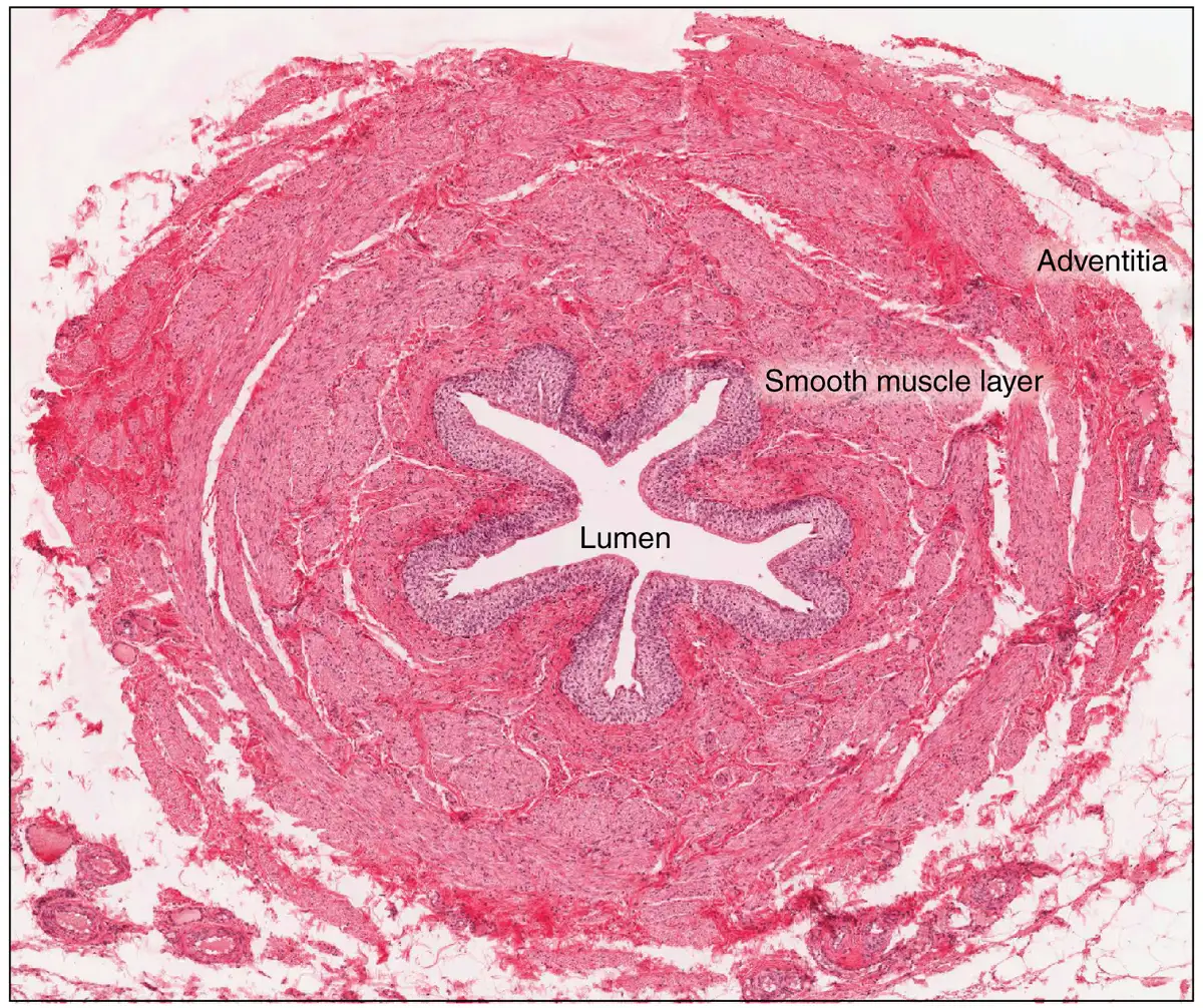
Form and structure
The wall has a mucosa lined by transitional epithelium (urothelium), smooth muscle (inner longitudinal, outer circular, plus an outer longitudinal layer in the lower third) and an adventitia. The oblique intramural course acts as a valve that closes as the bladder fills, preventing reflux.
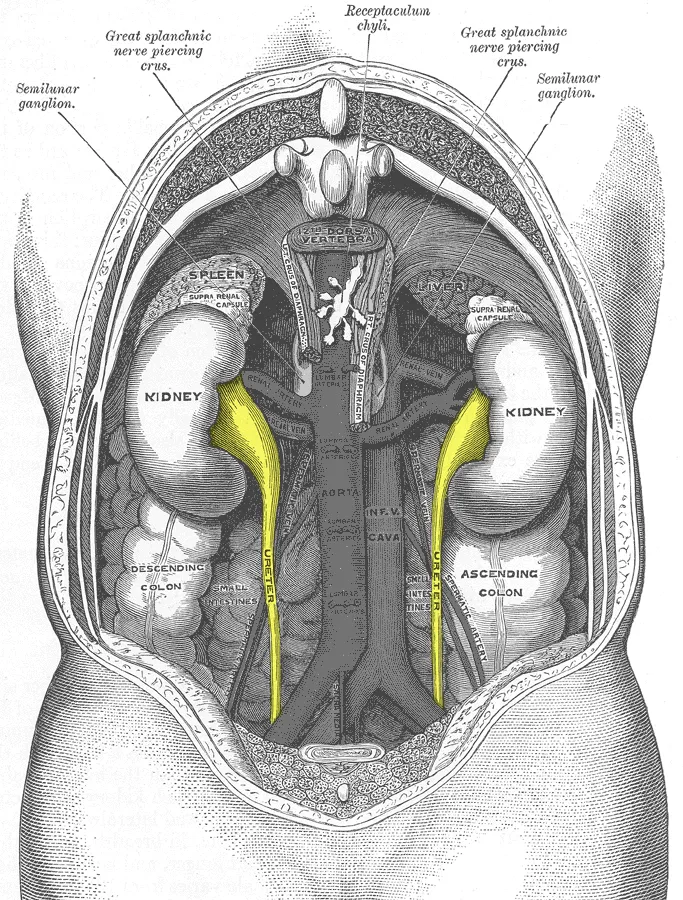
Vessels and nerves
The ureter receives segmental branches from the renal, gonadal, abdominal aortic, common iliac, internal iliac and vesical arteries, which anastomose longitudinally in the adventitia, so stripping it can cause ischaemia. Renal colic radiates from loin to groin along the T11–L2 segments.
Course, narrowings and peristalsis
The ureters are slender muscular tubes, 3–4 mm in diameter, derived from the ureteric bud, an outgrowth of the mesonephric duct. Each starts at the ureteropelvic junction, behind the renal vein and artery in the hilum at about L2, descends retroperitoneally in front of the psoas, enters the pelvis at the iliac bifurcation, follows the posterolateral pelvic wall and enters the bladder posterolaterally through the trigone.[1]
The ureter narrows at two points, the ureteropelvic and ureterovesical junctions, where renal stones can lodge and obstruct flow. At the iliac bifurcation it crosses the iliac vessels, lies in the cleft between the external and internal iliac arteries and is fixed there. Entering the pelvic brim it angles sharply backwards; this bend is not an anatomical narrowing, but long stones may struggle to pass it.[1]
Ureteral peristalsis is essentially myogenic: the wave starts in pacemaker cells of the renal pelvis and spreads from one smooth muscle cell to the next, the ureter acting as a functional syncytium, with nerves playing mainly a modulatory role. Each wave is driven by a plateau-type action potential. Ca²⁺ entry through L-type channels is the main activator source, and the blocker nifedipine abolishes both the action potential and the contraction. Urine flow rate sets the size and rhythm of the propelled boluses.[2][3]
Full-text reading (StatPearls)
The StatPearls chapters on this topic, reproduced verbatim. Their CC BY-NC-ND 4.0 licence allows unmodified copies only, so they are not translated or shortened.
Anatomy, Abdomen and Pelvis UreterLescay HA, Jiang J, Leslie SW, Tuma F · StatPearls 2024 · NBK532980 · 20k characters
Loading…
Lescay HA, Jiang J, Leslie SW, Tuma F. Anatomy, Abdomen and Pelvis Ureter. StatPearls [Internet]. Treasure Island (FL): StatPearls Publishing; 2024. NBK532980 · PMID 30422575 · CC BY-NC-ND 4.0. Reproduced verbatim, unmodified.
Organs in this system
References
Sources for the values and descriptions on this page, indexed in PubMed (US National Library of Medicine).
- [1]Anatomy, Abdomen and Pelvis UreterLescay HA et al., StatPearls 2024PMID 30422575
- [2]Ureteral motilityOsman F et al., Acta physiologica Hungarica 2009PMID 19942548
- [3]Excitation-Contraction Coupling in Ureteric Smooth Muscle: Mechanisms Driving Ureteric PeristalsisBurdyga T et al., Advances in experimental medicine and biology 2019PMID 31183824
- [4]Normal reference values for glomerular filtration rate: what do we really know?Delanaye P et al., 2012PMID 22802582Supports: Glomerular filtration rate