Suprarenal glands
Model
Layers
Z-Anatomy anatomical model based on BodyParts3D, licensed CC BY-SA 4.0.
Description
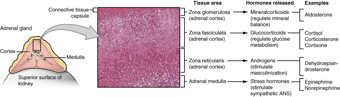
Paired retroperitoneal glands capping the upper poles of the kidneys, pyramidal on the right and crescentic on the left. The cortex has three zones secreting aldosterone (glomerulosa), cortisol (fasciculata) and androgens (reticularis); the medulla secretes adrenaline and noradrenaline under sympathetic control.
Parts
Each part has its own card; press “Show on the model” to fly to it and select it in the 3D model above.
- 01
Right and left glands
Glandula suprarenalis dextra et sinistra
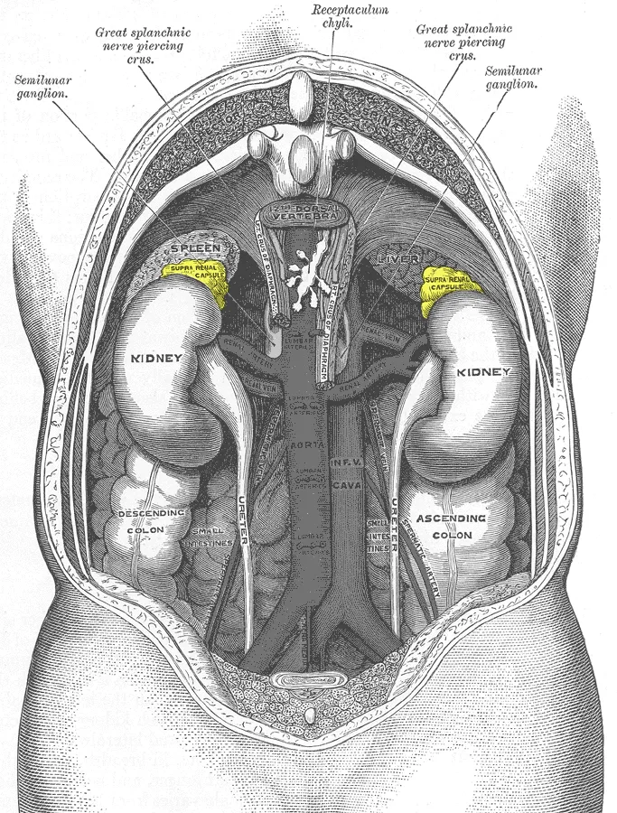
The right gland is pyramidal, on the upper pole of the right kidney behind the inferior vena cava; the left is crescentic, on the medial border of the upper pole of the left kidney near the aorta and pancreas. Each weighs 4–5 g and lies within the renal fascia in its own compartment, so it does not follow a ptotic kidney.
- 02
Zona glomerulosa
Zona glomerulosa
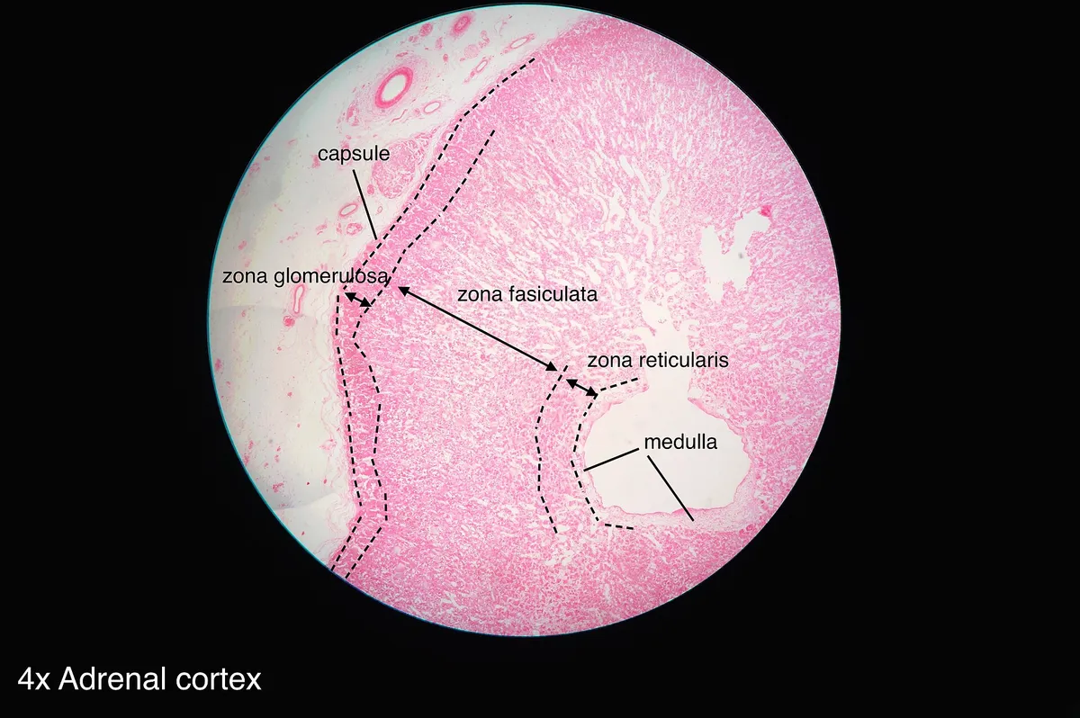
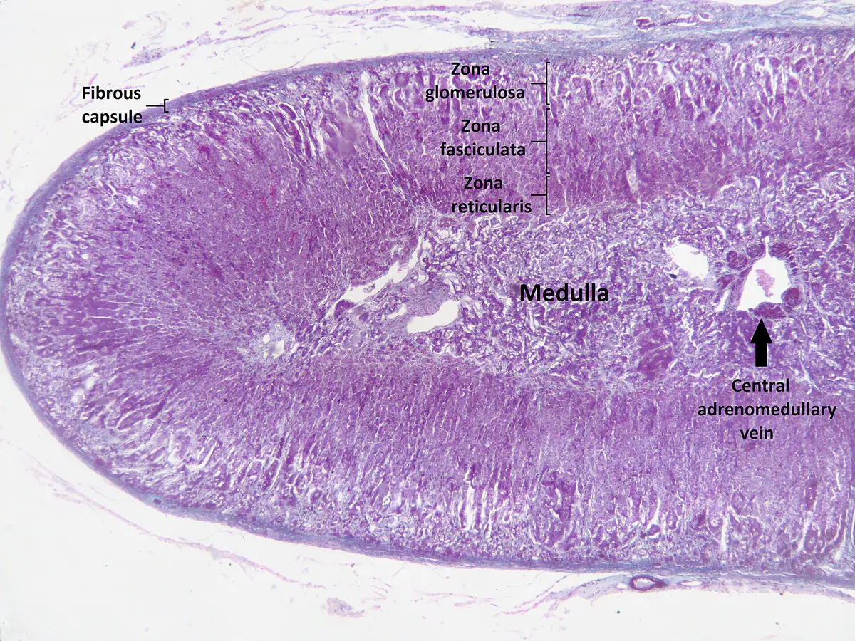
The thin outermost cortical layer (about 15% of the cortex), with cells in rounded clusters, secreting aldosterone in response to angiotensin II and plasma potassium. Aldosterone excess (Conn syndrome) causes hypertension and hypokalaemia.
- 03
Zona fasciculata
Zona fasciculata
The thickest layer (about 75% of the cortex), with lipid-laden cells in parallel columns secreting cortisol under ACTH control. Excess cortisol causes Cushing syndrome; prolonged steroid therapy atrophies this layer, so abrupt withdrawal can precipitate adrenal crisis.
- 04
Zona reticularis
Zona reticularis
The innermost cortical layer, with cells in anastomosing cords, secreting adrenal androgens (DHEA, androstenedione) and some cortisol. 21-hydroxylase deficiency diverts precursors to androgens, causing virilisation (congenital adrenal hyperplasia).
- 05
Adrenal medulla
Medulla glandulae suprarenalis
The central part of neural crest origin, made of chromaffin cells (equivalent to postganglionic sympathetic neurons) that receive preganglionic fibres directly through the splanchnic nerves and secrete adrenaline (about 80%) and noradrenaline. Phaeochromocytoma causes paroxysmal hypertension, sweating and palpitations.
- 06
Adrenal vessels
Arteriae et vena suprarenales
Three arterial sources (superior from the inferior phrenic, middle from the aorta, inferior from the renal artery) break into dozens of small branches; blood flows from cortex to medulla, so cortisol helps the medulla make adrenaline. A single vein drains each gland: the short right vein enters the inferior vena cava directly (easily torn at surgery), the left joins the left renal vein.
Position and relations
The suprarenal glands lie in the retroperitoneum, inside the renal fascia but separated from the kidney by a layer of fat, so they stay in place if the kidney is displaced. The right gland lies behind the inferior vena cava against the bare area of the liver; the left is related to the stomach across the omental bursa, the pancreas and the splenic vein. Both rest on the crura of the diaphragm.
Form and structure
The cortex (about 90% of the mass) is mesodermal and has three zones from outside in: the glomerulosa, secreting mineralocorticoids under the renin–angiotensin system, and the fasciculata and reticularis, secreting glucocorticoids and androgens under ACTH. The medulla derives from the neural crest and consists of chromaffin cells equivalent to postganglionic sympathetic neurons.
Vessels and nerves
Each gland has three arterial sources: the superior suprarenal artery (from the inferior phrenic), the middle (from the abdominal aorta) and the inferior (from the renal artery). By contrast there is a single vein: on the right it is short and enters the inferior vena cava directly, on the left it drains into the left renal vein. The medulla receives preganglionic sympathetic fibres directly from the splanchnic nerves.
Development
The suprarenal glands share an early origin with the kidneys and gonads; the adrenocortical primordium becomes individualised under many transcription factors and endocrine signals, and in humans it is massively remodelled around birth. Several factors that drive adrenocortical development also regulate the growth of adrenocortical tumours.[1]
The suprarenal glands help regulate blood pressure, metabolism, electrolyte balance and the stress response: the cortex secretes steroids from its three functional zones, and the medulla synthesises adrenaline, noradrenaline and dopamine under sympathetic control.[2]
Shape, cortical zones and their disorders
Each suprarenal gland is about 50 mm high, 30 mm wide and 10 mm thick and weighs about 5 g; the right is pyramidal, the left crescentic. At birth the gland is about one-third the size of the kidney but only one-thirtieth in adults, because the fetal cortex involutes after birth. The right gland lies below the liver, behind the inferior vena cava and in front of the diaphragm; the left lies medial to the spleen, above the splenic vessels, lateral to the abdominal aorta and in front of the diaphragm; both lie in the perirenal space within the renal fascia. The fat-rich cortex is yellow, the medulla reddish-brown, and a thick connective-tissue capsule surrounds the whole. The cortex uses cholesterol as the precursor of all its steroid hormones. The outer zona glomerulosa secretes aldosterone; hyperaldosteronism (Conn syndrome) causes hypertension resistant to three drugs, hypokalaemia and mild hypernatraemia. The zona fasciculata secretes cortisol under ACTH, regulating blood glucose through gluconeogenesis, immunity and metabolism; cortisol excess causes central obesity, striae, a buffalo hump, hyperglycaemia and poor wound healing. The zona reticularis secretes androgens; androgen excess causes precocious puberty in boys or virilisation in girls.[3]
The gland consists of two tissues of different embryonic origin. Around week 5 a cluster of cells (the adrenogonadal primordium) appears in the urogenital ridge; mesothelial cells invade the mesenchyme to form the fetal cortex and Leydig cells. Large acidophilic cells form the primitive cortex, then smaller cells migrate to surround them and form the definitive cortex; key factors include SF-1, CITED-2 and β-catenin. Around week 7, neural crest cells migrate along a ventral path into the primordium from its medial side and differentiate into chromaffin cells arranged centrally in cords and clusters, which the cortex gradually encloses. The chromaffin cells of the medulla cluster around blood vessels and synthesise adrenaline and noradrenaline, raising heart rate, contractility, metabolic rate and alertness in the “fight or flight” response. Phaeochromocytoma of the medulla belongs to multiple endocrine neoplasia type II together with medullary thyroid carcinoma.[3]
Zona glomerulosa
- Main hormone
- Aldosterone
- Control
- Renin–angiotensin, potassium
- Excess state
- Conn syndrome
Zona fasciculata
- Main hormone
- Cortisol
- Control
- ACTH
- Excess state
- Cushing syndrome
Zona reticularis
- Main hormone
- Androgens
- Control
- ACTH
- Excess state
- Precocious puberty, virilisation
Medulla
- Main hormone
- Adrenaline, noradrenaline
- Control
- Preganglionic sympathetic fibres
- Excess state
- Phaeochromocytoma
| Zone | Main hormone | Control | Excess state |
|---|---|---|---|
| Zona glomerulosa | Aldosterone | Renin–angiotensin, potassium | Conn syndrome |
| Zona fasciculata | Cortisol | ACTH | Cushing syndrome |
| Zona reticularis | Androgens | ACTH | Precocious puberty, virilisation |
| Medulla | Adrenaline, noradrenaline | Preganglionic sympathetic fibres | Phaeochromocytoma |
Full-text reading (StatPearls)
The StatPearls chapters on this topic, reproduced verbatim. Their CC BY-NC-ND 4.0 licence allows unmodified copies only, so they are not translated or shortened.
Anatomy, Abdomen and Pelvis: Adrenal Glands (Suprarenal Glands)Megha R, Wehrle CJ, Kashyap S, Leslie SW · StatPearls 2022 · NBK482264 · 13k characters
Loading…
Megha R, Wehrle CJ, Kashyap S, Leslie SW. Anatomy, Abdomen and Pelvis: Adrenal Glands (Suprarenal Glands). StatPearls [Internet]. Treasure Island (FL): StatPearls Publishing; 2022. NBK482264 · PMID 29489211 · CC BY-NC-ND 4.0. Reproduced verbatim, unmodified.
Organs in this system
References
Sources for the values and descriptions on this page, indexed in PubMed (US National Library of Medicine).
- [1]Adrenal cortex ontogenesisLalli E, Best practice & research. Clinical endocrinology & metabolism 2010PMID 21115154
- [2]Adrenal Anatomy and PhysiologyAl-Khanaty A et al., The Urologic clinics of North America 2025PMID 40250885
- [3]Anatomy, Abdomen and Pelvis: Adrenal Glands (Suprarenal Glands)Megha R et al., StatPearls 2022PMID 29489211
- [4]Physiology, Pituitary HormonesStatPearls (NCBI Bookshelf)PMID 32491488Supports: Pituitary and endocrine axes
- [5]Physiology, Anterior PituitaryStatPearls (NCBI Bookshelf)PMID 29763073