Muscle tissue
Model
Hover or tap to see names, click to read a description; click a name in the legend to fly to it. Procedurally built to histological proportions; colours follow illustration conventions.
Description
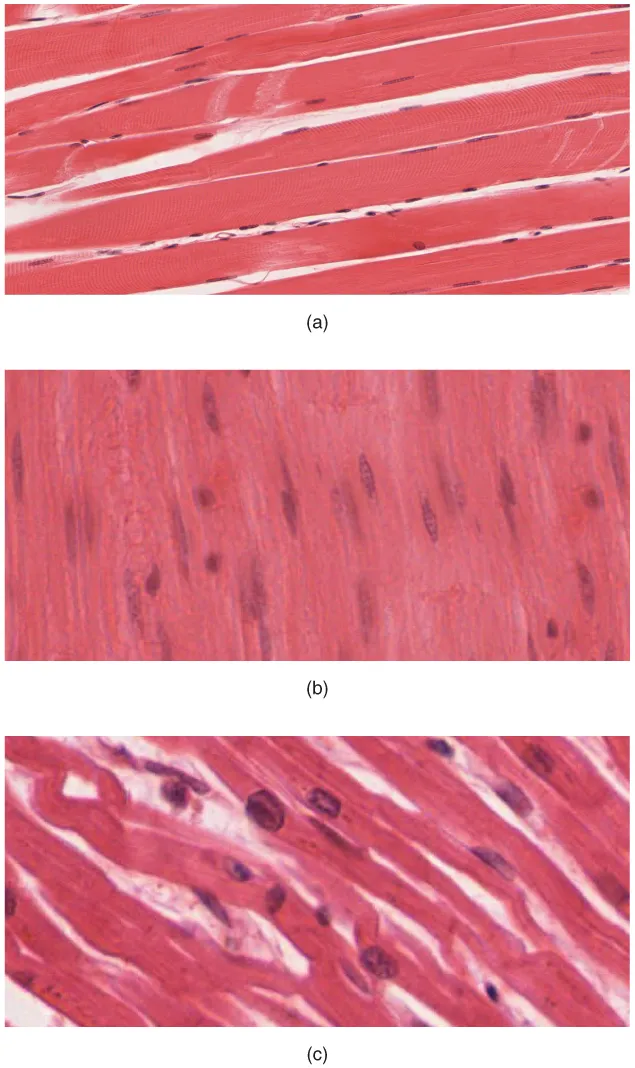
Tissue of elongated cells rich in actin and myosin filaments and able to contract. There are three kinds: skeletal muscle (multinucleate striated fibres under voluntary control), cardiac muscle (branched striated cells joined by intercalated discs, contracting automatically) and smooth muscle (spindle-shaped unstriated cells under involuntary control).
Skeletal muscle
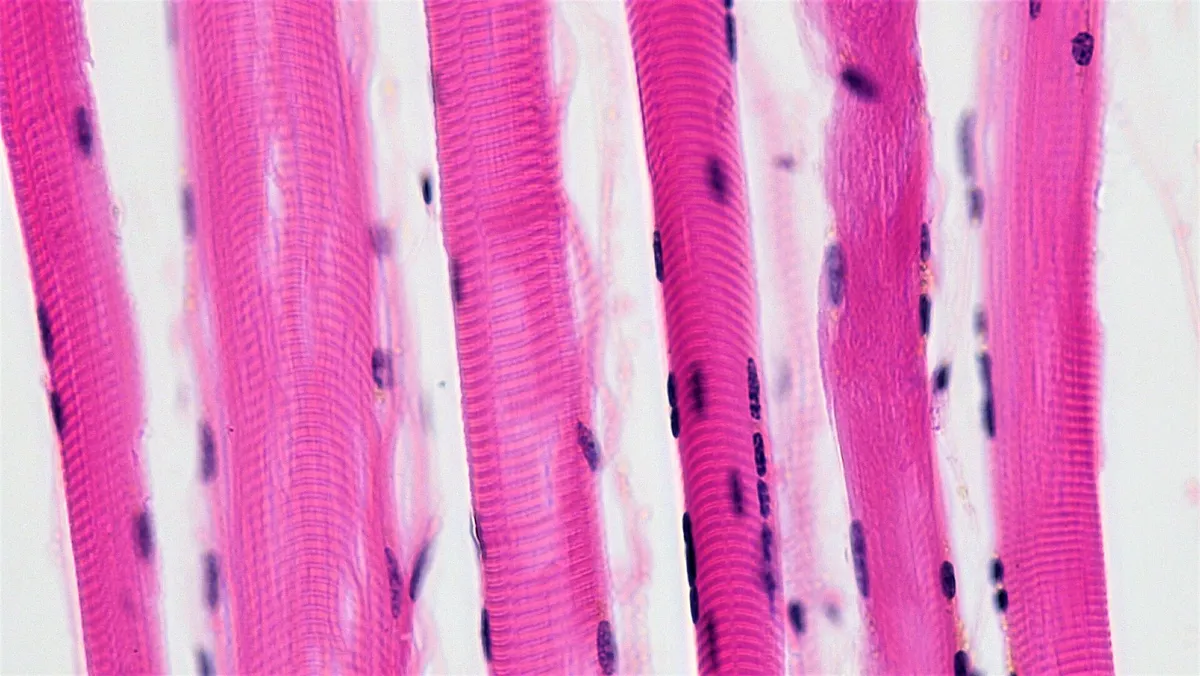
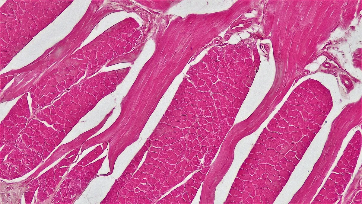
A skeletal muscle fibre is a cylindrical syncytium 10–100 µm wide and up to tens of centimetres long, formed when hundreds of myoblasts fuse in the embryo, so each fibre has hundreds of nuclei lying beneath the membrane. Connective tissue wraps muscle at three levels: epimysium around the whole muscle, perimysium around each fascicle and endomysium (with a basal lamina) around each fibre, and this collagen framework carries the force of contraction to the tendon. Between the sarcolemma and the basal lamina lie satellite cells, the muscle stem cells, normally quiescent and activated by injury or training.[1][2][3][4]
Fibres are divided into type I (slow-twitch, red, rich in mitochondria and myoglobin, fatigue-resistant, oxidative) and type II (fast-twitch, low in myoglobin, glycolytic, powerful but quick to tire), in proportions that differ between muscles and between endurance and power athletes. A motor neuron and all the fibres it supplies form a motor unit; force is graded by recruiting units from small to large and by raising firing frequency. Fatigue reflects several cellular mechanisms: accumulation of inorganic phosphate, reduced Ca²⁺ release from the sarcoplasmic reticulum and glycogen depletion.[5][6][7]
The sarcomere and contraction
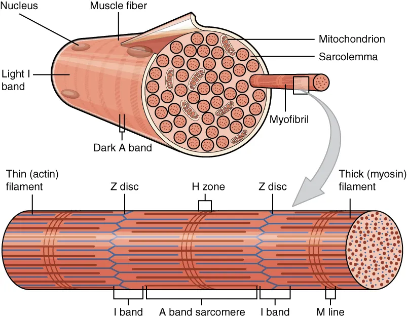
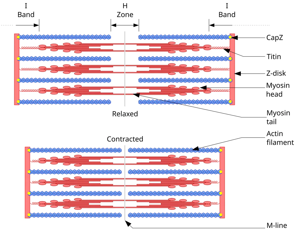
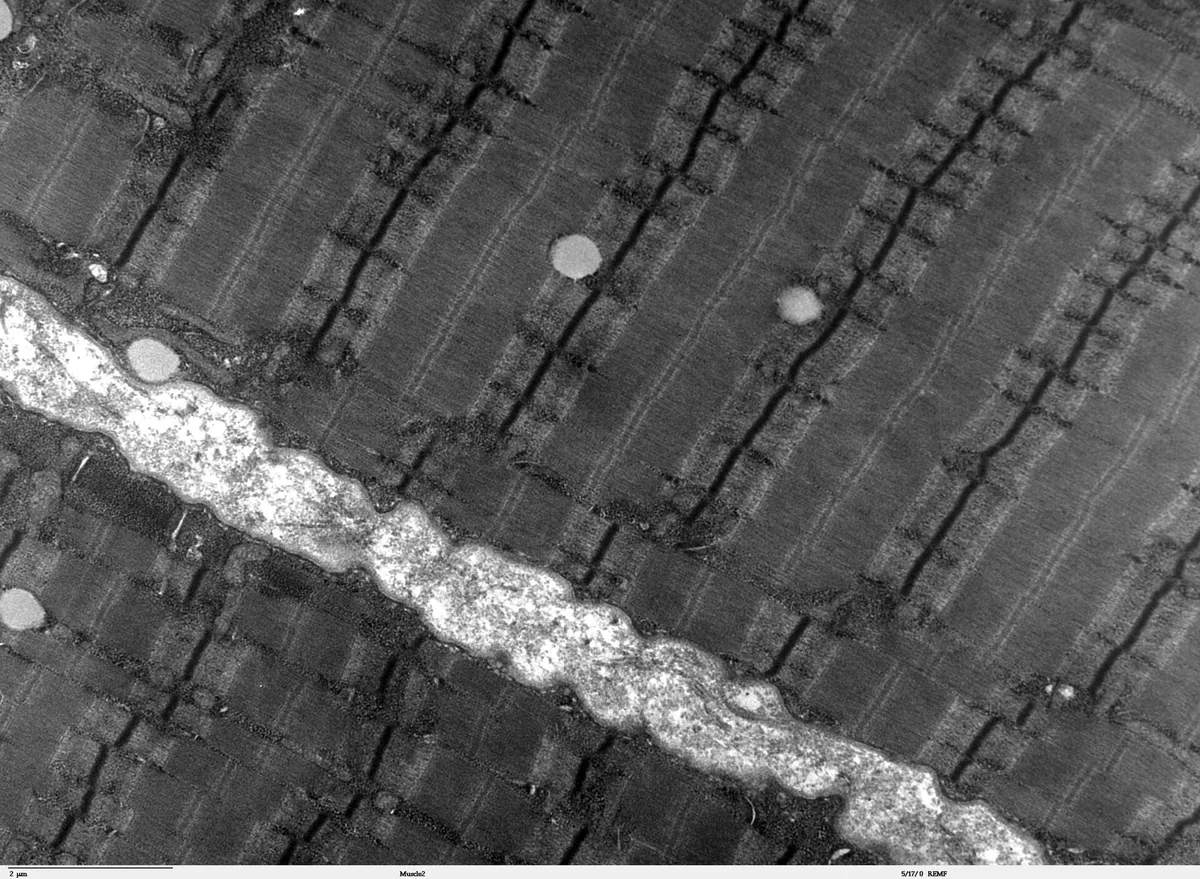
The fibre is packed with myofibrils, each a chain of sarcomeres about 2–2.5 µm long between two Z discs. Thin actin filaments (with tropomyosin and troponin) insert into the Z disc; thick myosin filaments sit in the middle, forming the dark A band, while the region of thin filaments alone is the pale I band. Titin, the largest protein in the body, runs from Z disc to M line like a spring that centres the thick filaments and provides passive tension, while nebulin rules thin-filament length. Register of sarcomeres across myofibrils produces the cross-striation seen under the microscope; desmin, the muscle intermediate filament, ties Z discs to one another and to the membrane.[2][8][9]
A nerve impulse at the neuromuscular junction releases acetylcholine, triggering an action potential that spreads along the membrane and down the T tubules. At the triad (one T tubule between two terminal cisternae of the sarcoplasmic reticulum), dihydropyridine receptors in the T tubule are mechanically coupled to RyR1 ryanodine receptors, opening them so Ca²⁺ floods the cytosol. Ca²⁺ binds troponin C and shifts tropomyosin off the binding sites on actin; myosin heads attach, perform their power stroke and pull the thin filaments towards the M line, then fresh ATP detaches the head so the cycle repeats. The sarcomere shortens while filament length stays constant. SERCA pumps return Ca²⁺ for relaxation; when ATP runs out after death the heads cannot detach, causing rigor mortis.[9][10][5]
Cardiac muscle
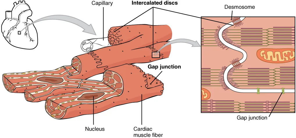
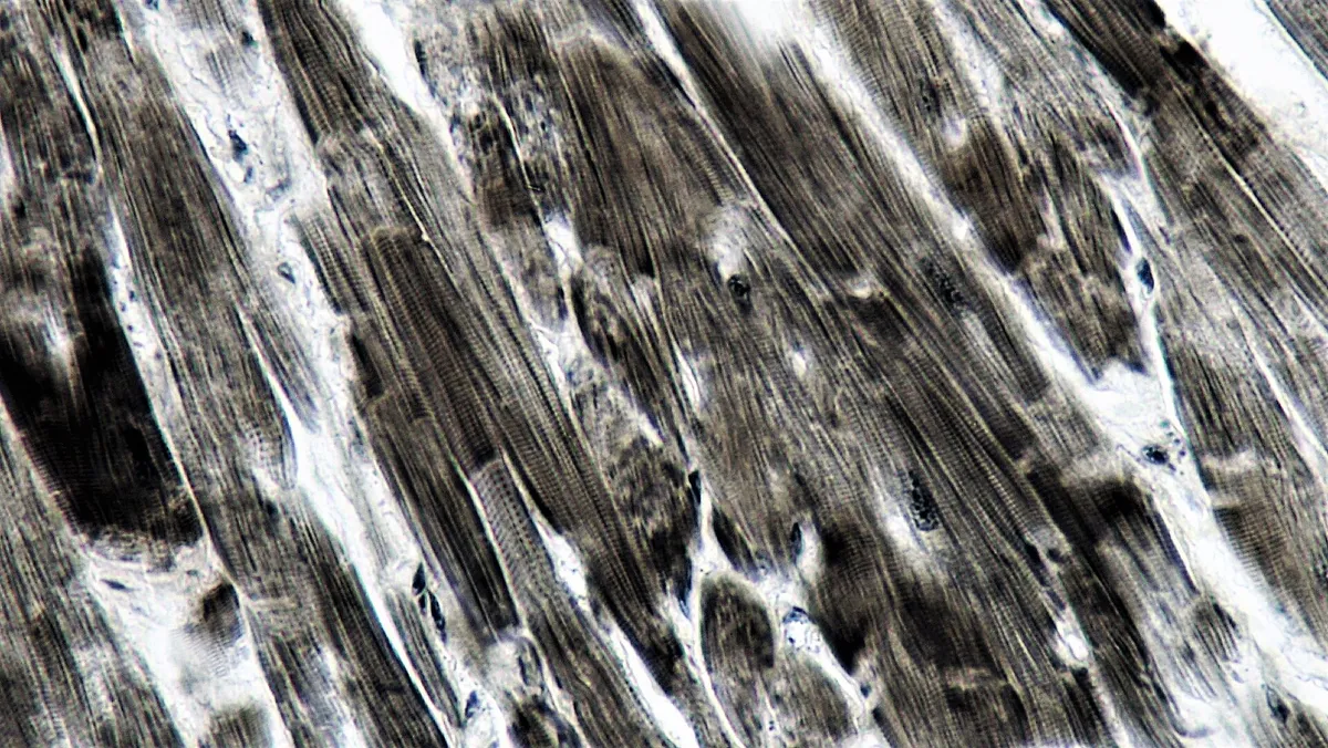
Cardiomyocytes are striated too, but they are individual branching cells about 100 µm long with one or two central nuclei. They join end to end at step-like intercalated discs: transverse parts contain adherens junctions and desmosomes that transmit force, longitudinal parts contain connexin-43 gap junctions that let the electrical impulse pass from cell to cell, so a whole chamber contracts as one. Mitochondria fill about a third of the cell, because the heart contracts ceaselessly and relies almost entirely on aerobic metabolism. Adult cardiomyocytes barely divide, so a myocardial infarct heals with fibrous scar rather than new muscle.[11][12]
Unlike skeletal muscle, cardiac excitation–contraction coupling relies on Ca²⁺-induced Ca²⁺ release: during the plateau of the action potential, L-type Ca²⁺ channels in the T tubule admit a small amount of Ca²⁺, which opens RyR2 channels of the sarcoplasmic reticulum and releases a much larger amount. The long plateau makes the refractory period last nearly the whole contraction, so cardiac muscle cannot be tetanised like skeletal muscle. Contractile force is tuned by Ca²⁺ per beat (sympathetic stimulation through β receptors increases Ca²⁺ entry and reuptake) and by starting sarcomere length (the Frank–Starling law). Ca²⁺ leak through RyR2 underlies many arrhythmias.[13][11]
Smooth muscle
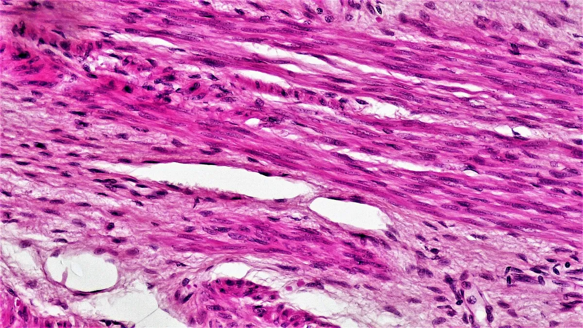
Smooth muscle cells are spindle-shaped, 20–500 µm long, with one central nucleus and no striations, because actin and myosin are not arranged in sarcomeres but criss-cross the cell, anchored to dense bodies in the cytoplasm and dense plaques on the membrane (the equivalent of Z discs). On contraction the cell shortens with a corkscrew twist. The membrane is studded with caveolae and the sarcoplasmic reticulum is sparse; Ca²⁺ entering from outside and from the reticulum binds calmodulin and activates myosin light-chain kinase, which phosphorylates myosin, in contrast to the troponin mechanism of striated muscle. A “latch” state keeps cross-bridges attached with very little ATP, letting vessels and sphincters hold tone for hours.[14][15]
Visceral (single-unit) smooth muscle (gut, uterus, ureter) consists of cells coupled by gap junctions that contract as a unit, often with spontaneous rhythm driven by interstitial cells of Cajal; multi-unit smooth muscle (iris, arrector pili, vas deferens) has individually innervated cells for fine control. Smooth muscle answers to autonomic nerves, hormones and local mediators, and it can divide. Vascular smooth muscle cells are especially plastic: in atherosclerosis they switch from a contractile to a synthetic phenotype, migrate into the intima and build the fibrous cap of the plaque; the K⁺ channels in their membrane are the target of many vasodilators.[15][16][17]
Cell shape
- Skeletal
- Long cylindrical syncytium
- Cardiac
- Branched cells
- Smooth
- Spindle
Nuclei
- Skeletal
- Many, peripheral
- Cardiac
- 1–2, central
- Smooth
- 1, central
Striations
- Skeletal
- Yes
- Cardiac
- Yes
- Smooth
- No
Cell–cell junctions
- Skeletal
- None
- Cardiac
- Intercalated discs
- Smooth
- Gap junctions (single-unit)
Ca²⁺ regulation
- Skeletal
- Troponin; RyR1 mechanically coupled
- Cardiac
- Troponin; Ca²⁺-induced Ca²⁺ release
- Smooth
- Calmodulin–MLCK
Control
- Skeletal
- Voluntary (motor nerves)
- Cardiac
- Intrinsic pacemaker, autonomic modulation
- Smooth
- Autonomic, hormonal, local
Regeneration
- Skeletal
- Good, via satellite cells
- Cardiac
- Very poor
- Smooth
- Good, cells divide
| Feature | Skeletal | Cardiac | Smooth |
|---|---|---|---|
| Cell shape | Long cylindrical syncytium | Branched cells | Spindle |
| Nuclei | Many, peripheral | 1–2, central | 1, central |
| Striations | Yes | Yes | No |
| Cell–cell junctions | None | Intercalated discs | Gap junctions (single-unit) |
| Ca²⁺ regulation | Troponin; RyR1 mechanically coupled | Troponin; Ca²⁺-induced Ca²⁺ release | Calmodulin–MLCK |
| Control | Voluntary (motor nerves) | Intrinsic pacemaker, autonomic modulation | Autonomic, hormonal, local |
| Regeneration | Good, via satellite cells | Very poor | Good, cells divide |
Full-text reading (StatPearls)
The StatPearls chapters on this topic, reproduced verbatim. Their CC BY-NC-ND 4.0 licence allows unmodified copies only, so they are not translated or shortened.
Physiology, Skeletal MuscleMcCuller C, Jessu R, Callahan AL · StatPearls 2023 · NBK537139 · 22k characters
Loading…
McCuller C, Jessu R, Callahan AL. Physiology, Skeletal Muscle. StatPearls [Internet]. Treasure Island (FL): StatPearls Publishing; 2023. NBK537139 · PMID 30725824 · CC BY-NC-ND 4.0. Reproduced verbatim, unmodified.
Physiology, Smooth MuscleHafen BB, Burns B · StatPearls 2023 · NBK526125 · 20k characters
Loading…
Hafen BB, Burns B. Physiology, Smooth Muscle. StatPearls [Internet]. Treasure Island (FL): StatPearls Publishing; 2023. NBK526125 · PMID 30252381 · CC BY-NC-ND 4.0. Reproduced verbatim, unmodified.
Physiology, Muscle ContractionGash MC, Kandle PF, Murray IV, Varacallo MA · StatPearls 2023 · NBK537140 · 19k characters
Loading…
Gash MC, Kandle PF, Murray IV, Varacallo MA. Physiology, Muscle Contraction. StatPearls [Internet]. Treasure Island (FL): StatPearls Publishing; 2023. NBK537140 · PMID 30725825 · CC BY-NC-ND 4.0. Reproduced verbatim, unmodified.
Anatomy, Smooth MuscleHafen BB, Shook M, Burns B · StatPearls 2023 · NBK532857 · 17k characters
Loading…
Hafen BB, Shook M, Burns B. Anatomy, Smooth Muscle. StatPearls [Internet]. Treasure Island (FL): StatPearls Publishing; 2023. NBK532857 · PMID 30422452 · CC BY-NC-ND 4.0. Reproduced verbatim, unmodified.
Physiology, Cardiac MuscleRipa R, George T, Shumway KR, Sattar Y · StatPearls 2023 · NBK572070 · 17k characters
Loading…
Ripa R, George T, Shumway KR, Sattar Y. Physiology, Cardiac Muscle. StatPearls [Internet]. Treasure Island (FL): StatPearls Publishing; 2023. NBK572070 · PMID 34283436 · CC BY-NC-ND 4.0. Reproduced verbatim, unmodified.
Physiology, Skeletal Muscle ContractionPham S, Puckett Y · StatPearls 2023 · NBK559006 · 14k characters
Loading…
Pham S, Puckett Y. Physiology, Skeletal Muscle Contraction. StatPearls [Internet]. Treasure Island (FL): StatPearls Publishing; 2023. NBK559006 · PMID 32644432 · CC BY-NC-ND 4.0. Reproduced verbatim, unmodified.
Anatomy, Thorax, Cardiac MuscleSaxton A, Tariq MA, Bordoni B · StatPearls 2023 · NBK535355 · 13k characters
Loading…
Saxton A, Tariq MA, Bordoni B. Anatomy, Thorax, Cardiac Muscle. StatPearls [Internet]. Treasure Island (FL): StatPearls Publishing; 2023. NBK535355 · PMID 30570976 · CC BY-NC-ND 4.0. Reproduced verbatim, unmodified.
Anatomy, Skeletal MuscleDave HD, Shook M, Varacallo MA · StatPearls 2023 · NBK537236 · 11k characters
Loading…
Dave HD, Shook M, Varacallo MA. Anatomy, Skeletal Muscle. StatPearls [Internet]. Treasure Island (FL): StatPearls Publishing; 2023. NBK537236 · PMID 30725921 · CC BY-NC-ND 4.0. Reproduced verbatim, unmodified.
Organs in this system
References
Sources for the values and descriptions on this page, indexed in PubMed (US National Library of Medicine).
- [1]Anatomy, Skeletal MuscleDave HD et al., StatPearls 2023PMID 30725921
- [2]MyofibersCretoiu D et al., Advances in experimental medicine and biology 2018PMID 30390246
- [3]Structure and function of the skeletal muscle extracellular matrixGillies AR et al., Muscle & nerve 2011PMID 21949456
- [4]Satellite Cells and Skeletal Muscle RegenerationDumont NA et al., Comprehensive physiology 2015PMID 26140708
- [5]Physiology, Skeletal MuscleMcCuller C et al., StatPearls 2023PMID 30725824
- [6]Skeletal muscle design to meet functional demandsLieber RL et al., Philosophical transactions of the Royal Society of London. Series B, Biological sciences 2011PMID 21502118
- [7]Skeletal muscle fatigue: cellular mechanismsAllen DG et al., Physiological reviews 2008PMID 18195089
- [8]New roles for desmin in the maintenance of muscle homeostasisAgnetti G et al., The FEBS journal 2022PMID 33825342
- [9]Physiology, Muscle ContractionGash MC et al., StatPearls 2023PMID 30725825
- [10]Proteins of the Triadic Excitation-Contraction Coupling Complex in Skeletal MuscleChang T et al., Cold Spring Harbor perspectives in biology 2025PMID 40097154
- [11]Physiology, Cardiac MuscleRipa R et al., StatPearls 2023PMID 34283436
- [12]Cardiomyocyte Maturation: New Phase in DevelopmentGuo Y et al., Circulation research 2020PMID 32271675
- [13]Calcium and Excitation-Contraction Coupling in the HeartEisner DA et al., Circulation research 2017PMID 28684623
- [14]Anatomy, Smooth MuscleHafen BB et al., StatPearls 2023PMID 30422452
- [15]Physiology, Smooth MuscleHafen BB et al., StatPearls 2023PMID 30252381
- [16]Vascular Smooth Muscle Cells in AtherosclerosisBennett MR et al., Circulation research 2016PMID 26892967
- [17]Potassium channels in vascular smooth muscle cellsGarrud TAC et al., Physiological reviews 2026PMID 41526008