Thoracic cage
Model
Layers
Z-Anatomy anatomical model based on BodyParts3D, licensed CC BY-SA 4.0.
Description
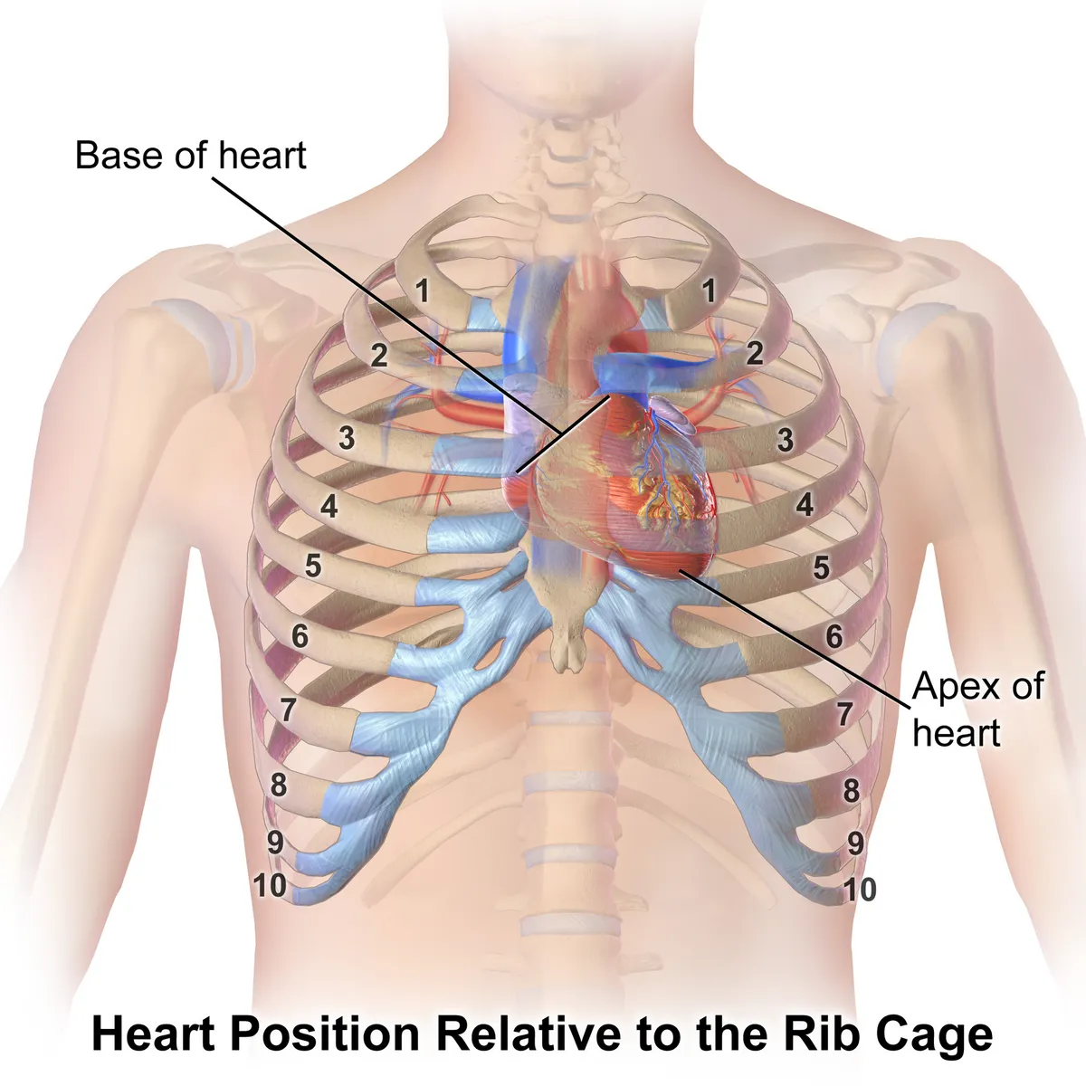
Formed by 12 pairs of ribs, the sternum and the 12 thoracic vertebrae; it protects the heart and lungs and takes part in the mechanics of breathing.
Parts
Each part has its own card; press “Show on the model” to fly to it and select it in the 3D model above.
- 01
Sternum
Sternum
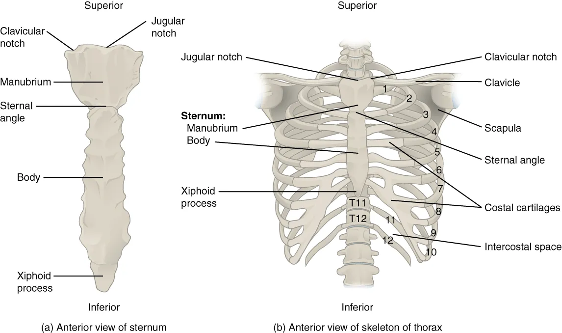
A flat bone in the midline of the chest made of the manubrium, body and xiphoid process. The manubrium articulates with the clavicles and first costal cartilages; its junction with the body forms the sternal angle (of Louis) at the second costal cartilage, the landmark for counting ribs and level with the tracheal bifurcation. The body is the site of chest compressions and bone marrow sampling.
- 02
True ribs (1–7)
Costae verae
The first seven pairs, joined directly to the sternum by their own costal cartilages. The first rib is the shortest, broadest and flattest and is grooved by the subclavian vessels; ribs 3 to 9 are typical, with a head (two articular facets), neck, tubercle and curved shaft whose costal groove on the lower border shelters the intercostal neurovascular bundle.
- 03
False ribs (8–10)
Costae spuriae
The cartilages of ribs 8, 9 and 10 join the cartilage above, forming the costal margin (arch). The costal margin is the palpation landmark for examining the liver, spleen and gallbladder.
- 04
Floating ribs (11–12)
Costae fluctuantes
The last two pairs are short and end freely in the abdominal wall muscles without reaching the sternum. The twelfth rib lies behind the kidney and pleura, a hazard in posterior approaches to the kidney.
- 05
Costal cartilages
Cartilagines costales
Hyaline cartilage joining the front ends of the ribs to the sternum or to each other, giving the cage the elasticity to expand in breathing and to absorb impacts. They may calcify with age; costochondritis (Tietze syndrome) causes chest pain easily mistaken for cardiac pain.
- 06
Thoracic inlet and outlet
Apertura thoracis superior et inferior
The inlet (bounded by T1, the first ribs and the top of the manubrium) passes the trachea, oesophagus, great vessels and nerves between neck and thorax; a cervical rib can compress the brachial plexus (thoracic outlet syndrome). The wider lower aperture is closed by the diaphragm.
- 07
Costovertebral joints
Articulationes costovertebrales
The head of each rib articulates with the bodies of two adjacent vertebrae and the tubercle with the transverse process. The axis through these joints sets the movement: upper ribs rise forwards like a pump handle (increasing the anteroposterior diameter), lower ribs swing outwards like a bucket handle (increasing the transverse diameter).
Form and structure
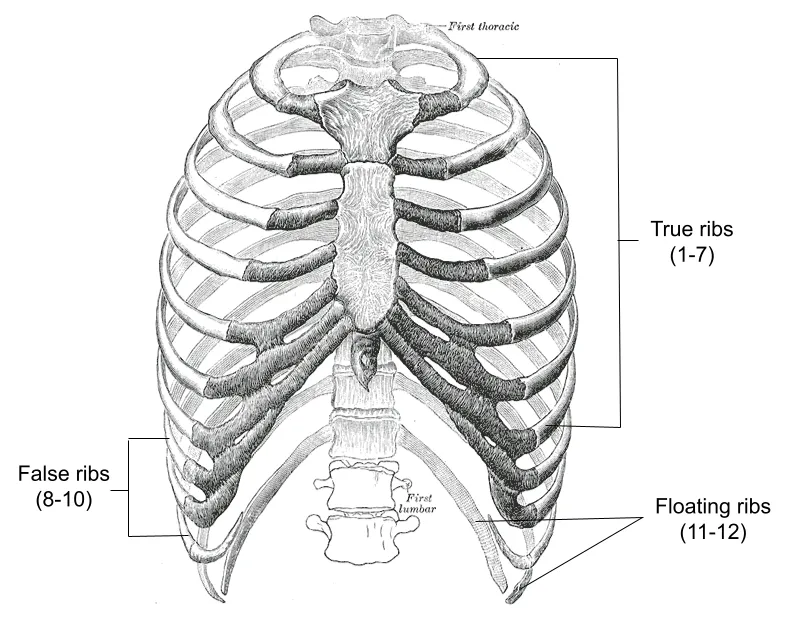
The thoracic cage consists of 12 thoracic vertebrae behind, 12 pairs of ribs at the sides and the sternum in front. The first seven pairs are true ribs, joined directly to the sternum by costal cartilages; ribs 8–10 are false ribs whose cartilages join the cartilage above; ribs 11–12 are floating ribs with free anterior ends.
Position and relations
The cage protects the heart, lungs and great vessels and takes part in breathing: on inspiration the intercostal muscles raise the ribs (“bucket-handle” and “pump-handle” movements) and, with the diaphragm, enlarge the thorax. The intercostal neurovascular bundle runs in the costal groove along the lower border of each rib, so pleural aspiration is done just above the upper border of the rib below.[1]
Respiratory mechanics
The diaphragm is the main inspiratory muscle and the landmark separating thorax from abdomen. Its contraction lowers the dome and lifts and widens the lower rib cage, while intercostal and accessory muscles move the ribs and sternum. Beyond breathing, it stabilises the lumbar spine during loading, aids venous and lymphatic return and takes part in swallowing, vomiting and the anti-reflux barrier.[2]
In intensive care, airway pressure and flow measured on the ventilator reflect lung and chest-wall compliance and airway resistance; ventilator graphics (pressure, flow and volume scalars and loops) reveal patient–ventilator asynchrony and help limit ventilator-induced lung injury.[3][4][5]
Pectus carinatum (keel chest) is a spectrum of progressive congenital protrusion of the sternum and/or costal cartilages; it may accompany Marfan syndrome, homocystinuria, Noonan syndrome or mitral valve prolapse.[6]
Typical and atypical ribs, vessels and nerves
Each rib articulates posteriorly with two thoracic vertebrae at the costovertebral joints, except the first, which articulates only with T1. True ribs (1–7) join the sternum directly through their costal cartilages at the sternocostal joints; the first sternocostal joint is a synarthrosis, and the first rib may also articulate with the clavicle at the costoclavicular joint. False ribs (8–10) join indirectly because their cartilages merge with the seventh costal cartilage. The classical floating ribs are 11 and 12, but recent cadaver studies show the costal margin is far more variable than textbooks describe: the tenth rib is unattached to the ninth in 46–59% of specimens, the tips of the eighth and ninth ribs are free in 58% and 92%, and the eighth rib reaches the sternum directly in about 10–42% depending on the study. Bridging interchondral joints are commonest between ribs 5 and 8 and spread forces from the costal margin across the chest wall. Atypical ribs are the first (short and wide, with two grooves and a single articular facet), the second (thin and long, with a tuberosity for serratus anterior), the tenth (one facet) and the eleventh and twelfth (one facet, no neck). The ribs protect the thoracic and mediastinal contents, give attachment to muscles, assist breathing and, after birth, remain a site of haematopoiesis because red marrow persists in flat bones.[7]
Anteriorly, ribs 1–7 receive anterior intercostal arteries from the internal thoracic artery and ribs 8–10 from the musculophrenic artery; posteriorly, the first two posterior intercostal arteries come from the superior intercostal artery of the costocervical trunk and the rest from the descending thoracic aorta. The eleventh rib receives the eleventh posterior intercostal artery and the twelfth the subcostal artery. Anterior intercostal veins drain to the internal thoracic and musculophrenic veins, posterior ones to the azygos and hemiazygos systems. The neurovascular bundle runs in the costal groove with the nerve lowest, but it is not always shielded: on CT of 298 arteries, the artery lay exposed in the intercostal space for the first 6 cm lateral to the spine, only 17% being covered by the rib above at 3 cm against 97% at 6 cm, with more variation in older patients and higher spaces, which matters for chest drains and thoracentesis. The intercostal nerves run between the internal and innermost intercostals and give lateral and anterior cutaneous branches, sensory branches to the parietal pleura, peritoneal branches (from nerves 7–11), a collateral branch along the upper border of the rib below, muscular branches and rami communicantes to the sympathetic ganglia. Nerves 7–9 also give a diaphragmatic branch and arborise at the costochondral junction, their position varying with how the eighth rib attaches, which matters for intercostal nerve blocks.[7]
True ribs
- Ribs
- 1–7
- Sternal connection
- Directly through costal cartilage
- Notes
- First sternocostal joint is a synarthrosis
False ribs
- Ribs
- 8–10
- Sternal connection
- Indirectly via the 7th cartilage
- Notes
- Rib 8 sometimes reaches the sternum directly
Floating ribs
- Ribs
- 11–12 (often also 10)
- Sternal connection
- None
- Notes
- Posterior vessels only
| Group | Ribs | Sternal connection | Notes |
|---|---|---|---|
| True ribs | 1–7 | Directly through costal cartilage | First sternocostal joint is a synarthrosis |
| False ribs | 8–10 | Indirectly via the 7th cartilage | Rib 8 sometimes reaches the sternum directly |
| Floating ribs | 11–12 (often also 10) | None | Posterior vessels only |
The sternum: parts, development and variants
The sternum is a partly T-shaped vertical bone in the middle of the anterior chest wall, made of the manubrium, body and xiphoid process. The quadrangular manubrium bears the suprasternal (jugular) notch above and clavicular notches on each side for the medial ends of the clavicles (sternoclavicular joints), and articulates with the first costal cartilages. The body, the longest part, is flat with lateral ridges where costal cartilages 3–7 articulate. The junction of manubrium and body forms the sternal angle (of Louis), because the two parts lie in slightly different planes; it lies level with the second costal cartilage and the T4–T5 disc and is the landmark for counting ribs. The triangular xiphoid process is highly variable in shape, largely cartilaginous until about 40 and fully calcified by about 60. Sternocleidomastoid, sternohyoid and sternothyroid attach to the manubrium; transversus thoracis and pectoralis major to the body; and the diaphragm and anterior abdominal muscles to the xiphoid. Blood comes mainly from sternal and perforating branches of the internal thoracic arteries, which matters for healing after median sternotomy, particularly when an internal thoracic artery has been harvested for coronary bypass.[8]
The sternum develops independently of the ribs from paired mesenchymal sternal bars beside the midline, which appear around week 6 from the parietal layer of lateral plate mesoderm, become precartilaginous, migrate and fuse craniocaudally into the sternal plate; the cartilage model has six sternebrae, the top one forming the manubrium, the bottom one the xiphoid and the middle four the body. Failure of midline fusion causes a sternal cleft (about 1 in 50,000–100,000 live births) that leaves the heart and great vessels unprotected, either isolated or with craniofacial haemangiomas. Incomplete fusion leaves a round sternal foramen in 2.5–13.8% of people, usually silent but a risk during sternal marrow biopsy or acupuncture, where the needle can reach the aorta or right ventricle with fatal results. A bifid xiphoid is a common variant; a misplaced sternal angle leads to miscounted ribs, with errors in examination, nerve blocks and needle decompression. The female sternal body is usually shorter and thinner. Pectus excavatum (funnel chest, 1 in 400–1,000 births) is posterior depression of the sternum and adjacent cartilages that reduces the sternovertebral distance; pectus carinatum is 4–13 times less common and less often affects heart and lung function.[8]
Muscles acting on the ribs, variants and chest-wall disorders
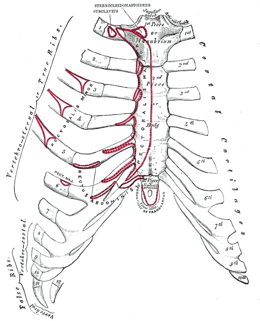
The external intercostals pass from the lower border of one rib to the upper border of the rib below and raise the ribs in inspiration; the internal and innermost intercostals, with transversus thoracis, depress the ribs and draw them in during forced expiration. The diaphragm (sternal, costal and lumbar parts inserting into the central tendon, supplied by the phrenic nerves) expands the lower rib cage and provides the main inspiratory force. Accessory inspiratory muscles include pectoralis major, pectoralis minor (raising ribs 3–5 when the scapula is fixed), serratus anterior and possibly latissimus dorsi; subclavius elevates the first rib. External oblique, rectus abdominis and quadratus lumborum depress the ribs and assist active expiration; serratus posterior superior and inferior raise or lower their ribs, but evidence does not support a respiratory role. The female rib cage is about 10% smaller but its ribs slope more steeply, so rib-cage muscles contribute more to quiet inspiration, whereas the wider lower rib cage of men is associated with a larger diaphragmatic contribution; the sexes begin to diverge in shape at about 12 years. Numerical and structural rib anomalies (cervical and lumbar ribs, bifid ribs, fusion, pseudarthrosis of the first rib) occur in 0.15–3.4% of people; supernumerary ribs make up about 30% of congenital rib anomalies, missing ribs 26% and segmentation defects and synostoses 20%.[7]
Rib fractures make up about 12% of all fractures and become commoner with age. The 2025 thoracic trauma guidelines recommend considering surgical stabilisation in every haemodynamically stable patient with flail chest, within 48–72 hours, and in patients with three or more severely displaced ipsilateral fractures of ribs 3–10 who respond poorly to ventilation or cannot be weaned; mortality is lower than with non-operative care (1.5% vs 2.7%, and 4.2% vs 10.1% with flail chest), but a 2026 randomised trial found that routine fixation did not shorten intensive-care stay and improved only pain and quality of life, supporting selective use. Slipping rib syndrome results from abnormal mobility of ribs 8–10, is diagnosed mainly with the hooking manoeuvre and dynamic ultrasound, and is typically recognised about 15 months late; vertical rib plating with bioabsorbable plates lowers recurrence from 17.1% to 3.4%. Chondrosarcoma is the commonest primary malignant bone tumour of the chest wall, and microscopically clear (R0) resection is the strongest predictor of survival. Pectus excavatum is now repaired mainly by the minimally invasive Nuss procedure, with the bar usually removed after 2–3 years, and sternal advancement improves cardiac output and exercise capacity. Anomalies of the first rib (elevation, fusion with the second) are a principal cause of thoracic outlet syndrome and can be treated by transaxillary first-rib resection.[7]
Full-text reading (StatPearls)
The StatPearls chapters on this topic, reproduced verbatim. Their CC BY-NC-ND 4.0 licence allows unmodified copies only, so they are not translated or shortened.
Anatomy, Thorax, RibsSafarini OA, Bordoni B · StatPearls 2026 · NBK538328 · 24k characters
Loading…
Safarini OA, Bordoni B. Anatomy, Thorax, Ribs. StatPearls [Internet]. Treasure Island (FL): StatPearls Publishing; 2026. NBK538328 · PMID 30855912 · CC BY-NC-ND 4.0. Reproduced verbatim, unmodified.
Anatomy, Thorax, SternumAltalib AA, Miao KH, Menezes RG · StatPearls 2023 · NBK541141 · 14k characters
Loading…
Altalib AA, Miao KH, Menezes RG. Anatomy, Thorax, Sternum. StatPearls [Internet]. Treasure Island (FL): StatPearls Publishing; 2023. NBK541141 · PMID 31082185 · CC BY-NC-ND 4.0. Reproduced verbatim, unmodified.
Organs in this system
References
Sources for the values and descriptions on this page, indexed in PubMed (US National Library of Medicine).
- [1]Monitoring diaphragm function in the ICUDres M et al., Current opinion in critical care 2020PMID 31876624
- [2]Network of breathing. Multifunctional role of the diaphragm: a reviewKocjan J et al., Advances in respiratory medicine 2017PMID 28871591
- [3]Respiratory MechanicsGertler R, Anesthesiology clinics 2021PMID 34392877
- [4]Ventilator Graphics: Scalars, Loops, & Secondary MeasuresDexter AM et al., Respiratory care 2020PMID 32457168
- [5]Ventilator-induced Lung InjuryBeitler JR et al., Clinics in chest medicine 2016PMID 27842744
- [6]Pectus carinatumRobicsek F et al., Thoracic surgery clinics 2010PMID 20974441
- [7]Anatomy, Thorax, RibsSafarini OA et al., StatPearls 2026PMID 30855912
- [8]Anatomy, Thorax, SternumAltalib AA et al., StatPearls 2023PMID 31082185
- [9]Anatomy, BonesStatPearls (NCBI Bookshelf)PMID 30725884Supports: Number of bones
- [10]Calcium metabolism in health and diseasePeacock M, 2010PMID 20089499Supports: Skeletal calcium store