Ovaries
Model
Human Reference Atlas (HuBMAP), CC BY 4.0, meshopt-compressed. Drag “Section” to look inside.
Description
Paired almond-shaped glands, about 3 × 2 × 1 cm, lying in the ovarian fossa on the lateral pelvic wall and attached to the broad ligament by the mesovarium. The cortex contains the follicles; the ovary produces oocytes and secretes oestrogens and progesterone.
Parts
Each part has its own card; press “Show on the model” to fly to it and select it in the 3D model above.
- 01
Surface epithelium and tunica albuginea
Epithelium superficiale; Tunica albuginea ovarii
An almond-shaped organ about 3 × 1.5 × 1 cm covered by a single layer of cuboidal epithelium (not true peritoneum) over a fibrous tunica albuginea. Many epithelial ovarian tumours were thought to arise here; many high-grade serous cancers are now thought to originate in the tubal fimbriae.
- 02
Cortex and follicles
Cortex ovarii; Folliculi ovarici
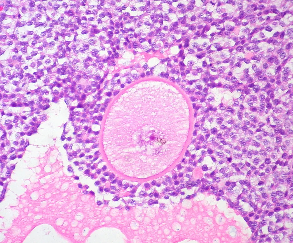
Holds follicles at every stage: primordial, primary, secondary and the dominant (Graafian, about 2 cm) follicle before ovulation, which then becomes the progesterone-secreting corpus luteum. The follicle count falls from 1–2 million at birth to about 400 000 at puberty.
- 03
Medulla and hilum
Medulla ovarii; Hilum ovarii
The central loose stroma carries coiled vessels, lymphatics and nerves that enter and leave at the hilum on the mesovarian border; hilus cells (homologous to Leydig cells) secrete androgens.
- 04
Ovarian ligaments
Ligamentum suspensorium ovarii; Ligamentum ovarii proprium; Mesovarium
The suspensory (infundibulopelvic) ligament carries the ovarian vessels down from the pelvic wall; the ligament of the ovary joins it to the uterine cornu; the mesovarium attaches it to the back of the broad ligament. A mobile ovary on a long pedicle can twist (torsion), an acute surgical emergency.
- 05
Ovarian vessels
Arteria et vena ovarica
The ovarian artery arises directly from the abdominal aorta at L2 and anastomoses with the ovarian branch of the uterine artery. The right ovarian vein drains into the inferior vena cava, the left into the left renal vein; lymph goes to para-aortic nodes.
Position and relations
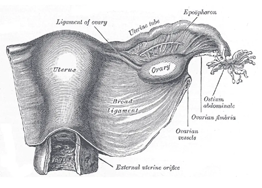
The ovary lies in the ovarian fossa on the lateral pelvic wall, bounded by the external iliac vessels above and by the ureter and internal iliac vessels behind; the obturator nerve runs just lateral, so ovarian disease may refer pain to the medial thigh. It is attached to the pelvic wall by the suspensory ligament of the ovary and to the uterus by the ovarian ligament.
Form and structure
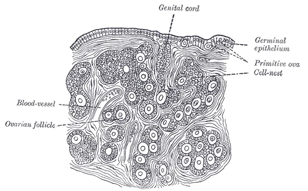
The ovary is covered by surface epithelium over a thin tunica albuginea. The cortex contains follicles at every stage (primordial, primary, secondary, antral), the corpus luteum after ovulation and the corpus albicans; the medulla contains loose connective tissue and vessels. The ovary is not covered by peritoneum, so the oocyte is released into the peritoneal cavity and captured by the fimbriated infundibulum of the tube.
Vessels and nerves
The ovarian artery arises from the abdominal aorta at about L2, runs in the suspensory ligament and anastomoses with the ovarian branch of the uterine artery. The right ovarian vein drains into the inferior vena cava and the left into the left renal vein. Lymph follows the ovarian vessels to the para-aortic nodes.
Physiology
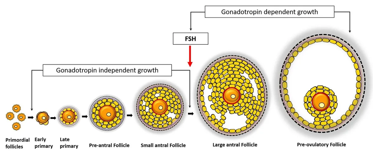
Each primordial follicle holds an oocyte arrested in prophase of meiosis I, surrounded by a flat layer of pre-granulosa cells. Most follicles stay dormant; only a selected group is activated to become secondary and then antral follicles, through two-way signalling between the oocyte and granulosa cells, much of it via TGF-β superfamily molecules.[1]
Besides follicles, the ovary contains a stroma of surface epithelium, tunica albuginea, rete ovarii, hilar cells, vessels, nerves, lymphatics and immune cells. Ovarian ageing is the progressive loss of both the number and the quality of oocytes, linked to chromosome mis-segregation, DNA damage and mitochondrial dysfunction; when early, it presents as diminished ovarian reserve or premature ovarian insufficiency.[2][3]
Relations, size, function and development
The ovary lies beside the uterine tube in the ovarian fossa, formed by the bifurcation into external and internal iliac arteries. In front is the medial umbilical ligament, behind are the ureter and internal iliac artery, above are the infundibulum of the tube and the suspensory (infundibulopelvic) ligament, and below is the uterotubal angle, reached by the proper ovarian ligament. The suspensory ligament carries the ovarian artery and vein and the sympathetic and parasympathetic plexuses; the proper ligament is a remnant of the gubernaculum and contains no vessels. The normal ovary is 2.0 cm wide, 3.5 cm long and 1.0 cm thick, about the size of a golf ball. Its volume changes with age: about 0.7 mL at 2 years, peaking at 7.7 mL at 20 and then slowly falling to about 2.8 mL at menopause. From outside in there is a germinal epithelium (simple cuboidal), a collagenous tunica albuginea, a cortex containing follicles of every size and maturity, and a medulla (hilum) of loose connective tissue with the major vessels.[4]
The ovary has two functions. The first is endocrine: from puberty, hypothalamic GnRH stimulates the anterior pituitary to release FSH and LH, forming the hypothalamic–pituitary–ovarian axis; FSH acts on granulosa cells to promote follicular growth, LH acts on theca cells to make androgens as precursors of oestradiol, and rising oestrogen produces the secondary sexual characteristics. The second is to house the oocytes, which begin developing in utero and then pause until puberty. Antral follicles average 2–9 mm and usually number fewer than 25; they grow during the cycle until a dominant follicle emerges and the others degenerate. The LH surge triggers ovulation; the corpus luteum takes the follicle’s place and secretes large amounts of progesterone with smaller amounts of inhibin A and oestradiol. If fertilisation occurs, the blastocyst secretes hCG, which maintains the corpus luteum until the placenta takes over; if not, it degenerates into the corpus albicans and progesterone withdrawal starts menstruation. Developmentally the ovary comes from intermediate mesoderm; germ cells from the endoderm of the yolk sac migrate through the hindgut to the gonadal ridge (formed by week 4), dividing on the way to become oogonia that are surrounded by cells destined to become granulosa cells. The ovary also descends, but stops in the abdomen because the gubernaculum is attached to the uterus.[4]
Vessels, nerves, variants and clinical significance
The ovary has a dual supply from the ovarian and uterine arteries. The ovarian artery arises from the abdominal aorta below the renal arteries (about L2), runs in the suspensory ligament into the mesovarium and may anastomose with the uterine artery in the broad ligament. The ovarian vein (on average 5 mm across) also drains the parametrium, cervix, mesosalpinx and pampiniform plexus and anastomoses with the para-ovarian, uterine, vesical, rectal and vulvar plexuses; the left drains into the left renal vein and the right directly into the inferior vena cava. Most lymph follows the ovarian vessels to para-aortic nodes around L2, but there are two other routes: through lateral vessels to hypogastric nodes and then to para-aortic nodes, and along the round ligament to external iliac and inguinal nodes; these are routes of metastasis for ovarian cancer. Sympathetic supply comes from the ovarian plexus (derived from the renal plexus and carried in the suspensory ligament) and from the superior ovarian nerve in the ovarian ligament; parasympathetic fibres come from the uterine (pelvic) plexus formed by the pelvic splanchnic nerves. At surgery the ureters, which lie close to the ovary and its ligaments, need particular care: clamping one by mistake can damage it and cause ipsilateral renal failure.[4]
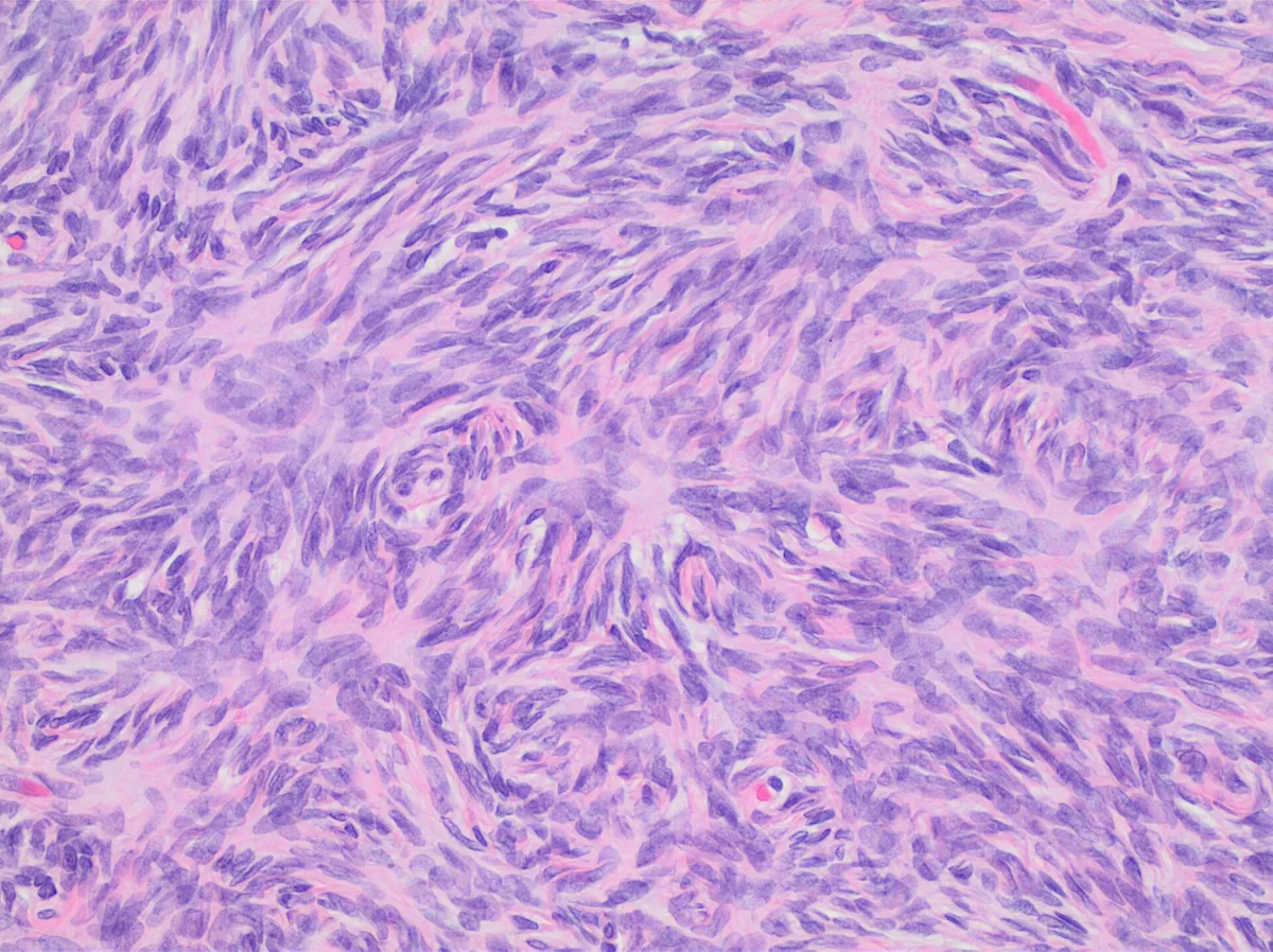
The commonest variant is the ovarian cyst: functional cysts arise naturally with the hormonal swings of the cycle, but persistent or very large cysts increase ovarian volume and the risk of torsion, and cysts of any size can cause pelvic pain or abnormal bleeding. An accessory ovary (uncommon) is a third ovary attached to the usual ligaments; a supernumerary ovary (extremely rare) is not attached to them but to the large bowel, bladder or rectum; both are usually small, found incidentally at surgery, often cystic, and should be removed because of a higher risk of neoplastic change. Some people have only one ovary. In Turner syndrome the ovaries develop normally at first but degenerate before birth into “streak” ovaries of wavy connective tissue with scattered groups of granulosa cells; instead of over two million oocytes there are very few, so most patients are infertile, although a small subset with a mosaic karyotype can conceive spontaneously. Premature ovarian failure causes infertility, menopausal symptoms and loss of oestrogen’s protection of bone and heart. Polycystic ovary syndrome presents with oligomenorrhoea, acne, hirsutism and infertility, with more than 25 follicles on high-resolution ultrasound; ovarian cancer is insidious and the third leading cause of cancer death in women.[4]
Full-text reading (StatPearls)
The StatPearls chapters on this topic, reproduced verbatim. Their CC BY-NC-ND 4.0 licence allows unmodified copies only, so they are not translated or shortened.
Anatomy, Abdomen and Pelvis, OvaryGibson E, Mahdy H · StatPearls 2023 · NBK545187 · 13k characters
Loading…
Gibson E, Mahdy H. Anatomy, Abdomen and Pelvis, Ovary. StatPearls [Internet]. Treasure Island (FL): StatPearls Publishing; 2023. NBK545187 · PMID 31424771 · CC BY-NC-ND 4.0. Reproduced verbatim, unmodified.
Anatomy, Abdomen and Pelvis, Ovary Corpus LuteumOliver R, Pillarisetty LS · StatPearls 2023 · NBK539704 · 8k characters
Loading…
Oliver R, Pillarisetty LS. Anatomy, Abdomen and Pelvis, Ovary Corpus Luteum. StatPearls [Internet]. Treasure Island (FL): StatPearls Publishing; 2023. NBK539704 · PMID 30969526 · CC BY-NC-ND 4.0. Reproduced verbatim, unmodified.
Organs in this system
References
Sources for the values and descriptions on this page, indexed in PubMed (US National Library of Medicine).
- [1]Ovarian FolliculogenesisRimon-Dahari N et al., Results and problems in cell differentiation 2016PMID 27300179
- [2]The ovarian stroma as a new frontierKinnear HM et al., Reproduction 2020PMID 32716007
- [3]Mechanisms of ovarian agingPark SU et al., Reproduction 2021PMID 33999842
- [4]Anatomy, Abdomen and Pelvis, OvaryGibson E et al., StatPearls 2023PMID 31424771