Trachea
Model
Layers
Z-Anatomy anatomical model based on BodyParts3D, licensed CC BY-SA 4.0.
Description
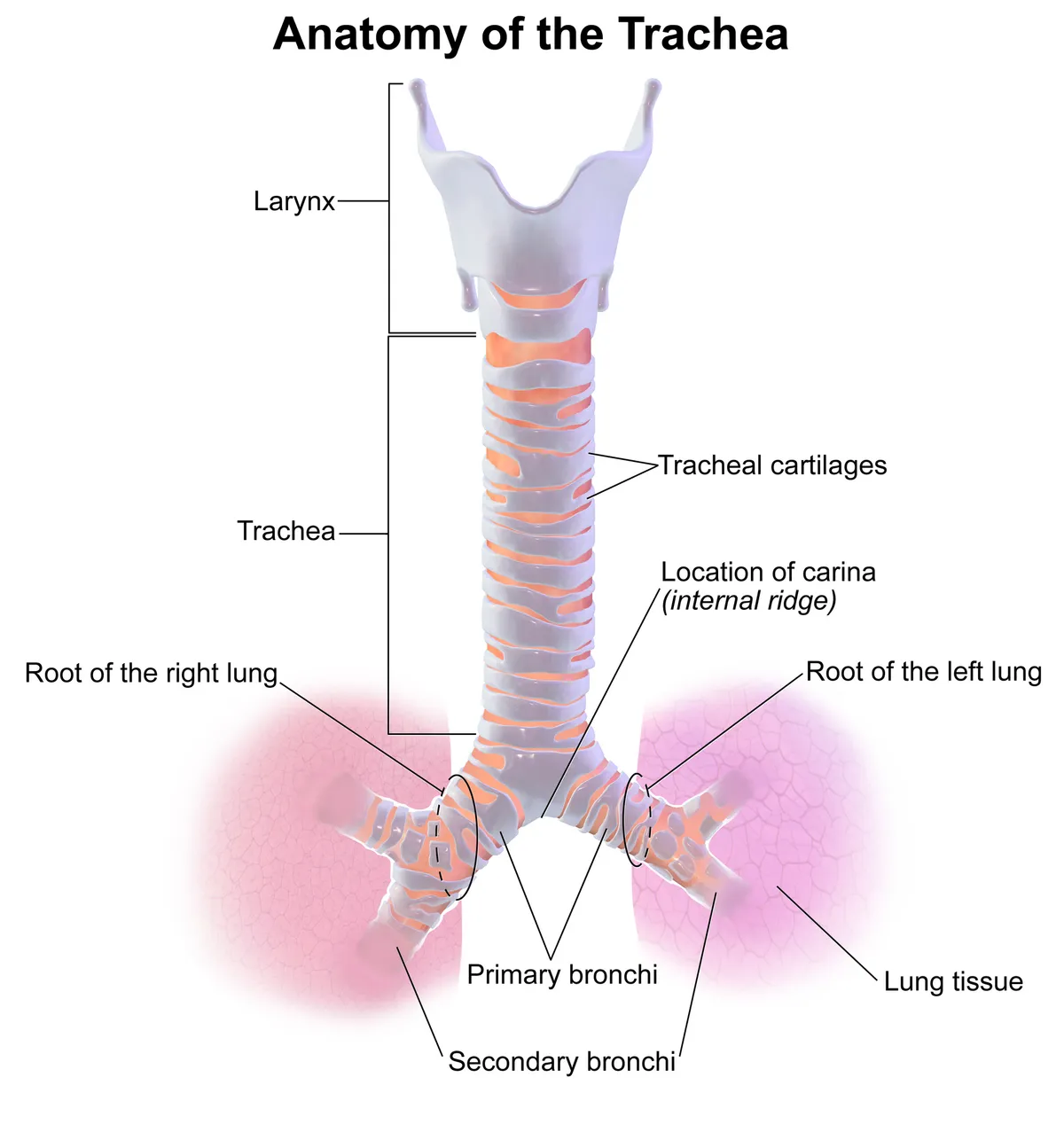
An airway about 10–12 cm long supported by 16–20 C-shaped cartilaginous rings; it bifurcates at the carina into the right and left main bronchi.
Parts
Each part has its own card; press “Show on the model” to fly to it and select it in the 3D model above.
- 01
Tracheal cartilages
Cartilagines tracheales
About 16–20 C-shaped hyaline cartilages open behind, joined by annular ligaments, keep the trachea patent. The trachea is about 10–12 cm long and 2 cm wide in adults, running from the lower border of the cricoid (C6) to its bifurcation.
- 02
Membranous wall and trachealis
Paries membranaceus; Musculus trachealis
Behind, the ends of the rings are joined by the smooth trachealis muscle and fibrous tissue, lying against the oesophagus; in coughing it contracts to narrow the lumen and speed airflow. This is where congenital tracheo-oesophageal fistulas usually occur.
- 03
Tracheal mucosa
Tunica mucosa tracheae
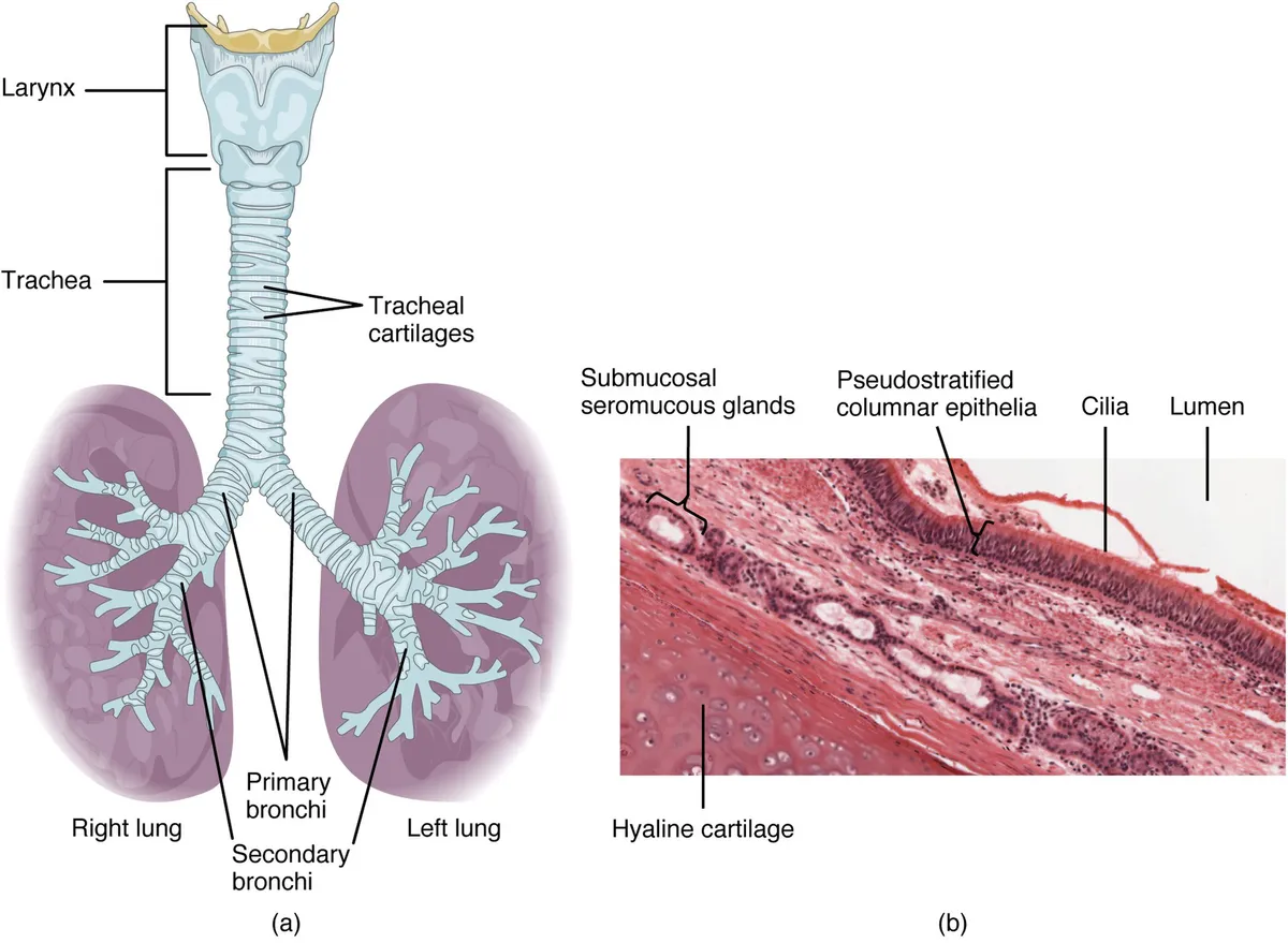
Ciliated pseudostratified columnar epithelium with goblet, basal and neuroendocrine cells, over a submucosa with seromucous glands. The mucociliary escalator carries dust and bacteria up to the pharynx; smoking destroys cilia and causes squamous metaplasia.
- 04
Cervical and thoracic parts
Pars cervicalis et thoracica tracheae
The cervical part is superficial: the thyroid isthmus crosses rings 2–4 (the tracheostomy zone), with the thyroid lobes and carotid sheaths at its sides. The thoracic part lies in the superior mediastinum, related to the aortic arch and brachiocephalic trunk in front and left, and to the oesophagus behind.
- 05
Bifurcation and carina
Bifurcatio tracheae; Carina tracheae
At the sternal angle (T4–T5) the trachea divides into the right and left main bronchi; the carina is the cartilaginous ridge between them, with mucosa highly sensitive to the cough reflex. The right main bronchus is shorter, wider and more vertical, so inhaled foreign bodies usually enter it.
Position and relations
The trachea begins at the lower border of the cricoid cartilage (C6), descends in front of the oesophagus through the neck and superior mediastinum, and divides into the two main bronchi at the sternal angle (T4–T5), where the carina lies. The right main bronchus is shorter, wider and more vertical than the left, so inhaled foreign bodies usually enter the right lung.
Form and structure
The trachea is about 10–12 cm long, held open by 16–20 C-shaped hyaline cartilage rings, open posteriorly and closed by the membranous wall containing the trachealis muscle. Its mucosa is pseudostratified ciliated columnar epithelium with goblet cells, forming the mucociliary escalator that sweeps dust and microbes up to the pharynx.[1]
Clinical correlations
Tracheobronchomalacia is excessive softness and collapse of the central airways on expiration, due to weakness of the cartilage rings or the posterior membranous wall. It is often misdiagnosed as asthma, bronchitis or chronic cough; diagnosis is usually confirmed by flexible bronchoscopy, and most cases are managed conservatively with non-invasive ventilation.[1]
Mucociliary clearance
Mucociliary clearance is the lung’s primary innate defence. Its components are the protective mucus layer, the airway surface liquid and the cilia of ciliated cells, which beat in metachronal waves to propel trapped pathogens and particles out of the airways. In primary ciliary dyskinesia the cilia are abnormal, clearance fails and chronic lung disease follows.[2]
Conducting-airway epithelium and submucosal glands secrete fluid, electrolytes, antimicrobial and anti-inflammatory proteins and mucus onto the surface; the mucociliary escalator removes material through ciliary action and cough. Mucins are released at a low baseline rate to sustain steady clearance and at a high stimulated rate from submucosal glands and goblet cells to sweep away larger particles in proximal airways. Failure at any step (secretion, cilia, cough) contributes to chronic lung diseases such as cystic fibrosis.[3][4][5]
The cervical trachea continues the larynx, the gateway between upper and lower airways; knowledge of the cartilages, muscles, mucosal spaces, neurovascular supply and lymphatics here is essential for cancer staging. Congenital laryngotracheo-oesophageal clefts, tracheobronchial stenosis and tracheobronchomalacia reflect incomplete separation of the trachea from the foregut.[6][7][1]
Full-text reading (StatPearls)
The StatPearls chapters on this topic, reproduced verbatim. Their CC BY-NC-ND 4.0 licence allows unmodified copies only, so they are not translated or shortened.
Anatomy, Head and Neck, TracheaDerricott H, Jimenez J, La Rose J · StatPearls 2026 · NBK448070 · 22k characters
Loading…
Derricott H, Jimenez J, La Rose J. Anatomy, Head and Neck, Trachea. StatPearls [Internet]. Treasure Island (FL): StatPearls Publishing; 2026. NBK448070 · PMID 28846296 · CC BY-NC-ND 4.0. Reproduced verbatim, unmodified.
Organs in this system
References
Sources for the values and descriptions on this page, indexed in PubMed (US National Library of Medicine).
- [1]TracheobronchomalaciaBiswas A et al., Disease-a-month 2017PMID 28549723
- [2]Cilia and Mucociliary ClearanceBustamante-Marin XM et al., Cold Spring Harbor perspectives in biology 2017PMID 27864314
- [3]Airway Epithelial Differentiation and Mucociliary ClearanceWhitsett JA, Annals of the American Thoracic Society 2018PMID 30431340
- [4]Airway Mucin SecretionJaramillo AM et al., Annals of the American Thoracic Society 2018PMID 30431339
- [5]Cystic fibrosisShteinberg M et al., Lancet 2021PMID 34090606
- [6]Anatomy of the Larynx and Cervical TracheaMcCullagh KL et al., Neuroimaging clinics of North America 2022PMID 36244725
- [7]Laryngotracheoesophageal cleftsStrychowsky JE et al., Seminars in pediatric surgery 2016PMID 27301597
- [8]The normal human lung: ultrastructure and morphometric estimation of diffusion capacityGehr P, Bachofen M, Weibel ER, 1978PMID 644146Supports: Alveolar surface area
- [9]Physiology, LungStatPearls (NCBI Bookshelf)PMID 31424761Supports: Respiratory rate